Articles published in Obstet Gynecol Sci are open-access, distributed under the terms of the Creative Commons Attribution Non-Commercial License (http://creativecommons.
org/licenses/by-nc/3.0/) which permits unrestricted non-commercial use, distribution, and reproduction in any medium, provided the original work is properly cited.
Copyright © 2015 Korean Society of Obstetrics and Gynecology
Case Report
Obstet Gynecol Sci 2015;58(1):65-68 http://dx.doi.org/10.5468/ogs.2015.58.1.65 pISSN 2287-8572 · eISSN 2287-8580
www.ogscience.org 65
Introduction
Epignathus or oral teratoma is an extremely rare congenital tumor, which arises in the sphenoid region of the palate or pharynx, occurring in 1 in 35,000 to 1 in 200,000 live births [1]. The tumor fills the oral cavity and hence is associated with a high mortality rate owing to severe airway obstruction, es- pecially in the neonatal period [1-3]. Usually, this tumor was detected in late second trimester or third trimester [2,3], but technical developments in three-dimensional (3D) ultrasonog- raphy and magnetic resonance imaging (MRI) have enabled early diagnosis and detailed characterization of the tumor [4-6].
Herein, we present a case of epignathus affecting 1 fetus in a twin pregnancy. The epignathus in this case was associated with multiple congenital malformation including cleft palate, bifid tongue, bifid uvula, congenital heart defect, and bilateral inguinal hernias. To the best of our knowledge, this constel- lation of congenital malformations has not been reported previously. In addition, we examined the clinical value of 3D ultrasound and MRI during antenatal counseling and the es- tablishment of peripartum management.
Case report
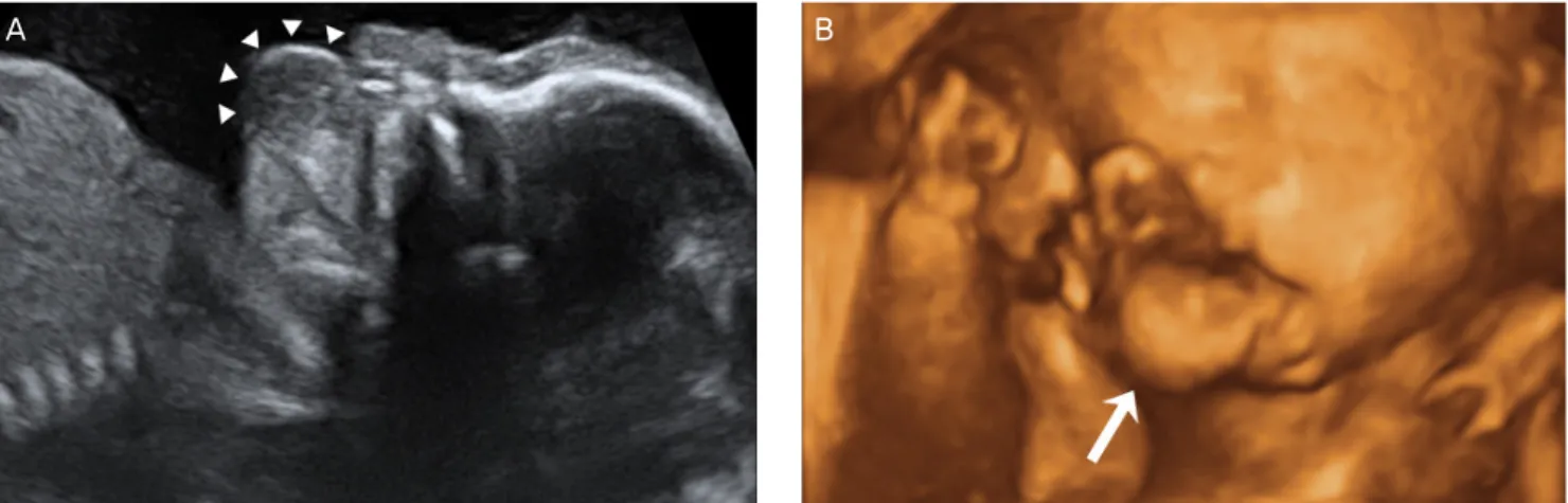
A 28-year-old woman pregnant with twins was transferred to our institution for abnormal findings on ultrasonography in one twin’s oral cavity at 20 weeks 6/7 days of gestation. In a 2D so- nogram, one fetus in the dichorionic-diamniotic twin pregnancy had a solid and cystic mass measuring 13×9 mm that originated in the mouth, extended outside the mouth, and showed no vas- cularization on color Doppler examination. Using 3D ultrasonog- raphy in surface-rendering mode, we confirmed the presence of an oral tumor, distinguishable from the lips (Fig. 1). Then,
Received: 2014.5.31. Revised: 2014.7.25. Accepted: 2014.7.31.
Corresponding author: In Yang Park
Department of Obstetrics and Gynecology, The Catholic University of Korea College of Medicine, 222 Banpo-daero, Seocho-gu, Seoul 137- 701, Korea
Tel: +82-2-2258-2813 Fax: +82-2-595-1549 E-mail: [email protected]
Prenatal diagnosis of epignathus with multiple
malformations in one fetus of a twin pregnancy using three-dimensional ultrasonography and magnetic
resonance imaging
Na Rae Moon, Jae Young Min, Yeon Hee Kim, Sae Kyung Choi, Jong Chul Shin, In Yang Park
Department of Obstetrics and Gynecology, The Catholic University of Korea College of Medicine, Seoul, Korea
Epignathus is an extremely rare type of congenital teratoma arising in the oral cavity. Although it is a benign tumor, it is associated with high mortality and morbidity rates because of severe airway obstruction and other malformations.
We present a case of epignathus affecting one fetus in a twin pregnancy. The tumor was associated with multiple congenital malformations including cleft palate, bifid tongue, bifid uvula, congenital heart defect, and bilateral inguinal hernias. The diagnostic value of three-dimensional ultrasonography and magnetic resonance imaging was explored with respect to antenatal counseling and peripartum management.
Keywords: Epignathus; Magnetic resonance imaging; Malformation; Prenatal; Three-dimensional ultrasonography
www.ogscience.org 66
Vol. 58, No. 1, 2015
using the 3D sonogram, we explained the perinatal risks related to airway obstruction caused by the fetal lesion to the mother and her family. She required further antenatal work-up for pre- cise delineation of the mass and further counseling regarding whether the mass could be resected after delivery. She refused to undergo anantenatal chromosomal test because of the risk to the other twin through amniocentesis.
To examine the relationship between the tumor and sur- rounding structures, we performed a fetal MRI. A sagittal HASTE T2-weighted MRI scan showed a hyperintense mass with a stalk projecting from the palate and upper lip, measuring 16×10 mm in size in the lower-positioned fetus. There was no evidence of central nervous system, CNS-related anomaly and
intracranial invasion of the tumor. A multidisciplinary medical team comprising an obstetrician, otolaryngologist, neonatolo- gist, and radiologist discussed the diagnosis and management of the tumor; they planned to perform elective surgery after birth rather than the ex utero intrapartum treatment (EXIT) pro- cedure, considering that endotracheal intubation of the new- born would be possible after birth according to the fetal MRI scan.
The size of the lesion had increased to 22×17 mm at 32 weeks of gestation. Swallowing difficulty and polyhydramnios were not noted. At 33 weeks 5/7 days of gestation, an emer- gency cesarean section was performed because of premature rupture of the amniotic membrane. The male neonate weigh- Fig. 1. Antenatal image of the tumor. A round mass (arrow heads) protruding from the oral cavity was found on two-dimensional ultrasonography (A), and three- dimensional image (B).
A B
Fig. 2. Sagittal HASTE T2-weighted magnetic resonance image at 21 weeks 6/7 days of gestation showing a 16×10 mm hyperintense mass (arrows) with a stalk (ar- rowhead) projecting from the palate and upper lip (UL) (A). A pedunculated tumor originated from the mouth floor in the newborn just after delivery (B). T, tongue; LL, lower lip.
A B
www.ogscience.org 67 Na Rae Moon, et al. Prenatal imaging in epignathus
ing 1,745 g with the oral mass was delivered first; his Apgar scores were 5 at 1 minute and 6 at 5 minutes. The neonate was immediately endotracheally intubated. The oral mass was whitish, soft, and movable, and measured approximately 20×25 mm (Fig. 2A). It appeared to originate from the hard palate and a fibrous band was observed between the mass and the tongue base. The mass was accompanied by complete cleft palate, bifid uvula, and bifid tongue. Bilateral inguinal hernias were also found on physical examination. Moreover, a small ventricular septal defect and patent ductus arterio- sus were observed on echocardiography. The chromosome analysis of the affected neonate showed normal karyotype (46, XY). The second baby weighing 2,090 g had no gross anomaly. At 10 days after birth, the tumor was excised and the tongue was reconstructed (Fig. 2B). The resected tissue measured 40×23 mm and microscopic examination showed the presence of some hair follicles with mature adipocytes and keratinized squamous epithelium, consistent with mature tera- toma. A bilateral inguinal herniotomy was performed on day 73 at the age of 13 months; the baby’s condition was good.
Discussion
Epignathus is known to be associated with other malforma- tions in 6% of all epignathus cases [3,7]. Cleft palate is the most common malformation, as the tumor prevents closure of the palate [7]. Other associated malformations are bifid tongue or nose, glossoptosis, diaphragmatic hernia, nasopharyngeal teratoma, and an inguinal hernia [7,8]. Bifidity and glossoptosis indicate impaired fusion of the primitive tongue buds and an- terior positioning of the tongue because of very early develop- ment of the tumor. Moreover, functional disorders secondary to glossoptosis can also hinder the growth of the jaw, resulting in mandibular microretrognathia. The case presented here was that of epignathus accompanied by multiple malformations, a cleft palate, bifid tongue, bifid uvula, congenital heart defects, and bilateral inguinal hernias, without chromosomal defect.
In some studies, epignathi have been found to be associated with chromosomal abnormalities, early embryonic defects, or fetal syndrome [7,9,10]; however, to the best of our knowl- edge, no study in the literature has reported recurrent fetal epignathus.
Epignathus can present in a variety of ways. High levels of maternal serum α-fetoprotein indicate the presence of a tumor that can be confirmed by ultrasonography [10]. Epignathus
could also be identified by 2D ultrasonography, as seen in this case. The antenatal finding of epignathus is an anterior or bidi- rectional organoid facial mass, which is partially solid and cystic [4]. Polyhydramnios occurs in approximately 30% of the cases, secondary to obstruction of the mouth of the fetus and swal- lowing difficulty because of the local mass [7,11,12]. In the case presented here, polyhydramnios was not found, probably because of the relatively small size of the intraoral stalk part of the tumor, and partially accompanying cleft palate.
3D ultrasonography could facilitate prenatal diagnosis and help in planning delivery, through the various 3D software programs such as the surface-rendering mode, the “multislice”
view, the “reverse face” view, and tomographic mode [4-6].
Reconstructed images from the 3D volume data can provide information about the size, location, composition, and exten- sion of the tumor. The resolution is limited, however, when examining the intraoral mass [6]. In the case discussed here, we used 3D ultrasonography in the surface-rendering mode, and the resultant sonograms were used to explain to the pa- tient what fetal lesions are, but they were not very helpful in revealing any anatomical relationship of the lesion with the surrounding tissues because of the interference of the other twin and perioral structures.
Fetal MRI, a complementary diagnostic tool for epignathus [13], might be especially useful in identifying the need for an EXIT procedure to be performed on a fetus. MRI is helpful in ensuring airway patency by establishing a relationship be- tween the mass and airway structures. In addition, this modal- ity is useful in the assessment of the intracranial invasion that contributes to the poor prognosis of the patients. In this case, we were able to view an enhanced characterization of the tu- mor and ensure airway patency of the fetus through the MRI scans, which informed peripartum management.
If tracheal obstruction is prenatally suspected, physicians should consider a cesarean section and subsequent intubation by tracheostomy before the umbilical cord is clamped, as pre- scribed in the EXIT procedure, or resection of the tumor mass while the newborn is on placental support [7]. Immediate in- tubation and oxygenation, however, might fail because of the huge size of the tumor, and surgical removal is difficult owing to modification of anatomical landmarks [3,14]. Some authors have suggested that a short EXIT procedure limited to securing the airway of the fetus followed by early surgery is preferable to confirmative resection under placental support [15].
Prognosis depends on the size and location of the tumor, the rate of growth, associated polyhydramnios, and the de-
www.ogscience.org 68
Vol. 58, No. 1, 2015
gree of intracranial spread [7]. Malignant degeneration rarely occurs in cases of epignathus teratoma. Polyhydramnios, commonly caused by swallowing difficulties, is an important factor for prognosis because it indicates the severity of airway obstruction and the extent of tumor invasion; polyhydramnios is also associated with an increased risk of preterm labor, pre- term premature rupture of membranes and further pulmonary hypoplasia, and respiratory distress in the neonate.
In conclusion, this report described the case of a single twin with epignathus associated with inguinal hernia and other malformations. According to the studies in the related literature, although the images reconstructed from the 3D ul- trasonography data are valuable in the diagnosis of tumors of the head and neck, the usefulness of these data were limited in the case herein because of low resolution. Fetal MRI helped determine if special management of the tumor was necessary for securing the airway.
Conflict of interest
No potential conflict of interest relevant to this article was reported.
References
1. Gull I, Wolman I, Har-Toov J, Amster R, Schreiber L, Less- ing JB, et al. Antenatal sonographic diagnosis of epigna- thus at 15 weeks of pregnancy. Ultrasound Obstet Gyne- col 1999;13:271-3.
2. Levine AB, Alvarez M, Wedgwood J, Berkowitz RL, Holz- man I. Contemporary management of a potentially lethal fetal anomaly: a successful perinatal approach to epigna- thus. Obstet Gynecol 1990;76:962-6.
3. Tonni G, Centini G, Inaudi P, Rosignoli L, Ginanneschi C, De Felice C. Prenatal diagnosis of severe epignathus in a twin: case report and review of the literature. Cleft Palate Craniofac J 2010;47:421-5.
4. Takagi MM, Bussamra LC, Araujo Junior E, Drummond CL, Herbst SR, Nardozza LM, et al. Prenatal diagnosis of a large epignathus teratoma using two-dimensional and three-dimensional ultrasound: correlation with pathologi-
cal findings. Cleft Palate Craniofac J 2014;51:350-3.
5. Dar P, Rosenthal J, Factor S, Dubiosso R, Murthy AS. First- trimester diagnosis of fetal epignathus with 2- and 3-di- mensional sonography. J Ultrasound Med 2009;28:1743-6.
6. Allen LM. Prenatal 3-dimensional imaging techniques in the sonographic evaluation of an oral mass: comparison with postnatal imaging modalities. J Ultrasound Med 2011;30:561-8.
7. Tonni G, De Felice C, Centini G, Ginanneschi C. Cervi- cal and oral teratoma in the fetus: a systematic review of etiology, pathology, diagnosis, treatment and prognosis.
Arch Gynecol Obstet 2010;282:355-61.
8. Pasupathy M, Narayanan PV, Mani V, Adenwalla HS. A case report of nasopharyngeal teratoma with a cleft pal- ate and an inguinal hernia. J Plast Reconstr Aesthet Surg 2011;64:1525-7.
9. Witters I, Moerman P, Louwagie D, Van Assche FA, Mi- geon BR, Fryns JP. Second trimester prenatal diagnosis of epignathus teratoma in ring X chromosome mo- saicism with inactive ring X chromosome. Ann Genet 2001;44:179-82.
10. Yapar EG, Ekici E, Gokmen O. Sonographic diagnosis of epignathus (oral teratoma), prosencephaly, meromelia and oligohydramnios in a fetus with trisomy 13. Clin Dysmor- phol 1995;4:266-71.
11. Smart PJ, Schwarz C, Kelsey A. Ultrasonographic and bio- chemical abnormalities associated with the prenatal diag- nosis of epignathus. Prenat Diagn 1990;10:327-32.
12. Ekici E, Soysal M, Kara S, Dogan M, Gokmen O. Prenatal diagnosis of epignathus causing acute polyhydramnios.
Acta Obstet Gynecol Scand 1996;75:498-501.
13. Abendstein B, Auer A, Pumpel R, Mark E, Desch B, Tscharf J. Epignathus: prenatal diagnosis by sonography and mag- netic resonance imaging. Ultraschall Med 1999;20:207-11.
14. Sichel JY, Eliashar R, Yatsiv I, Moshe Gomori J, Nadjari M, Springer C, et al. A multidisciplinary team approach for management of a giant congenital cervical teratoma. Int J Pediatr Otorhinolaryngol 2002;65:241-7.
15. Noah MM, Norton ME, Sandberg P, Esakoff T, Farrell J, Al- banese CT. Short-term maternal outcomes that are associ- ated with the EXIT procedure, as compared with cesarean delivery. Am J Obstet Gynecol 2002;186:773-7.