Original Articles Korean Circulation J 1999;;;;29((((9))))::::913-918
승모판 혈류의 E 파와 폐정맥혈류의 D파의 최대속도 간의 시간 간격과 승모판 도플러 지수들과의 관계
가톨릭대학교 의과대학 내과학교실
전두수·이만영·박지원·김용주·임효영·강동헌 이길환·김종진·채장성·김재형·홍순조·최규보
Correlation of the Time Interval from the Peak of Mitral E Wave to the Peak of Pulmonary Venous D Wave with Mitral Doppler Indexes
Doo-Soo Jeon, MD, Man-Young Lee, MD, Ji-Won Park, MD, Yong-Ju Kim, MD, Hyou-Young Rhim, MD, Dong-Hun Kang, MD, Gil-Hwan Lee, MD, Jong-Jin Kim, MD, Jang-Seong Chae, MD, Jae-Hyung Kim, MD, Soon-Jo Hong, MD and Kyu-Bo Choi, MD Department of Internal Medicine, Catholic University Medical College, Seoul, Korea
ABSTRACT
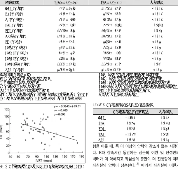
Background:Pulmonary venous diastolic flow follows the pattern of mitral flow and is dependent on the pre- ssure difference between the pulmonary vein and the left atrium (LA). The magnitude of the decrease in LA pressure in early diastole depends on both the volume of the blood leaving the LA and the stiffness of the left ventricle (LV) and the LA. Relaxation process is known to govern early diastolic compliance. We hypothesized that in patients with decreased early diastolic compliance due to LV relaxation abnormality, there may be rapid rise in LV and LA pressure, resulting in early peak of pulmonary venous D wave as early LV diastolic filling progress. This study was undertaken to define this hypothesis and to examine the relation of the time interval between E wave peak and D wave peak to mitral doppler indexes. Method:Patients with significant mitral or aortic valvular disease, or patients with LV ejection fraction below 60%, or patients who have pseudonormal or restrictive LV filling pattern on mitral and pulmonary venous Doppler, were excluded from this study. Mitral Doppler indexes including peak E velocity, peak A velocity, E wave acceleration time (EAT) and deceleration time (EDT) were measured. E/A ratio was calculated. The isovolumic relaxation time from aortic valve closure (Ac) to the onset of E wave , the time interval from Ac to the peak of E wave (AcE), the time interval from Ac to the peak of D wave, and the diastolic time from Ac to R of electrocardiogram (AcR) were measured by the pulsed wave Doppler and phonocardiography. The time interval from the peak of E wave to the peak of D wave (ED) was calculated by the subtraction of AcE from AcD. Results:1) ED is significantly shorter in patients with E/A<1 than those with E/A≥1 (58.9±27.4 msec versus 74.7±17.2 msec, p<0.05). 2) ED correlated with IVRT (r=-0.400, p<0.01), AcR (r=0.414, p<0.01), but not with E/A ratio, EDT, or EAT. 3) Multivariate lin- ear regression analysis with all the previously mentioned variables showed that IVRT, AcR, and EAT were independent determinants of the ED. Conclusion:This study demonstrates that the ED is shortened in patients
논문접수일:1999년 5월 28일심사완료일:1999년 9월 1일
교신저자:전두수, 403-720 인천광역시 부평구 부평6동 665번지 가톨릭대학교 의과대학 내과학교실 전화:(032) 510-5500・전송:(032) 510-5683
E-mail:[email protected]