• 검색 결과가 없습니다.

Apoptotic Effects of A Cisplatin and Eugenol Co-treatment of G361 Human Melanoma Cells

N/A
N/A
Protected

Academic year: 2021

Share "Apoptotic Effects of A Cisplatin and Eugenol Co-treatment of G361 Human Melanoma Cells"

Copied!
8
0
0

로드 중.... (전체 텍스트 보기)

전체 글

(1)

Copyright ⓒ 2011, The Korean Academy of Oral Biology

155

Journal of Oral Biology

Apoptotic Effects of A Cisplatin and Eugenol Co-treatment of G361 Human Melanoma Cells

Jun-Young Park, Jae-Beom Jo, In-Ryoung Kim, Gyoo-Cheon Kim, Hyun-Ho Kwak, and Bong-Soo Park*

Department of Oral Anatomy, School of Dentistry, Pusan National University (received July 19, 2011 ; revised August 26, 2011 ; accepted September 2, 2011)

Eugenol (4-allyl-2-methoxyphenol) is a naturally occurring phenolic compound that is widely used in dentistry as a com- ponent of zinc oxide eugenol cement that is commonly applied to the mouth environment. Cisplatin is one of the most potent known anticancer agents and shows significant clinical activity against a variety of solid tumors. This study was undertaken to investigate the synergistic apoptotic effects of co-treatments with eugenol and cisplatin on human melanoma (G361) cells.

To investigate whether this co-treatment efficiently reduces the viability of G361 cells compared with each single treatment, an MTT assay was conducted. The induction and augmentation of apoptosis were confirmed by DNA electrophoresis, Hoechst staining and an analysis of DNA hypoploidy. Western blot analysis and immunofluorescent staining were also performed to evaluate the expression levels and the translocation of apoptosis-related proteins following this co-treatment. Fur- thermore, proteasome activity and mitochondrial membrane potential (MMP) changes were also assayed. The results indicated that a co-treatment with eugenol and cisplatin induced multiple pathways and processes associated with an apoptotic response in G361 cells including nuclear conden- sation, DNA fragmentation, a reduction in MMP and proteasome activity, the increase and decrease of Bax and Bcl-2, a decreased DNA content, the release of cytochrome c into the cytosol, the translocation of AIF and DFF40 (CAD) into the nucleus, and the activation of caspase-9, caspase-7, caspase-3, PARP and DFF45 (ICAD). In contrast, separate treatments of 300 µM eugenol or 3 µM cisplatin for 24 h did not induce apoptosis. Our present data thus suggest that a combination therapy of eugenol and cisplatin is a potential treatment strategy for human melanoma.

Key words: eugenol, cisplatin, apoptosis, G361 human melanoma cell line

Introduction

Cells undergoing apoptosis usually develop characteristic changes, including nuclear condensation and degradation of DNA into oligonucleosomal fragments (Williams, 1991).

Apoptotic cell death is thought to result ultimately from the proteolytic actions of caspase (Yuan, 1996) and alterations in mitochondrial function play a key role in the regulation of apoptosis (Susin et al., 1999). Moreover, the proteasome system has been shown to be implicated as a negative or positive mediator of apoptosis. The proteasome pathway is known to work mostly in upstream of mitochondrial alterations and caspase activation (Orlowski, 1999).

Malignant melanoma is such one of the most important cutaneous malignancy that it has been responsible for the majority of mortality from skin disease. Numbers of patients suffering from malignant melanoma have increased in recent years. Furthermore, malignant melanoma cells have been reported to be highly resistant to chemotherapeutic agents (Shibuya et al., 2003).

Cisplatin is one of the most potent anticancer agents showing significant clinical activity against a variety of solid tumors belonging to a class of platinum containing anti cancer compounds (Wang et al., 2004). It is a representative of anticancer drug used to treat certain types of head and neck cancer, cervical carcinoma, lung cancer, neurologic cancers, and a wide variety of other cancers. However, its resistance remains a significant barrier to the survival of cancer patient.

Eugenol (4-allyl-2-methoxyphenol) is a natural phenolic substance that is the main component of clove oil and it is widely used not only in dentistry as a componet of zinc oxide eugenol cement and is applied to the mouth environment, but

*Corresponding author: Bong-Soo Park, Department of Oral

Anatomy, School of Dentistry, Pusan National University, Yangsan,

626-870, Korea. Tel: +82-51-510-8242, Fax: +82-51-510-8241

E-mail :[email protected]

(2)

also as a fragrant and flavoring agent in various cosmetics and food products (Markowitz et al., 1992). Eugenol-related com- pounds not only act as antioxidant and also as prooxindant, which may be involved in the cytotoxicity induction. They enhanced the generation of tissue damaging free radicals (Suzuki et al., 1985; Wright et al., 1995; Satoh et al., 1998).

Recent studies have demonstrated that the co-treatment of an antitumor agent with an anti-cancer natural product can be one of the potential therapeutic strategies reducing the extent and severity of cancer treatment-related toxicity (Adhami et al., 2007; Mai et al., 2007; Song et al., 2007; Vinall et al., 2007;

Lee et al., 2008). In spite that usefulness of cancer therapy combining already known chemotherapeutic reagent and anti- cancer natural product can be expected, there is few studies on evaluating the significance of co-treatment of eugenol and cisplatin on human melanoma cell line is still scarce. Here we have investigated the synergistic apoptotic effect of co-treat- ment with one of natural phenolic compound, eugenol, and an anticancer drug, cisplatin, on human melanoma (G361) cell line.

Materials and Methods

Reagents

The following reagents were obtained commercially: Mouse monoclonal anti-human caspase-3, caspase-7, caspase-9, poly (ADP-ribose) polymerase (PARP), cytochrome c, apoptosis- inducing factor (AIF) antibodies, and rabbit polyclonal anti- human DFF40 (CAD), DFF45 (ICAD), β-actin antibodies, and FITC-conjugated goat anti-mouse and anti-rabbit IgGs were from Santa Cruz Biotechnology (Santa Cruz, CA, USA);

HRP-conjugated sheep anti-mouse and anti-rabbit IgGs were from Amersham GE Healthcare (Little Chalfront, UK). 5,5',- 6,6'-tetrachloro-1,1',3,3'-tetraethylbenzimidazol carbocyanine iodide (JC-1) was from Molecular Probes (Eugene, OR, USA).

ApoScreenTM Annexin V-FITC Apoptosis Kit was from Beckman coulter (Fullerton, CA, USA). Suc-LLVY-AMC was from Calbiochem (Darmstadt, Germany). Dulbecco's modi- fied Eagle's medium (DMEM) and FBS were from Gibco (Gaithersburg, MD, USA). Dimethyl sulfoxide (DMSO), eugenol, cisplatin, Hoechst 33342, RNase A, proteinase K, aprotinin, leupeptin, PMSF, thiazolyl blue tetrazolium bromide and propidium iodide (PI) were from Sigma (St. Louis, MO, USA); SuperSignal West Pico enhanced chemiluminescence Western blotting detection reagent was from Pierce (Rockford, IL, USA).

Cell culture

The G361 human melanoma cell line was purchased from ATCC (Rockville, MD, USA). Cells were maintained at 37

o

C with 5% CO

2

in air atmosphere in RPMI1640 with 4 mM L- glutamine, 1.5 g/l sodium bicarbonate, 4.5 g/l glucose and 1.0 mM sodium pyruvate supplemented with 10% FBS.

MTT assay

G361 cells were placed in a 96-well plate and were incubated for 24 h. The cells were treated with 300 µM of eugenol, 3 µg/

ml of cisplatin or co-treated with eugenol and cisplatin for 24 h.

Then cells were treated with 500 µg/ml of thiazolyl blue tetrazolium bromide (MTT solution), and incubated at 37

o

C with 5% CO

2

for 4 h. After aspirating the medium, formed formazan crystals were dissolved in DMSO. Cell viability was measured by an ELISA reader (Tecan, Männedorf, Switzer- land) at 570 nm excitatory emission wavelength. The concen- tration of DMSO used in this study had no effect on G361 cell proliferation in a preliminary study.

Hoechst staining

Cells were centrifuged onto a clean, fat-free glass slide with a cytocentrifuge. The samples were stained in 4 µg/ml Hoechst 33342 for 30 min at 37

o

C and fixed for 10 min with 4%

paraformaldehyde.

DNA electrophoresis

2 × 10

6

cells were resuspended in 1.5 mL of lysis buffer [10 mM Tris (pH 7.5), 10 mM EDTA (pH 8.0), 10 mM NaCl and 0.5% SDS] into which proteinase K (200 µg/ml) was added. After samples were incubated overnight at 48

o

C, 200 µl of ice cold 5 M NaCl was added and the supernatant containing fragmented DNA was collected after centrifugation. The DNA was then precipitated overnight at −20

o

C in 50% isopropanol and Rnase A-treated for 1 h at 37

o

C. The DNA from 10

6

cells (15 µl) was equally loaded on each lane of 2% agarose gels in Tris-acetic acid/EDTA buffer containing 0.5 µg/ml ethidium bromide at 50 mA for 1.5 h.

Proteasome activity

Cells were collected and lysed in proteasome buffer [10 mM Tris-HCl, pH 7.5, 1 mM EDTA, 2 mM ATP, 20% glycerol, and 4 mM dithiothreitol (DTT)], sonicated, and then centri- fuged at 13,000 g at 4

o

C for 10 min. The supernatant (20 µg of protein) was incubated with proteasome activity buffer [0.05 M Tris-HCl, pH 8.0, 0.5 mM EDTA, 50 µM Suc-LLVY- AMC] for 1 h at 37

o

C. The fluorescence intensity of each solution was measured by a modular fluorimetric system (Spex Edison, NJ, USA) at 380 nm excitation and 460 nm emission wavelengths. All readings were standardized based on the fluo- rescence intensity of an equal volume of free AMC solution (50 µM).

Western blot analysis

Cells (2 × 10

6

) were washed twice in ice-cold PBS, re-

suspended in 200 µl ice-cold solubilizing buffer [300 mM

NaCl, 50 mM Tris-Cl (pH 7.6), 0.5% Triton X-100, 2 mM

PMSF, 2 µl/ml aprotinin and 2 µl/ml leupeptin] and incubated

at 4

o

C for 30 min. The lysates were centrifuged at 14,000 rpm

for 15 min at 4

o

C. Protein concentrations of cell lysates were

(3)

determined with Bradford protein assay (Bio-Rad, Richmond, CA, USA) and 50 µg of proteins were loaded onto 7.5-15%

SDS/PAGE. The gels were transferred to Nitrocellulose mem- brane (Amersham GE Healthcare) and reacted with primary antibody. Immunocomplexes were detected using SuperSignal West Pico enhanced chemiluminescence substrate and imaged with Alpha Imager HP (Alpha Innotech, Santa Clara, USA).

Assay of mitochondrial membrane potential (MMP) JC-1 was added directly to the cell culture medium (1 µM final concentration) and incubated for 15 min. The medium was then replaced with PBS. Flow cytometry to measure MMP was performed on a CYTOMICS FC500 flow cytometry (Beckman Coulter, Brea, CA, USA). Data were acquired and analyzed using CXP software version 2.2.

Immunofluorescent staining

Cells were cytocentrifuged and fixed for 10 min in 4%

paraformaldehyde, incubated with each primary antibody for 1 h, washed 3 times for 5 min, and then incubated with FITC- conjugated secondary antibody for 1 h at room temperature.

Cells were mounted with PBS. Fluorescent images were observed and analyzed under Zeiss LSM 510 laser-scanning confocal microscope (Göettingen, Germany).

Quantification of DNA hypoploidy and cell cycle phase by flow cytometry

After treatment for 24 h, cells were harvested by trypsiniz- ation and ice cold 95% ethanol with 0.5% Tween 20 was added to the cell suspensions to a final concentration of 70%

ethanol. Fixed cells were pelleted, and washed in 1% BSA- PBS solution. Cells were resuspended in 1 ml PBS containing 20 µg/ml RNase A, incubated at 4

o

C for 30 min, washed once with BSA-PBS, and resuspended in PI solution (10 µg/ml).

After cells were incubated at 4

o

C for 5 min in the dark, DNA content were measured on a CYTOMICS FC500 flow cytom- etry system (Beckman Coulter, FL, CA, USA) and data was analyzed using the Multicycle software which allowed a simul- taneous estimation of cell-cycle parameters and apoptosis.

Annexin V-FITC/PI assay

After treatment for 24 h, cells were collected and washed twice in ice-cold PBS and resuspended in 300 µl of binding buffer at 2 × 10

5

cells/ml. The samples were incubated with 5 µl of Annexin V-FITC and 5 µl propidium iodide in the dark for 15 min at room temperature. Finally, samples were analyzed by flow cytometry and evaluated based on the percentage of cells for Annexin V positive.

Statistical analysis

Three independent experiments were performed for each experimental group and each experiment was performed in triplicate. The results of the experimental and control groups were compared for statistical significance (p < 0.001, 0.01 and 0.05) using paired t-test statistical method by SPSS for Win

12.0 for summary data.

Results

Co-treatment with cisplatin and eugenol significantly reduced the viability of G361 cells

To investigate reduced viability of G361 cells by the co-treat- ment with cisplatin and eugenol, MTT assay was conducted.

Single treatment with either cisplatin of 3 µg/ml or eugenol of 300 µM for 24 h slightly reduced the viability of G361 cells (cisplatin, 92.8%; eugenol, 91.0%). In contrast, co-treatment with cisplatin and eugenol remarkably reduced the cell viability to 28.8% compared to the treatment with single reagent (Fig. 1).

Morphological and biochemical changes in G361 cells after co-treatment with cisplatin and eugenol

To explore whether the nuclear condensation and fragmen- tation were induced by the co-treatment, Hoechst staining, a hallmark of apoptosis, was conducted. Hoechst staining showed slight nuclear condensation by single treatment with either cisplatin or eugenol compared to the control group.

However, co-treatment with cisplatin and eugenol showed a variety of condensed and fragmented nuclei (Fig. 2A & 2B).

To investigate that DNA fragmentation was induced by the co-treatment, DNA electrophoresis was conducted. DNA elec- trophoresis did not show a ladder pattern in DNA fragments by the single treatment with either cisplatin or eugenol whereas a ladder pattern in DNA fragments was observed in cells with co-treatment (Fig. 2C).

The co-treatment with cisplatin and eugenol induced the degradation of caspase-3, caspase-7 and PARP, and produced the processed caspase-7 20 kDa, PARP 85 kDa, and DFF45 (ICAD) 30 kDa and 11 kDa cleaved products though cleavage of caspase-9 was insignificant (Fig. 3A & Fig. 3B). The

Fig. 1. Co-treatment with cisplatin and eugenol significantly reduced

cell viability in G361 cells. Cell viability was determined by MTT

assay (Cis + Eu, p < 0.001). Three independent assays were per-

formed. Values are means ± SD of triplicates of each experiment

(Cis, cells treated with 3 µg/ml cisplatin for 24 h; Eu, cells treated

with 300 µM eugenol for 24 h; Cis + Eu, cells treated with 3 µg/ml

cisplatin plus 300 µM eugenol for 24 h).

(4)

confocal microscopy showed that DFF40 (CAD) was located in cytosol by the single treatment with either cisplatin or eugenol whereas DFF40 (CAD) was evidently translocated into the nuclei by the co-treatment (Fig. 3C).

Proteasome activity in G361 cells after co-treatment with cisplatin and eugenol

Though single treatment with cisplatin or eugenol reduced proteasome activity compared to the control group, the dif- ference was not so significant. The co-treatment with cisplatin and eugenol remarkably reduced proteasome activity com- pared to the single treatment (Fig 3D).

Mitochondrial events were closely associated with apoptosis by co-treatment with cisplatin and eugenol of G361 cells

Through the co-treatment, the expression level of anti- apoptotic protein, Bcl-2 slightly decreased and that of apoptotic protein, Bax significantly increased (Fig. 4A). Single treatment with either cisplatin or eugenol reduced slightly MMP com- pared to control group. The co-treatment with cisplatin and

eugenol showed a loss of MMP vs the single treatment (Fig.

4B). The confocal microscopy showed that AIF was located at mitochondria by the single treatment with either cisplatin or eugenol whereas AIF was evidently translocated onto nuclei by the co-treatment (Fig. 4C). Interestingly cytochrome c was located at mitochondria by the single treatment with either cisplatin or eugenol whereas cytochrome c was evidently released into the cytosol by the co-treatment (Fig 4D).

Augmentation of apoptosis by co-treatment with cisplatin and eugenol was demonstrated by the decrease in DNA content of G361 cells

The flow cytometry showed that co-treatment with cisplatin and eugenol remarkably increased apoptotic cells with DNA hypoploidy compared to the single treatment (Fig. 5).

Fig. 2. Co-treatment with cisplatin and eugenol showed numerous condensed and fragmented nuclei in G361 cells compared to the single treatment. (A) Immunofluorescent micrographs showing nuclear morphology after Hoechst staining. Scale bar, 10 µm. (B) The values below micrographs are the mean ± SD of the means of apoptotic cells as determined by Hoechst staining. The results pre- sented are representative of three independent experiments (Cis + Eu, p < 0.001). (C) Co-treatment with cisplatin and eugenol effi- ciently showed DNA fragmentation in G361 cells. DNA fragmen- tation analysis was determined by the agarose gel electrophoresis.

Whereas single reagent treated cells showed no DNA fragmenta- tion, co-treated cells exhibited DNA degradation characteristic of apoptosis with a ladder pattern of DNA fragments (Cis, cells treated with 3 µg/ml cisplatin for 24 h; Eu, cells treated with 300 µM eugenol for 24 h; Cis + Eu, cells treated with 3 µg/ml cis- platin plus 300 µM eugenol for 24 h).

Fig. 3. Western blot analyses (A and B) of caspase-9, caspase-3, caspase-7, PARP and DFF45, confocal microscopy (C), and protea- some activity assay in G361 cells co-treated of cisplatin and eugenol (D). (A) The co-treatment of cisplatin and eugenol in G361 cells remarkably induced caspase-3, caspase-7 and PARP degrada- tions and produced the processed caspase-3 17 kDa, caspase-7 20 kDa, and PARP 85 kDa cleaved products. β-actin, a loading control. (B) The co-treatment remarkably induced DFF45 (ICAD) degradation and produced the processed DFF45 30 kDa and 11 kDa cleaved product. β-actin, a loading control. (C) The confocal microscopy showed that DFF40 (CAD) was translocated onto the nuclei in the co-treatment of cisplatin and eugenol. Scale bar, 10 µm. (D) Co-treatment with cisplatin and eugenol showed signif- icantly the reduction of proteasome activity in G361 cells compared to the single treatment (Cis + Eu, p < 0.001). Three independent assays were performed. Values are means ± SD of triplicates of each experiment (Cis, cells treated with 3 µg/ml cisplatin for 24 h;

Eu, cells treated with 300 µM eugenol for 24 h; Cis + Eu, cells

treated with 3 µg/ml cisplatin plus 300 µM eugenol for 24 h).

(5)

Augmentation of late stage apoptosis by co-treatment with cisplatin and eugenol was demonstrated by Annexin V-FITC/PI assay in G361 cells

Annexin V-FITC/PI assay was performed to quantitate apoptotic cells induced by the reagents. As shown in Fig. 6, the co-treatment with cisplatin and eugenol remarkably increased the percentage of late stage apopotic cells compared to the single treatment (Table 1/Fig. 6).

Discussion

Using natural products as herbal remedies attracts a growing public interest. Moreover, as the pharmacological mechanism of herbal compound become known, herbal medicine is more Fig. 4. Demonstration of mitochondrial events in G361 cells after

co-treatment with cisplatin. (A) Western blot analysis showing that co-treatment with cisplatin and eugenol induced up-regulation of the BAX and down-regulation of the Bcl-2. The levels of β-actin were used as an internal standard for quantifying Bcl-2 and BAX expression. (B) Co-treatment with cisplatin and eugenol showed remarkably the loss of MMP (∆Ψm) compared to the single treat- memt (Cis + Eu, p < 0.001). MMP was measured by JC-1 with flow cytometry. (C) The confocal microscopy showed that AIF was evidently translocated onto nuclei in G361 cells when co- treated with cisplatin and eugenol. Scale bar, 10 µm. (D) The con- focal microscopy showed that cytochrome c was evidently released to the cytosol in G361 cells when co-treated with cisplatin and eugenol. Scale bar, 10 µm (Cis, cells treated with 3 µg/ml cisplatin for 24 h; Eu, cells treated with 300 µM eugenol for 24 h; Cis + Eu, cells treated with 3 µg/ml cisplatin plus 300 µM eugenol for 24 h).

Fig. 5. The kinetic analysis of the effect of co-treatment on the G361 cell cycle progression and the induction of apoptosis. Co- treatment remarkably showed the increase of apoptotic cells with DNA hypoploidy compared to the single treatment (Cis + Eu, p < 0.001) (Cis, cells treated with 3 µg/ml cisplatin for 24 h; Eu, cells treated with 300 µM eugenol for 24 h; Cis + Eu, cells treated with 3 µg/ml cisplatin plus 300 µM eugenol for 24 h).

Fig. 6. Annexin V-FITC/PI assay. Cells were subjected to Annexin

V-FITC/PI staining and analyzed by flow cytometry. Co-treatment

showed remarkably the increase of apoptosis in late stage cells

compared to the single treatment (Late stage; Cis + Eu, p < 0.001)

(Cis, cells treated with 3 µg/ml cisplatin for 24 h; Eu, cells treated

with 300 µM eugenol for 24 h; Cis + Eu, cells treated with 3 µg/ml

cisplatin plus 300 µM eugenol for 24 h).

(6)

and more popular in health care professionals and the public.

A number of studies has elucidated that individual herbal medicines extracted from herbal plants have a number of pharmacological activities, e.g. anti-allergic, anti-pyretic, anal- gesic, anti-inflammatory and anticancer effects (Kim et al., 1998; Kim et al., 1999; Park et al., 2001; Na et al., 2002; Kim et al., 2003). Furthermore, recent studies have focused on inhibition of melanogenesis by herbal medicines (Im et al., 2003; Dattner, 2004; Lee et al., 2006). In spite of numerous in vitro and in vivo studies, the mode of action of most herbal medicines remains elusive.

Eugenol is a naturally occurring phenolic compound used as a food flavour and fragrance agent and the main component of oil of clove and the essential oils or extracts of numerous plants (IARC Monographs, 1985; Zhu, 1996). It have been identified that eugenol inhibited immediate hypersensitivity by blocking the release of histamine and induced apoptosis from mast cells in vivo and in vitro (Kim et al., 1997, Shin et al., 1997).

Since the 1970s, the neutral, square planar, coordination complex cis-diamminedichloroplatinum (II) (cisplatin) has become more and more popular in the treatment of cancer.

Cisplatin, which is a well-known DNA-damaging agent and one of the most active cancer treatment agents available, is widely used for the treatment of many malignancies, including testicular, ovarian, bladder, cervical, head and neck, and small-cell and non-small-cell lung cancers. Unfortunately, however, cisplatin causes many untoward side effects (Cooley et al.. 1994; Gonzales et al., 2001). It have been reported that cisplatin-induced cell death is involved in apoptotic- and cell cycle-regulating pathways (Seki et al., 2000; Del Bello et al., 2003; Zhou et al., 2004; Fox et al., 2005; Yde and Issinqer, 2006; García-Berrocal JR et al., 2007; Gaqnon et al., 2008).

Moreover, the synergistic antitumor effect of the combination treatment with cisplatin and antitumor agents or natural products have also been demonstrated (Kondo et al., 2006;

Mohammad et al., 2006; Iwase et al., 2007; Pisano et al., 2007).

If the co-treatment with a natural phenolic compound, eugenol and a representative of anticancer drug, cisplatin which has severe toxicity and resistance in the chemotherapy shows a synergistic antitumor effect, it could be a more fundamental therapeutic strategy for cancer chemotherapy.

Proteasome is a fundamental non-lysosomal tool that cells use to process or degrade a variety of short-living proteins. It was reported that proteolysis mediated by ubiquitin-proteasome system has been reported to be implicated in the regulation of apoptosis (Drexler et al., 2000). The proteasome pathway is mostly known to work at the upstream of the mitochondrial

alterations and caspase activation (Orlowski, 1999). In this study, the co-treatment with cisplatin and eugenol on G361 melanoma cells causes the significant reduction of proteasome acitivity compared to the single treatment.

Mitochondria plays an important role in the induction of the mitochondrial permeability transition and also plays a key part in the regulation of apoptosis (Kroemer et al., 1997;

Green and Reed, 1998; Susin et al., 1999). Outer mitochondrial membrane becomes permeable to intermembrane space pro- teins such as cytochrome c and AIF (apoptosis inducing factor) during apoptosis (Golab et al., 2000). Release of cytochrome c and disruption of mitochondrial membrane potential (MMP) are known as features in apoptosis triggered by proteasome inhibition (Wagenknecht et al., 2000; Marshansky et al., 2001).

On induction of apoptosis, AIF translocates to the nucleus, resulting in chromatin condensation and large-scale DNA fragmentation (Daugas et al., 2000). This study evidently showed that co-treatment with cisplatin and eugenol in G361 cells results in remarkable decrease of MMP, increase and decrease of Bax and Bcl-2, respectivly. The release of cyto- chrome c into cytosol and translocation of AIF onto nuclei were also observed in cells which were co-treated with cisplatin and eugenol whereas the single treatment did not show these patterns.

A common final event of apoptosis is the nuclear con- densation, which is controlled by caspases, DFF, and PARP.

Caspases, the cysteinyl aspartate-specific intracellular pro- teinase, play an essential role during apoptotic death (Acehan et al., 2002). Once activated, the effector caspases (caspase-3, caspase-6 or caspase-7) are responsible for the proteolytic cleavage of a broad spectrum of cellular targets, ultimately leading to cell death. The known cellular substrates include structural components (such as actin and nuclear lamin), inhibitors of deoxyribonuclease (such as DFF45 or ICAD) and DNA repair proteins (such as PARP) (Gross et al., 1999; Porter, 1999). In apoptotic cells, activation of DFF40 (CAD), also a substrate of caspase-3, occurs with the cleavage of DFF45 (ICAD). Once DFF40 is activated and released from the complex of DFF45 and DFF40, it can translocate to the nucleus and then degrade chromosomal DNA and produce DNA fragmentation (Cheng AC, 2007). This study demonstrates that co-treatment with cisplatin and eugenol in G361 cells results in the degradation and the cleavage of caspase-3, caspase-7, PARP and DFF45 (ICAD), and also the translocation of DFF40 (CAD) onto nuclei whereas the single treatment does not. Interestingly, cleavage of caspase-9 was not prominent in cells co-treated with the reagents, in spite of cells exhibiting degradation of caspase-3 and -7 prominently. This finding implies that apoptotic response induced by co-treatment with cisplatin and engenol is caspase-9 independent and combined treatment of those reagents dose not activate caspase-9 activity.

Taken collectively, combination therapy with cisplatin and eugenol could be considered, in the future, as an alternative therapeutic strategy for human melanoma (G361). Its clinical Table 1. Summary of the fractions of cells in each stage of apoptosis

Normal Early apoptosis Late apoptosis Others

Con 91.56 3.99 2.59 1.87

Cis 90.74 5.62 2.56 1.08

Eu 91.60 3.91 3.61 0.88

Cis + Eu 29.84 1.58 58.71 9.88

(7)

application awaits further extensive studies.

Acknowledgement

This work was supported by for a 2-Year Research Grant of Pusan National University.

References

Acehan D, Jiang X, Morgan DG, Heuser JE, Wang X, Akey CW.

Three-dimensional structure of the apoptosome: Implications for assembly, procaspase-9 binding, and activation. Mol Cell.

2002;9:423-32.

Adhami VM, Malik A, Zaman N, Sarfaraz S, Siddiqui IA, Sved DN, Afaq F, Pasha FS, Saleem M, Mukhata H. Combined inhibitory effects of green tea polyphenols and selective cyclooxygenase-2 inhibitors on the growth of human prostate cancer cells both invitro and in vivo. Clin Cancer Res.

2007;13:1611-9.

Cheng AC, Jian CB, Huang YT, Lai CS, Hsu PC, Pan MH.

Induction of apoptosis by Uncaria tomentosa through reactive oxygen species production, cytochrome c release, and caspases activation in human leukemia cells. Food Chem Toxicol.

2007;45:2206-18.

Cooley ME, Davis LE, DeStefano M, Abrahm J. Cisplatin: a clinical review. Part I--Current uses of cisplatin and admini- stration guidelines. Cancer Nurs. 1994;17:173-84.

Dattner AM. Herbal and complementary medicine in derma- tology. Dermatol Clin. 2004;22:325-32.

Daugas E, Susin SA, Zamzami N, Ferri KF, Irinopoulou T, Larochette N, Prevost MC, Leber B, Andrews D, Penninger J, Kroemer G. Mitochondrio-nuclear translocation of AIF in apoptosis and necrosis. FASEB J. 2000;14:729-39.

Del Bello B, Valentini MA, Comporti M, Maellaro E. Cisplatin- induced apoptosis in melanoma cells: role of caspase-3 and caspase-7 in Apaf-1 proteolytic cleavage and in execution of the degradative phases. Ann N Y Acad Sci. 2003;1010:200-4.

Drexler HC, Risau W, Konerding MA. Inhibition of proteasome function induces programmed cell death in proliferating endothelial cells. FASEB J. 2000;14:65-77.

Fox SA, Kusmiaty, Loh SSW, Dharmarajan AM, Garlepp MJ.

Cisplatin and TNF-alpha downregulate transcription of Bcl- xL in murine malignant mesothelioma cells. Biochem Biophys Res Commun. 2005;337:983-91.

Gaqnon V, Van Themsche C, Turner S, Leblanc V, Asselin E. Akt and XIAP regulate the sensitivity of human uterine cancer cells to cisplatin, doxorubicin and taxol. Apoptosis. 2008;13:259-71.

García-Berrocal JR, Nevado J, Ramírez-Camacho R, Sanz R, González-García JA, Sánchez-Rodríguez C, Cantos B, España P, Verdaguer JM, Trinidad Cabezas A. The anticancer drug cisplatin induces an intrinsic apoptotic pathway inside the inner ear. Br J Pharmacol. 2007;152:1012-20.

Golab J, Stoklosa T, Czajka A, Dabrowska A, Jakobisiak M, Zagozdzon R, Wojcik C, Marczak M, Wilk S. Synergistic antitumor effects of a selective proteasome inhibitor and

TNF in mice. Anticancer Res. 2000;20:1717-21.

Gonzalez VM, Fuertes MA, Alonso C, Perez JM. Is cisplatin- induced cell death always produced by apoptosis? Mol Pharmacol. 2001;59:657-63.

Green DR, Reed JC. Mitochondria and apoptosis. Science.

1998;281:1309-12.

Gross A, McDonnell JM, Korsmeyer SJ. BCL-2 family members and the mitochondria in apoptosis. Genes Dev. 1999;13:1899- 911.

IARC Monographs on the Evaluation of Carcinogenic Risks to Humans. Allyl Compounds, Aldehydes, Epoxides and Peroxides.

1985;36:75.

Im SJ, Kim KN, Yun YG, Lee JC, Mun YJ, Kim JH, Woo WH.

Effect of Radix Ginseng and Radix Trichosanthis on the melanogenesis. Biol Pharm Bull. 2003;26:849-53.

Iwase M, Yoshiba S, Uchid M, Takaoka S, Kurihara Y, Ito D, Hatori M, Shintani S. Enhanced susceptibility to apoptosis of oral squamous cell carcinoma cells subjected to combined treatment with anticancer drugs and phosphatidylinositol 3- kinase inhibitors. Int J Oncol. 2007;31:1141-7.

Kim HM, Lee EH, Hong SH, Song HJ, Shin MK, Kim SH, Shin TY. Effect of Syzygium aromaticum extract on immediate hypersensitivity in rats. J Ethnopharmacol. 1998;60:125-31.

Kim HM, Lee EH, Kim CY, Chung JG, Kim SH, Lim JP, Shin TY. Antianaphylactic properties of eugenol. Pharmacol Res.

1997;36:475-80.

Kim HM. Yi JM, Lim KS. Magnoliae flos inhibits mast cell- dependent immediate-type allergic reactions. Pharmacol Res. 1999;39:107-11.

Kim JH, Bae HR, Park BS, Lee JM, Ahn HB, Rho JH, Yoo KW, Park WC, Rho SH, Yoon HS, Yoo YH. Early mitochondrial hyperpolarization and intracellular alkalinization in lactacystin- induced apoptosis of retinal pigment epithelial cells. J Pharmacol Exp Ther. 2003;305:474-81.

Kondo K, Yamasaki S, Inoue N, Suqie T, Teratani N, Kan T, Shimada Y. Prospective antitumor effects of the combination of tumor necrosis factor-related apoptosis-inducing ligand (TRAIL) and cisplatin against esophageal squamous cell carcinoma. Surg Today. 2006;36:966-74.

Kroemer G, Zamzami N, Susin SA. Mitochondrial control of apoptosis. Immunol Today. 1997;18:44-51.

Lee KJ, Kim JY, Choi JH, Kim HG, Chung YC, Roh SH, Jeong HG. Inhibition of tumor invasion and metastasis by aqueous extract of the radix of Platycodon grandiflorum. Food Chem Toxicol. 2006;44:1890-6.

Lee SH, Ryu Jk, Lee KY, Woo SM, Park JK, Yoo JW, Kim YT, Yoo YB. Enhanced anti-tumor effect of combination therapy with gemcitabine and apigenin in pancreatic cancer. Cancer Lett. 2008;259:39-49.

Mai Z, Blackbum GL, Zhou JR. Genistein sensitizes inhibitory effect of tamoxifen on the growth of estrogen receptor-positive and HER2-overexpressing human breast cancer cells. Mol Carcinog. 2007;46:534-42.

Markowitz K, Moynihan M, Liu M, Kim S. Biologic properties of eugenol and zinc oxide-eugenol. Oral Surg Oral Med Oral Pathol. 1992;73:729-37.

Marshansky V, Wang X, Bertrand R, Luo H, Duguid W,

Chinnadurai G, Kanaan N, Vu MD, Wu J. Proteasomes modu-

(8)

late balance among proapoptotic and antiapoptotic Bcl-2 family members and compromise functioning of the electron transport chain in leukemic cells. J Immunol. 2001;166:3130-42.

Mohammad RM, Banerjee S, Li Y, Aboukameel A, Kucuk O, Sarkar FH. Cisplatin-induced antitumor activity is potentiated by the soy isoflavone genistein in BxPC-3 pancreatic tumor xenografts. Cancer. 2006;106:1260-8.

Na HJ, Jeong HJ, Bae H, Kim YB, Park ST, Yun YG, Kim HM.

Tongkyutang inhibits mast cell-dependent allergic reactions and inflammatory cytokines secretion. Clin Chim Acta.

2002;319:35-41.

Orlowski RZ. The role of the ubiquitin-proteasome pathway in apoptosis. Cell Death Differ. 1999;6:303-13.

Park HI, Jeong MH, Lim YJ, Park BS, Kim GC, Lee YM, Kim HM, Yoo KS, Yoo YH. Szygium aromaticum (L.) Merr. Et Perry (Myrtaceae) flower bud induces apoptosis of p815 mastocytoma cell line. Life Sci. 2001;69:553-66.

Pisano C, Vesci L, Foderà R, Ferrara FF, Rossi C, De Cesare M, Zuco V, Pratesi G, Supino R, Zunino F. Antitumor activity of the combination of synthetic retinoid ST1926 and cisplatin in ovarian carcinoma models. Ann Oncol. 2007;18:1500-5.

Porter AG. Protein translocation in apoptosis. Trends Cell Biol.

1999;9:394-401.

Satoh K, Sakagami H, Yokoe I, Kochi M, Fujisawa S. Interaction between eugenol-related compounds and radicals. Anticancer Res. 1998;18:425-8.

Seki K, Yoshikawa H, Shiiki K, Hamada Y, Akamatsu N, Tasaka K. Cisplatin (CDDP) specifically induces apoptosis via sequential activation of caspase-8, 3 and -6 in osteosarcoma.

Cancer Chemother Pharmacol. 2000;45:199-206.

Shibuya H, Kato Y, Saito M, Isobe T, Tsuboi R, Koga M, Toyota H, Mizuguchi J. Induction of apoptosis and/or necrosis following exposure to antitumour agents in a melanoma cell line, probably through modulation of Bcl-2 family proteins.

Melanoma Research. 2003;13:457-64.

Shin BK, Lee EH, Kim HM. suppression of L-histidine decar- boxylase mRNA expression by methyleugenol. Biochem Biophys Res Commun. 1997;232:188-91.

Song MQ, Zhu JS, Chen JL, Wamg L, Da W, Zhu L, Zhang WP.

Synergistic effect of oxymatrine and angiogenesis inhibitor

NM-3 on modulating apoptosis in human gastric cancer cells.

World J Gastroenterol. 2007;13:1788-93.

Susin SA, Lorenzo HK, Zamzami N, Marzo I, Snow BE, Brothers GM, Mangion J, Jacotot E, Costantini P, Loeffler M, Larochette N, Goodlett DR, Aebersold R, Siderovski DP, Penninger JM, Kroemer G. Molecular characterization of mitochondrial apoptosis-inducing factor. Nature. 1999;397:

441-6.

Suzuki Y, Sugiyama K, Furuta H. Eugenol-mediated superoxide generation and cytotoxicity in guinea pig neutrophils. Jpn J Pharmacol. 1985;39:381-6.

Vinall RL, Hwa k, Ghosh P, Pan CX, Lava PN Jr, de vere White RW. Combination treatment of prostate cancer cell lines with bioactive soy isoflavones and perifosine causes increased growth arrest and/or apoptosis. Clin Cancer Res. 2007;13:

6204-16.

Wagenknecht B, Hermisson M, Groscurth P, Liston P, Krammer PH, Weller M. Proteasome inhibitor-induced apoptosis of glioma cells involves the processing of multiple caspases and cytochrome c release. J Neurochem. 2000;75:2288-97.

Wang G, Reed E, Li QQ. Molecular basis of cellular response to cisplatin chemotherapy in non-small cell lung cancer (Review). Oncol Rep. 2004;12:955-65.

Williams GT. Programmed cell death: Apoptosis and oncogene- sis. Cell. 1991;65:1097-8.

Wright SE, Baron DA, Heffner JE. Intravenous eugenol causes hemorrhagic lung edema in rats: proposed oxidant mechanisms.

J Lab Clin Med. 1995;125:257-64.

Yuan J. Evolutionary conservation of a genetic pathway of programmed cell death. J Cell Biochem. 1996;60:4-11.

Yde CW, Issinqer OG. Enhancing cisplatin sensitivity in MCF- 7 human breast cancer cells by down-regulation of Bcl-2 and cyclin D1. Int J Oncol. 2006;29:1397-404.

Zhou H, Kato A, Yasuda H, Miyaji T, Fujigaki Y, Yamamoto T, Yonemura K, Hishida A. The induction of cell cycle regu- latory and DNA repair proteins in cisplatin-induced acute renal failure. Toxicol Appl Pharmacol. 2004;200:111-20.

Zhu YP. Materia Medica: Chemistry, Pharmacology and Appli- cation. 1st ed., Amsterdam, Harwood Academic Publishers.

1996;1-3.

수치

Fig. 1. Co-treatment with cisplatin and eugenol significantly reduced cell viability in G361 cells
Fig. 3. Western blot analyses (A and B) of caspase-9, caspase-3, caspase-7, PARP and DFF45, confocal microscopy (C), and  protea-some activity assay in G361 cells co-treated of cisplatin and eugenol (D)
Fig. 5. The kinetic analysis of the effect of co-treatment on the G361 cell cycle progression and the induction of apoptosis

참조

관련 문서

Taken together, our results show that the activity of withaferin A to modulate multiple components in apoptotic response of human leukemia cells and raise the

Also, the cells were pre-treated with Chaga extracts(125 ㎍/㎖) for 4 h then incubated in the presence of cisplatin (20 μ M) for indicated time periods. Total RNA was then isolated

Results using flow cytometric analysis indicated that treatment with lapathoside A increased the number of apoptotic cells in pancreatic cancer cell lines.. These

S100A8 and S100A9 suppressed the cell survival, and this suppression was found to be associated with apoptosis because they increased the number of apoptotic cells in a dose-

response, both recovery and destruction are induced by redox and cell death signaling pathways to damaged cells and tissues. 4) Such chronic inflammatory response is

Our results indicated that HEABG induced apoptotic cell death and the effect is mediated through a signaling cascade of extrinsic as well as intrinsic caspase pathways..

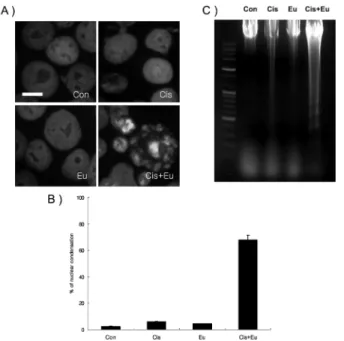
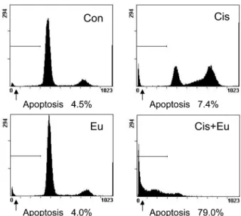
In our current study, we examined whether the cytotoxic effects of eugenol upon human melanoma G361 cells are associated with cell cycle arrest and apoptosis using a range