Apoptotic Effect of Co-Treatment with Chios Gum Mastic and Eugenol on SCC25 Human Tongue Squamous Cell
Carcinoma Cell Line
Hyeon-Jin Sohn, D.D.S.,M.S.D., Byeong-Ho Yea, D.D.S.,M.S.D., In-Ryoung Kim, M.S.,Ph.D., Bong-Soo Park, D.D.S.,M.S.D.,Ph.D., Sung-Hee Jeong1, D.D.S.,M.S.D.,Ph.D., Yong-Woo Ahn1, D.D.S.,M.S.D.,Ph.D.,
Myung-Yun Ko1, D.D.S.,M.S.D.,Ph.D.
Department of Oral Anatomy, School of Dentistry, Pusan National University
1Department of Oral Medicine, School of Dentistry, Pusan National University
Eugenol (4-allyl-2-methoxyphenol) is a natural phenolic constituent extensively used in dentistry as a component of zinc oxide eugenol cement and is applied to the mouth environment. Chios gum mastic (CGM) is a resinous exudate obtained from the stem and the main leaves ofPistacia lenticulus tree native to Mediterranean areas. This study was undertaken to investigate the synergistic apoptotic effect of co-treatment with a natural product, CGM and natural phenolic compound, eugenol on SCC25 human tongue squamous cell carcinoma cell line.
To investigate whether the co-treatment with eugenol and CGM compared to each single treatment efficiently reduces the viability of SCC25 cells, MTT assay was conducted. Induction and augmentation of apoptosis were confirmed by Hoechst staining, TUNEL staining and DNA hypoploidy. Westen blot analysis and immunofluorescent staining were performed to study the alterations of the expression level and the translocation of apoptosis-related proteins in co-treatment.
In this study, co-treatment of with eugenol and CGM on SCC25 cells showed several lines of apoptotic manifestation such as nuclear condensations, DNA fragmentation, the increase and decrease of Bax and Bcl-2, decrease of DNA content, the release of cytochrome c into cytosol, translocation of AIF and DFF40 (CAD) onto nuclei, and activation of caspase-3, caspase-6 caspase-7, caspase-9, PARP, Lamin A/C and DFF45 (ICAD) whereas each single treated SCC25 cells did not show or very slightly these patterns.
Although the single treatment of 40 μg/ml CGM and 0.5 mM eugenol for 24 h did not induce apoptosis, the co-treatment of these reagents prominently induced apoptosis. Therefore our data provide the possibility that combination therapy with CGM and eugenol could be considered as a novel therapeutic strategy for human oral squamous cell carcinoma.
Key words : Apoptosis, Chios gum mastic (CGM), Eugenol, Human tongue squamous cell carcinoma cell line
Corresponding author : Myung-Yun, Ko
Professor, Department of Oral Medicine, Schoool of Dentistry, Pusan National University, Beomeo-Ri, Mulgeum-Eup, Yangsan-Si, Gyeongsangnam-Do, 626-870, Korea
Received: 2011-07-15 Accepted: 2011-08-20
* This work was supported by for a 2-Year Research Grant of Pusan National University.
Ⅰ. INTRODUCTION
Apoptosis is an essential physiological process required for embryonic development, regulation of immune responses and maintenance of tissue homeostasis. However, apoptosis is also implicated in a wide range of pathological conditions, including immunological diseases, allergy and cancer.1,2) The induction of apoptosis leads to specific morpho- logical and biochemical changes, including cell blebbing, exposure of cell surface phosphati- dylserine, cell size reduction including cell shrinkage, chromatin condensation and internu- cleosomal cleavage of genomic DNA.3,4)
Carcinoma of the oral cavity, especially oral squamous cell carcinoma (OSCC), are one of the most leading causes of cancer related death and affect nearly 500,000 patients annually world-wide.
And OSCC is one of the most malignancies that remain incurable with current therapies.5) OSCC patients are treated by classical modalities of treatment consisting of surgery, radiotherapy, and/or chemotherapy. But OSCC still shows noticeable mortality rates. therefore new therapeutic approaches have been investigated and the most promising one is obtained from natural agents with known anti-cancer effects.
The plantPistiacia lentiscus L. var. Chia. grows almost only in the South region of Chios Island, Greece, and produces a resin, known as Chios gum mastic (CGM). It is gained from the stem and leaves ofPistacia lentiscustrees and has been widely used for centuries in Mediterranean and Middle Eastern countries, both as a dietary supplement and herbal remedy.6,7)
Eugenol (4-allyl-2-methoxyphenol) is a natural phenolic constituent of clove oil, cinnamon, basil, and nutmeg which is used as a food flavor and fragrance agent.8) It is extensively used in dentistry as a component of zinc oxide eugenol cement and is applied to the mouth environment.9) Ghosh et al.8) showed that 125 mg/kg body weight of eugenol caused significant delay in tumor development and no sign of metastasis in B16 melanoma xenograft
model. Eugenol-related compounds not only act as antioxidant, but also as prooxidant, which may be involved in the cytotoxicity induction. High concentration of their enhanced the generation of tissue damaging free radicals.10-13)
Recent studies have demonstrated that the co-treatment of an antitumor agent with an anti-cancer natural product can be one of the potential therapeutic strategies reducing the extent and severity of cancer treatment-related toxicity.
14-18) Up to now, there is no report about the
synergistic apoptotic effects of co-treatment with CGM and eugenol on human tongue sqaumous carcinoma cell lines. Therefore, this study was undertaken to investigate the synergistic apoptotic effect of co-treatment with a natural product, CGM and natural phenolic compound, eugenol on SCC25 human tongue squamous cell carcinoma cell line.
Ⅱ. MATERIALS AND METHODS 1. Reagents
Chios gum mastic resin was obtained from Mastic Korea (Seoul, Korea). The following reagents were obtained commercially: TUNEL reaction mixture was purchased from Boehringer Mannheim (Mannheim, Germany); Dulbecco’s modified Eagle’s medium : Nutrient Mixture F-12 (Ham) (1:1) (D-MEM/F-12) and fetal bovine serum (FBS) were purchased from Gibco (Gaithersburg, MD, USA);
Eugenol, Dimethyl sulfoxide (DMSO), Hoechst 33342, RNase A, proteinase K, aprotinin, leupeptin, phenylmethylsulfonyl fluoride (PMSF), thiazolyl blue tetrazolium bromide, collagenase and propidium iodide (PI) were purchased from Sigma (St. Louis, MO, USA); SuperSignal West Pico enhanced chemiluminescence Western blotting detection reagent was purchased from Pierce (Rockford, IL, USA). Rabbit polyclonal anti-human AIF antibody and Anti-Caspase 6 were purchased from Upstate (NY, USA); caspase-9, caspase-7, caspase-3, Bax, Bcl-2, cytochrome c, DFF45 (ICAD), poly(ADP- ribose) polymerase (PARP) antibodies, Lamin A/C,
rabbit polyclonal anti-human β-actin antibody, FITC-conjugated goat anti-mouse and anti-rabbit IgGs were purchased from Santa Cruz Biotechnology (Santa Cruz, CA, USA); rabbit polyclonal anti-human DFF40 (CAD) antibody was purchased from Stressgen (San Diego, USA);
HRP-conjugated sheep anti-mouse and anti-rabbit IgGs were purchased from Amersham GE Healthcare (Little Chalfont, UK).
2. Cell culture
SCC25 human tongue squamous cell carcinoma cell line was purchased from the ATCC (Rockville, MD, USA). This cell was maintained at 37℃ with 5% CO2in air atmosphere in D-MEM/F-12 medium with 4 mM L-glutamine, 1.5 μg/L sodium bicarbonate, 4.5 g/L glucose and 1.0 mM sodium pyruvate supplemented with 10% fetal bovine serum (FBS).
3. treatment with CGM and eugenol for co- treatment
The stock solution of CGM (100 mg/ml) made by dissolving in DMSO were kept frozen at -20℃ until use. The concentrations of DMSO (0.01-0.1%
[vol/vol]) used in this study, both as a vehicle for CGM and as a control, had no effect on SCC25 cells proliferation in our preliminary studies. And stock solution of eugenol (10 mM) just made by dissolving in D-MEM/F-12 medium before treatment. Cell was cultured for 24 h. The original medium was removed and that washed with phosphate-buffered saline (PBS). It was changed that the fresh medium on the plates. When 40 μg/ml of CGM and 0.5 mM of eugenol were each treated for 24 h, slight cell death occurred in MTT assay, consequently this single concentration was utilized for further assessment of apoptosis for co-treatment.
4. MTT assay
Cells were cultured in a 96-well plate and
incubated for 24 h. Then cells treated with 40 μg/ml of CGM and/or 0.5 mM of eugenol for 24 h, and then cells were treated with 500 μg/ml of MTT stock solution. After the cells were incubated at 37℃ with 5% CO2 for 4 h. The medium was aspirated and formed formazan crystals were dissolved in the mixture solution of DMSO and absolute ethanol (1 : 1). Cell viability was monitored on a ELISA reader (Sunrise Remote Control, Tecan, Austria) at 570 nm excitatory emission wavelength.
5. Hoechst staining
Cells were harvested and cytocentrifuged onto a clean, fat-free glass slide with a cytocentrifuge.
Cells were stained in 4 μg/ml Hoechst 33342 for 10 min at 37℃ in the dark and washed twice in PBS.
The slides were mounted with glycerol. The samples were observed and photographed under an epifluorescence microscope (Carl Zeiss, German).
The number of cells that showed condensed or fragmented nuclei was determined by a blinded observer from a random sampling of 3 × 10² cells per experiment. Three independent experiments were conducted.
6. TUNEL technique
To identify apoptotic cells by terminal deoxynucleotidyl transferase (TDT) - mediated dUTP nick and labelling (TUNEL), An In SituCell Death Detection Kit was used as recommended by the manufacturer. Cells were harvested after treatment of CGM and/or eugenol on 60 mm culture dishes. The cell suspension was centrifuged onto a clean fat-free glass slide with a cytocentrifuge.
After fixing with 4% paraformaldehyde for 1 h, washing with PBS and permeabilizing with 0.1%
Triton X-100 solution for 2 min on ice, cells were added with reaction mixture for 1 h at 37℃. Total cell number, at least 300 cells from each group, was counted under DIC optics and the percentage of TUNEL positive cells were calculated and photographed under epifluorescence microscope
(Carl Zeiss, German).
7. Western blot analysis
Cells were plated at a density of 2 x 10⁶cells in 100 mm culture dishes. Cells treated with CGM were washed twice with ice-cold PBS and centrifuged at 2,000 rpm for 10 min. Total cell proteins were lysed with a RIPA buffer [300 mM NaCl, 50 mM Tris-HCl (pH 7.6), 0.5% TritonX-100, 2 mM PMSF, 2 μg/ml aprotinin and 2 μg/ml leupeptin] and incubated at 4°C for 1 h. The lysates were centrifuged at 14,000 revolutions per min for 15 min at 4°C, and sodium dodecyl sulfate (SDS) and sodium deoxycholic acid (0.2% final concentration) were added. Protein concentrations of cell lysates were determined with Bradford protein assay (Bio-Rad, Richmond, CA, USA) and BSA was used as a protein standard. A sample of 50 μg protein in each well was separated and it was loaded onto 7.5-15% SDS/PAGE. The gels were transferred to Nitrocellulose membrane (Amersham Pharmacia Biotech, Piscataway, UK) and reacted with each antibody. Immunostaining with antibodies was performed using SuperSignal West Pico enhanced chemiluminescence substrate and detected with Alpha Imager HP (Alpha Innotech, San Leandro, USA). Equivalent protein loading was confirmed by Ponceau S staining.
8. Immunofluorescent staining
Cells were placed on slides by cytocentrifuge and fixed for 10 min in 4% paraformaldehyde. After blocking nonspecific binding with 3% bovine serum albumin, the cells were incubated with a primary antibody at a dilution of 1 : 100 for 1 h. After the incubation, the cells were washed 3 each for 5 min, and then incubated with FITC-conjugated secondary antibody at a dilution of 1 : 100 for 1 h at room temperature. Fluorescent images were observed and analyzed under Zeiss LSM 510 laser-scanning confocal microscope (Göettingen, Germany).
9. Flow cytometry analysis
Cells were seeded into a 6-well plate at 1 × 10⁶ cells/ml and incubated overnight. Cells treated with CGM and/or eugenol were incubated for 24 h. Cells were harvested. The harvested cells were washed with PBS containing 1% bovine serum albumin and centrifuged at 2,000 rpm for 10 min. The cells were resuspended ice-cold 95% ethanol with 0.5% Tween 20 to a final concentration of 70% ethanol. Fixed cells were pelleted, and washed in 1% BSA-PBS solution. Cells were resuspended in 1 ml PBS containing 20 μg/ml RNase A, incubated at 4℃ for 30 min, washed once with BSA-PBS, and resuspended in PI solution (10 μg/ml). After cells were incubated at 4℃ for 5 min in the dark, DNA content were measured on a CYTOMICS FC500 flow cytometry system (Beckman Coulter, FL, CA, USA) and data was analyzed using the Multicycle software which allowed a simultaneous estimation of apoptosis.
Ⅲ. RESULTS
1. Co-treatment with CGM and eugenol augmented the reduction in viability of SCC25 cells.
Single treatment of CGM at 40 μg/ml or eugenol at 0.5 mM for 24 h reduced the viability of SCC25 cells slightly (CGM, 86.7%; eugenol, 94.0%).
Co-treatment with CGM and eugenol significantly reduced the to 22.8% cell viability vs single treatment (Fig. 1).
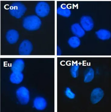
2. Co-treatment with CGM and eugenol augmented the nuclear condensation in SCC25 cells.
To study a nuclear condensation induced by the co-treatment, Hoechst staining by which a hallmark of apoptosis was conducted. Hoechst staining showed a nuclear condensation by the single treatment of CGM and eugenol. Co-treatment with
Fig. 1. Co-treatment with CGM and eugenol significantly reduced the cell viability in SCC25 cells. Cell viability was determined by MTT assay. Three independent assays were performed. Values are means ± SD of triplicates of each experiment (CGM, cells treated with 40 μg/ml Chios gum mastic for 24 h; Eu, cells treated with 0.5 mM eugenol for 24 h; CGM+Eu, cells treated with 40 μg/ml Chios gum mastic plus 0.5 mM eugenol for 24 h).
Fig. 2. Immunofluorescent micrographs showing nuclear morphology after Hoechst staining. Co-treatment with CGM and eugenol showed numerous condensed nuclei in SCC25 cells compared to the single treatment (CGM, cells treated with 40 μg/ml Chios gum mastic for 24 h; Eu, cells treated with 0.5 mM eugenol for 24 h;
CGM+ Eu, cells treated with 40 μg/ml Chios gum mastic plus 0.5 mM eugenol for 24 h).
CGM and eugenol showed an increased nuclear condensation vs the single treatment (Fig. 2).
3. Co-treatment with CGM and eugenol showed the DNA fragmentation in SCC25 cells.
DNA fragmentation which is the biochemical hallmark of apoptosis, was demonstrated by TUNEL technique. The TUNEL positive SCC25 cells in the control cells did not show and in single treatment cells a few showed. But the number of TUNEL positive SCC25 cells in co-treatment cells were remarkably increased (Fig. 3).
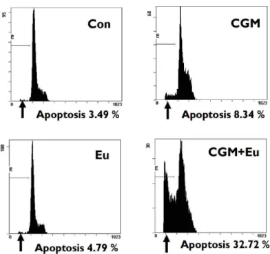
4. Augmentation of apoptosis by the co-treatment with CGM and eugenol was demonstrated by the decrease in a DNA content of SCC25 cells.
The flow cytometry showed that the co-treatment with CGM and eugenol significantly increased
Fig. 3. Immunofluorescent micrographs showing TUNEL positive cells. Co-treatment with CGM and eugenol showed numerous TUNEL positive cells in SCC25 cells compared to the single treatment (CGM, cells treated with 40 μg/ml Chios gum mastic for 24 h; Eu, cells treated with 0.5 mM eugenol for 24 h; CGM+Eu, cells treated with 40 μg/ml Chios gum mastic plus 0.5 mM eugenol for 24 h).
Fig. 4. The kinetic analysis of the effect of co-treatment on the induction of apoptosis. Co-treatment showed remarkably the increase of apoptotic cells with DNA hypoploidy compared to the single treatment (CGM, cells treated with 40 μg/ml Chios gum mastic for 24 h; Eu, cells treated with 0.5 mM eugenol for 24 h;
CGM+Eu, cells treated with 40 μg/ml Chios gum mastic plus 0.5 mM eugenol for 24 h).
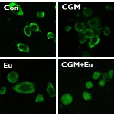
Fig. 5. The confocal microscopy showed that AIF was obviously translocated onto nuclei in SCC25 cells when co-treated with CGM and eugenol (CGM, cells treated with 40 μg/ml Chios gum mastic for 24 h; Eu, cells treated with 0.5 mM eugenol for 24 h; CGM+Eu, cells treated with 40 μg/ml Chios gum mastic plus 0.5 mM eugenol for 24 h).
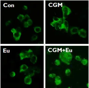
Fig. 6. The confocal microscopy showed that cytochrome c was obviously released into cytosol in SCC25 cells when co-treated with CGM and eugenol (CGM, cells treated with 40 μg/ml Chios gum mastic for 24 h; Eu, cells treated with 0.5 mM eugenol for 24 h; CGM+Eu, cells treated with 40 μg/ml Chios gum mastic plus 0.5 mM eugenol for 24 h).
Fig. 7. Western blot analysis showing that co-treatment with CGM and eugenol induced up-regulation of the Bax and down-regulation of the Bcl-2.
The levels of β-actin were used as an internal standard for quantifying Bcl-2 and Bax expression (CGM, cells treated with 40 μg/ml Chios gum mastic for 24 h; Eu, cells treated with 0.5 mM eugenol for 24 h; CGM+Eu, cells treated with 40 μg/ml Chios gum mastic plus 0.5 mM eugenol for 24 h).
apoptotic cells with DNA hypoploidy compared to the example of single treatment (Fig. 4).
5. Co-treatment with CGM and eugenol caused the translocation of AIF from mitochondria onto the nucleus.
In the single treatment of CGM or eugenol, AIF was located at mitochondria whereas in the co-treatment, AIF was obviously translocated onto the nuclei in SCC25 cells (Fig. 5).
6. Co-treatment with CGM and eugenol caused the release of cytochrome c from mitochondria into the cytosol.
In the single treatment of CGM or eugenol, cytochrome c was located at mitochondria whereas in the co-treatment, cytochrome c was obviously release into the cytosol in SCC25 cells (Fig. 6).
7. Augmentation of apoptosis by co-treatment with CGM and eugenol was demonstrated by Western blot assay.
Bcl-2 has a function of antiapoptosis, whereas
Fig. 8. Western blot analysis showing that the co- treatment with CGM and eugenol in SCC25 cells remarkably induced Caspase-3, Caspase -6, Caspase-7, Caspase-9, PARP and Lamin A/C degradations and produced Caspase-6 34 kDa cleaved products. β-actin, a loading control (CGM, cells treated with 40 μg/ml Chios gum mastic for 24 h; Eu, cells treated with 0.5 mM eugenol for 24 h; CGM+Eu, cells treated with 40 μg/ml Chios gum mastic plus 0.5 mM eugenol for 24 h).
Bax promotes apoptosis. Bcl-2 remarkably decreased in the single treatment of CGM or eugenol and disappear in the co-treatment. Bax significantly increased by the co-treatment (Fig. 7).
The co-treatment with CGM and eugenol induced the degradation of caspase-3, caspase-6, caspase-7, caspase-9, PARP and lamin A/C, and produced caspase-6 34 kDa (appeared in single treatment of CGM or eugenol), and DFF45 (ICAD) 11 kDa cleaved products (Fig. 8 and Fig. 9).
Fig. 9. Western blot analysis showing the co-treated with CGM and eugenol in SCC25 cells remarkably produced DFF45 11 kDa cleaved product. β-actin, a loading control (CGM, cells treated with 40 μg/ml Chios gum mastic for 24 h; Eu, cells treated with 0.5 mM eugenol for 24 h; CGM+Eu, cells treated with 40 μg/ml Chios gum mastic plus 0.5 mM eugenol for 24 h).
Fig. 10. The confocal microscopy showed that DFF40 (CAD) was translocated onto the nuclei in the co-treatment of CGM and eugenol (CGM, cells treated with 40 μg/ml Chios gum mastic for 24 h; Eu, cells treated with 0.5 mM eugenol for 24 h; CGM+Eu, cells treated with 40 μg/ml Chios gum mastic plus 0.5 mM eugenol for 24 h).
8. Co-treatment with CGM and eugenol caused the translocation of DFF40 (CAD) from cytosol onto the nuclei.
In the single treatment of CGM or eugenol, DFF40 (CAD) was located at cytosol whereas in the co-treatment, DFF40 (CAD) was obviously translocated onto the nuclei in SCC25 cells (Fig. 10).
Ⅳ. DISCUSSION
Public attention on natural products as herbal remedies continues to grow. Moreover, the revelation of the pharmacological mechanisms of herbal plant compounds is contributing to their acceptance by healthcare professionals and the public. A number of studies has elucidated that individual herbal medicines extracted from herbal plants have a number of pharmacological activities, e.g. anti-allergic, anti-pyretic, analgesic, anti- inflammatory and anticancer effects.19-23) In spite of numerous in vitro and in vivostudies, the mode of
action of most herbal medicines remains elusive.
Chios gum mastic (CGM) is a resinous exudate obtained from the stem and the main leaves of Pistacia lenticulus tree native to Mediterranean areas. Previous studies have demonstrated that CGM is effective in the treatment of benign gastric and duodenal ulcers and it have definite antibacterial activity against Helicobacter pylori.24-27) It has also been shown to have antimicrobial properties.28) Recently, He et al.7) have demonstrated that CGM treatment inhibited the proliferation of androgen- independent prostate cancer cells apparently through modulation of the NF-κB target gene. In addition, CGM has also been shown to contain compounds that can induce in vitro apoptosis of human colon cancer cells through caspase-dependent pathways.6) Eugenol is a naturally occurring phenolic compound used as a food flavour and fragrance agent and the main component of oil of clove and the essential oils or extracts of numerous plants.29,30) It have been identified that eugenol inhibited immediate hypersensitivity by inhibiting the release of histamine and induced apoptosis from mast cells in vivo and in vitro.31,32)
If the co-treatment with a natural phenolic compond, eugenol and a natural product CGM which has severe toxicity and resistance in the chemotherapy shows a synergistic antitumor effect, it could be a more fundamental therapeutic strategy for cancer chemotherapy.
During apoptosis, apoptotic cells undergo specific morphological changes such as the cell blebbing, reduction of cell size, cell shrinkage, chromatin condensation and DNA fragmentation.3,4) In the results of Hoechst stain and TUNEL assay, the co-treatment with CGM and eugenol showed a large number of apoptotic hallmarks such as condensed nuclei and numerous TUNEL-positive cells.
whereas the single treatment did not show these patterns.
Because mitochondria play a crucial role in apoptosis, the induction of the mitochondrial permeability transition plays a key part in the regulation of apoptosis.33-36) The mitochondrial
pathway can also be triggered by various intracellular and extracellular stress signals, which result in activation of pro-apoptotic proteins, including Bax and Bak, or inactivation of anti-apoptotic Bcl-2 family members, such as Bcl-2 and Bcl-xL.36)As a result of activation/inactivation of Bcl-2 family proteins, changes in mitochondrial membrane lead to the dissipation of inner membrane potential and permeabilization of the outer mitochondrial membrane (OMM). This in turn induces the release of various proapoptotic proteins, such as cytochrome c, Smac/Diablo, endonuclease G and AIF.37,38) The present study showed the co-treatment with CGM and eugenol in SCC25 result in increase BAX and decrease of Bcl-2.
whereas the single treatment slightly showed these pattern.
It has been reported that the pro-apoptotic Bcl-2 family in isolated mitochondria induces cytochrome c release, the loss of mitochondrial membrane potential and results in AIF release.39,40) Cytochrome c release is known to contribute to apoptosis triggered by proteasome inhibition.41,42) Generally, cytochrome c is released into the cytosol during apoptosis, where it binds to Apaf-1. This cytochrome c/Apaf-1 complex (apoptosome) promotes the autoactivation of procaspase-9 to caspase-9. Caspase-9 then acts on procaspase-3 to initiate a caspase activation cascade.43,44) Released AIF through pro-apoptotic Bcl-2 family activation induces its translocation to the nucleus, resulting in chromatin condensation and large-scale DNA fragmentation.45) In the present study, the co-treatment CGM and eugenol in SCC25 also induced translocation of AIF from mitochondria into nuclei, cytochrome c release from mitochondria into the cytosol. whereas the single treatment did not show.
A common final event of apoptosis is nuclear condensation, which is controlled by caspases, DFF (DNA fragmentation factor), and PARP. Caspases, the aspartate-specific intracellular cysteine protease, play an essential role during apoptotic death.46)Once activated, the effector caspases (caspase-3,
caspase-6 or caspase-7) are responsible for the proteolytic cleavage of a broad spectrum of cellular targets, leading ultimately to cell death. The known cellular substrates include structural components (such as actin and nuclear lamin), inhibitors of deoxyribonuclease (such as DFF45 or ICAD) and DNA repair proteins (such as PARP).47,48) In apoptotic cells, activation of DFF40 (CAD), also a substrate of caspase-3, occurs with the cleavage of DFF45 (ICAD). Once DFF40 is activated and released from the complex of DFF45 and DFF40, it can translocate to the nucleus and then degrade chromosomal DNA and produce DNA fragmentation.49) Furthermore, in apoptotic cells, the degradation of the lamin A/C, a substrate of caspase-6 was sometimes occurred. In this study, the co-treatment with CGM and eugenol in SCC25 cells result in cleavage of caspase-6, DFF45 (ICAD) and degradation of caspase-3, caspase-7, caspase-9, PARP and Lamin A/C. Furthermore, confocal microscopy showed that the co-treatment with CGM and eugenol led to the translocation of DFF40 (CAD) from cytosol onto nuclei in SCC25 cells.
whereas the single treatment did not show or slightly showed.
In this study, SCC25 cells co-treated with CGM and eugenol showed several lines of apoptotic manifestation such as nuclear condensations, DNA fragmentation, the content, the release of cytochrome c into cytosol, translocation of AIF and DFF40 (CAD) onto nuclei, the activation of caspase-3, 6, 7, 9, PARP, Lamin A/C and DFF45 (ICAD), the up-regulation of BAX whereas each single treated SCC25 cells did not show or slightly showed.
In conclusion, combination therapy with CGM and eugenol, could be considered, in the future, as an alternative therapeutic strategy for human oral squamous cell carcinoma. Its clinical application awaits further extensive studies.
V. CONCLUSION
Eugenol (4-allyl-2-methoxyphenol) is a natural
phenolic constituent extensively used in dentistry as a component of zinc oxide eugenol cement and is applied to the mouth environment. Chios gum mastic (CGM) is a resinous exudate obtained from the stem and the main leaves of Pistacia lenticulus tree native to Mediterranean areas. This study was undertaken to investigate the synergistic apoptotic effect of co-treatment with a natural product, CGM and natural phenolic compound, eugenol on SCC25 human tongue squamous cell carcinoma cell line.
To investigate whether the co-treatment with eugenol and CGM compared to each single treatment efficiently reduces the viability of G361 cells, MTT assay was conducted. Induction and augmentation of apoptosis were confirmed by Hoechst staining, TUNEL staining and DNA hypoploidy. Westen blot analysis and immuno- fluorescent staining were performed to study the alterations of the expression level and the translocation of apoptosis-related proteins in co-treatment.
In this study, co-treatment of with eugenol and CGM on SCC25 cells showed several lines of apoptotic manifestation such as nuclear conden- sations, DNA fragmentation, the increase and decrease of Bax and Bcl-2, decrease of DNA content, the release of cytochrome c into cytosol, translocation of AIF and DFF40 (CAD) onto nuclei, and activation of caspase-3, caspase-6 caspase-7, caspase-9, PARP, Lamin A/C and DFF45 (ICAD) whereas each single treated SCC25 cells did not show or very slightly these patterns.
Although the single treatment of 40 μg/ml CGM and 0.5 mM eugenol for 24 h did not induce apoptosis, the co-treatment of these reagents prominently induced apoptosis. Therefore our data provide the possibility that combination therapy with CGM and eugenol could be considered as a novel therapeutic strategy for human oral squamous cell carcinoma.
REFERENCES
1. Carson DA and Ribeiro JM. Apoptosis and disease.
Lancet 1993;341:1251-1254.
2. Ohta K and Yamashita N. Apoptosis of eosinophils and lymphocytes in allergic inflammation. J Allergy Clin Immunol 1999;104:14-21.
3. Wyllie AH, Kerr JF, Currie AR. Cell death: the significance of apoptosis. Int Rev Cytol 1980;68:
251-306.46. Acehan D, Jiang X, Morgan DG, Heuser JE, Wang X, Akey CW. Three-dimensional structure of the apoptosome: Implications for assembly, procaspase-9 binding, and activation. Mol Cell 2002;9:423-432.
4. Williams GT. Programmed cell death: apoptosis and oncogenesis. Cell 1991;65:1097-1098.
5. Shen J, Huang C, Jiang L, Gao F, Wang Z, Zhang Y, Bai J, Zhou H, Chen O. Enhancement of cisplatin induced apoptosis by suberoylanilide hydroxamic acid in human oral squamous cell carcinoma cell lines.
Biochem Pharmacol 2007;73:1901-1909.
6. Balan KV, Prince J, Han Z, Dimas K, Cladaras M, Wyche JH, Sitaras NM, Pantazis P. Antiproliferative activity and induction of apoptosis in human colon cancer cells treated in vitro with constituents of a product derived from Pistacia lentiscus L. var. chia.
Phytomedicine 2007;14:263-272.
7. He M, Li A, Xu CS, Wang SL, Zhang MJ, Gu H, Yang YQ, Tao HH. Mechanisms of antiprostate cancer by gum mastic: NF-kappaB signal as target. Acta Pharmacol Sin 2007;28:446-452.
8. Ghosh R, Nadiminty N, Fitzpatrick JE, Alworth WL, Slaga TJ, Kumar AP. Eugenol causes melanoma growth suppression through inhibition of E2F1 transcriptional activity. J Biol Chem 2005;280:
5812-5819.
9. Markowitz K, Moynihan M, Liu M, Kim S. Biologic properties of eugenol and zinc oxide-eugenol. Oral Surg Oral Med Oral Pathol 1992;73:729-737.
10. Suzuki Y, Sugiyama K, Furuta H. Eugenol-mediated superoxide generation and cytotoxicity in guinea pig neutrophils. Jpn J Pharmacol 1985;39:381-386.
11. Wright SE, Baron DA, Heffner JE. Intravenous eugenol causes hemorrhagic lung edema in rats:
proposed oxidant mechanisms. J Lab Clin Med 1995;125:257-264.
12. David CT, Rola B, Robert CB. Comparative Toxicity of Eugenol and Its Quinone Methide Metabolite in Cultured Liver Cells Using Kinetic Fluorescence Bioassays. Toxicology and applied pharmacology 1998;149:55-63.
13. Satoh K, Sakagami H, Yokoe I, Kochi M, Fujisawa S.
Interaction between eugenol-related compounds and
radicals. Anticancer Res 1998;18:425-428.
14. Adhami VM, Malik A, Zaman N, Sarfaraz S, Siddiqui IA, Sved DN, Afaq F, Pasha FS, Saleem M, Mukhata H. Combined inhibitory effects of green tea polyphenols and selective cyclooxygenase-2 inhibitors on the growth of human prostate cancer cells both invitro and in vivo. Clin Cancer Res 2007;13:1611- 1619.
15. Mai Z, Blackbum GL, Zhou JR. Genistein sensitizes inhibitory effect of tamoxifen on the growth of estrogen receptor-positive and HER2-overexpressing human breast cancer cells. Mol Carcinog 2007;46:
534-542.
16. Song MQ, Zhu JS, Chen JL, Wamg L, Da W, Zhu L, Zhang WP. Synergistic effect of oxymatrine and angiogenesis inhibitor NM-3 on modulating apoptosis in human gastric cancer cells. World J Gastroenterol 2007;13:1788-1793.
17. Vinall RL, Hwa k, Ghosh P, Pan CX, Lava PN Jr, de vere White RW. Combination treatment of prostate cancer cell lines with bioactive soy isoflavones and perifosine causes increased growth arrest and/or apoptosis. Clin Cancer Res 2007;13:6204-6216.
18. Lee SH, Ryu Jk, Lee KY, Woo SM, Park JK, Yoo JW, Kim YT, Yoo YB. Enhanced anti-tumor effect of combination therapy with gemcitabine and apigenin in pancreatic cancer. Cancer Lett 2008;259:39-49.
19. Kim HM, Lee EH, Hong SH, Song HJ, Shin MK, Kim SH, Shin TY. Effect of Syzygium aromaticum extract on immediate hypersensitivity in rats. J Ethno- pharmacol 1998;60:125-131.
20. Kim HM. Yi JM, Lim KS. Magnoliae flos inhibits mast cell-dependent immediate-type allergic reactions.
Pharmacol Res 1999;39:107-111.
21. Park HI, Jeong MH, Lim YJ, Park BS, Kim GC, Lee YM, Kim HM, Yoo KS, Yoo YH. Szygium aromaticum (L.) Merr. Et Perry (Myrtaceae) flower bud induces apoptosis of p815 mastocytoma cell line.
Life Sci 2001;69:553-566.
22. Na HJ, Jeong HJ, Bae H, Kim YB, Park ST, Yun YG, Kim HM. Tongkyutang inhibits mast cell-dependent allergic reactions and inflammatory cytokines secretion. Clin Chim Acta 2002;319:35-41.
23. Kim JH, Bae HR, Park BS, Lee JM, Ahn HB, Rho JH, Yoo KW, Park WC, Rho SH, Yoon HS, Yoo YH. Early mitochondrial hyperpolarization and intracellular alkalinization in lactacystin-induced apoptosis of retinal pigment epithelial cells. J Pharmacol Exp Ther 2003;305:474-481.
24. Al-Habbal MJ, Al-Habbal Z, Huwez FU. A double- blind controlled clinical trial of mastic and placebo in the treatment of duodenal ulcer. Clin Exp Pharmacol Physiol 1984;11:541-544.
25. Al-Said MS, Ageel AM, Parmar NS, Tariq M.
Evaluation of mastic, a crude drug obtained from Pistacia lentiscus for gastric and duodenal anti-ulcer activity. J Ethnopharmacol 1986;15:271-278.
26. Huwez FU and Al-Habbal MJ. Mastic in treatment of benign gastric ulcers. Gastroenterol Jpn 1986;21:
273-274.
27. Hawez FU, Thirlwell D, Cockayne A, Ala’Aldeen PA.
Mastic gum kills Helicobacter pylori. N Engl J Med 1998;339:1946.
28. Aksoy A, Duran N, Koksai F. In vitro and in vivo antimicrobial effects of mastic chewing gum against Streptococcus mutans and mutans streptococci. Arch Oral Biol 2006;51:476-481.
29. IARC Monographs on the Evaluation of Carcinogenic Risks to Humans. Allyl Compounds, Aldehydes, Epoxides and Peroxides. 1985;36:75.
30. Zhu YP. Materia Medica: Cemistry, Pharmacology and Application. 1st ed Amsterdam Harwood Academic Publishers 1996;1-3.
31. Kim HM, Lee EH, Kim CY, Chung JG, Kim SH, Lim JP, Shin TY. Antianaphylactic properties of eugenol.
Pharmacol Res 1997;36:475-480.
32. Shin BK, Lee EH, Kim HM. suppression of L-histidine decarboxylase mRNA expression by methyleugenol.
Biochem Biophys Res Commun 1997;232:188-191.
33. Kroemer G, Zamzami N, Susin SA. Mitochondrial control of apoptosis. Immunol Today 1997;18:44-51.
34. Green DR and Reed JC. Mitochondria and apoptosis.
Science 1998;281:1309-1312.
35. Susin SA, Lorenzo HK, Zamzami N, Marzo I, Snow BE, Brothers GM, Mangion J, Jacotot E, Costantini P, Loeffler M, Larochette N, Goodlett DR, Aebersold R, Siderovski DP, Penninger JM, Kroemer G. Molecular characterization of mitochondrial apoptosis-inducing factor. Nature 1999;397:441-446.
36. Orrenius S. Mitochondrial regulation of apoptotic cell death. Toxicol Lett 2004;149:19-23.
37. Hengartner MO. The biochemistry of apoptosis.
Nature 2000;407:770-776.
38. Barczyk K, Kreuter M, Pryjma J, Booy EP, Maddika S, Ghavami S, Berdel WE, Roth J, Los M. Serum cytochrome c indicates in vivo apoptosis and can serve as a prognostic marker during cancer therapy.
Int J Cancer 2005;116:167-173.
39. Hunter JJ and Parslow TG. A peptide sequence from Bax that converts Bcl-2 into an activator of apoptosis.
J Biol Chem 1996;271:8521-8524.
40. Narita M, Shimizu S, Ito T, Chittenden T, Lutz RJ, Matsuda H, Tsujimoto Y. Bax interacts with the permeability transition pore to induce permeability transition and cytochrome c release in isolated mitochondria. Proc Natl Acad Sci U S A 1998;95:
14681-14686.
41. Wagenknecht B, Hermission M, Groscurth P, Liston P, Krammer PH, Weller M. Proteasome inhibitor- induced apoptosis of glioma cells involves the processing of multiple caspases and cytochrome c release. J Neurochem 2000;75:2288-2297.
42. Marshansky V, Wang X, Bertrand R, Luo H, Duguid W, Chinnadurai G, Kanaan N, Vu MD, Wu J.
Proteasomes modulate balance among proapoptotic and antiapoptotic Bcl-2 family members and compromise functioning of the electron transport chain in leukemic cells. J Immunol 2001;166:3130-3142.
43. Li P, Nijhawan D, Budihardjo I, Srinivascula SM, Ahmad M, Alnemri ES, Wang X. Cytochrome c and dATP-dependent formation of Apaf-1/caspase-9 complex initiates an apoptotic protease cascade. Cell 1997;91:479-489.
44. Zou H, Li Y, Liu X, Wang, X. An APAF-1, cytochrome c multimeric complex is a functional apoptosome that activates procaspase-9. J Biol Chem 1999;274:11549- 11556.
45. Douglas E, Susin SA, Zamzami N, Ferri KF, Irinopoulou T, Larochette N, Prevost MC, Leber B, Andrews D, Penninger J, Kroemer G. Mitochondrio- nuclear translocation of AIF in apoptosis and necrosis.
FASEB J 2000;14:729-739.
46. Acehan D, Jiang X, Morgan DG, Heuser JE, Wang X, Akey CW. Three-dimensional structure of the apoptosome: Implications for assembly, procaspase-9 binding, and activation. Mol Cell 2002;9:423-432.
47. Gross A, McDonnell JM, Korsmeyer SJ. BCL-2 family members and the mitochondria in apoptosis. Genes Dev 1999;13:1899-1911.
48. Porter AG. Protein translocation in apoptosis. Trends Cell Biol 1999;9:394-401.
49. Cheng AC, Jian CB, Huang YT, Lai CS, Hsu PC, Pan MH. Induction of apoptosis by Uncaria tomentosa through reactive oxygen species production, cytochrome c release, and caspases activation in human leukemia cells. Food Chem Toxicol 2007;45:
2206-2218.
국문초록
사람혀편평세포암종세포에서 Chios gum mastic과 eugenol의 병용처리가 미치는 세포자멸사 효과에 관한 연구
부산대학교 치의학전문대학원 구강해부학교실,1부산대학교 치의학전문대학원 구강내과학교실
손현진․예병호․김인령․박봉수․정성희1․안용우1․고명연1
Chios gum mastic (CGM)은 그리이스 키오스 섬에서만 자생하는Pistiacia lentiscus L. var. Chia. 의 잎과 줄기로부터 추출되어진 식물성 수지이며, 과거 수세기 동안 지중해와 중동 지역 국가들에서 음식 첨가물과 위궤양, 십이지장궤양 등의 민간 치료약재로서 사용되어져 왔다. 정향나무에서 추출하는 페놀화합물인 eugenol은 zinc oxide eugenol의 구성성분으로 치과치료를 위해 많이 사용되고 있다. 본 연구는 사람혀편평세포암종세포(SCC25 cells)에서 천연물질인 CGM과 eugenol을 병용처리한 후 세포자멸사 효과가 있는지를 알아보기 위해 수행하였다.
CGM과 eugenol의 병용처리가 단독처리에 비해서 효과적인 세포생존율 감소가 있는지 확인하기 위하여 MTT법을 시행하 였고, 세포자멸사의 유도와 증가를 알기 위하여 Hoechst 염색법, TUNEL 염색법, DNA hypoploidy법을 사용하였다. 그리고 세포자멸사에 관계하는 단백질의 발현 변화와 세포내에서의 이동을 밝혀내기 위하여 Western blot 분석과 면역형광염색법을 수행하였다.
본 연구에서는 CGM과 eugenol이 병용처리된 SCC25 세포에서 핵의 농축, DNA분절, Bax의 증가와 Bcl-2의 감소, DNA양 의 감소, cytochrome c의 세포질로의 유리, AIF와 DFF40 (CAD)의 핵으로의 이동, caspase-3, caspase-6, caspase-7, caspase-9, PARP, Lamin A/C 그리고 DFF45 (ICAD)의 활성화와 같은 다양한 세포자멸사 증거를 보였다. 반면에 CGM과 eugenol이 각각 단독 처리된 SCC25 세포에서는 세포자멸사 현상이 미미하였다.
24시간 동안 40 μg/ml의 CGM과 0.5 mM의 eugenol을 각기 단독처리 한 결과에서는 세포자멸사를 거의 유도하지 못했으 나, 병용처리 한 결과에는 아주 탁월한 세포자멸사의 유도를 보였다. 그러므로 본 실험결과는 사람구강편평세포암종 환자들 에게 CGM과 eugenol의 병용요법이 새로운 치료전략으로서의 가능성을 줄 수 있다고 생각한다.
Key words : 세포자멸사, chios gum mastic (CGM), eugenol, 사람혀편평세포암종세포