약학회 지 제 48 권 제 2 호 147~152 (2004)
Yakhak Hoeji Vol. 48,No. 2 藥 學 舍 확
인체 흑색종 세포 (SK-MEL-28 Cell Line) 에서 Cisplatin, Heptaplatin, 그리고 Sunpla 에 의한 Apoptosis 으| 유도
최 수 라
•
명 평 근#
충남대학교 약학대학 임상생화학실,형질전환복제돼지센터 (Received March 2,2004; Revised April 9, 2004)
Induction of Apoptosis by Cisplatin, Heptaplatin and Sunpla in Human Melanoma (SK-MEL-28) Cell Line
Su-La C ho i and P yung-K eun M yung#
Lab of Clinical Biochemistry, Research Center for Transgenic Cloned Pigs, College of Pharmacy, Chungnam National University, Daejon 305-764,Korea
Abstract — A wide variety of cancer chemotherapeutic agents have
been
shown to induce programmedcell
death (PCD, APOPTOSIS) in various tumor cell lines in vitro. cis-Malonato [(4R,5R)-4,5-bis(aminomethyl)-2-isoprpopy 1-1,3-dioxolane]platinum(II) (heptaplatin), which is a new drug approved by KFDA in 1999, in a novel platinum-based antitumor agent with clinical potential against stomach cancer and the 3rd generation of the cisplatin. This study was performed to know how heptaplatin and cisplatin and sunpla (mixture of heptaplatin and mannitol) affect on SK-MEL-28 cell line, and how they induce the apoptosis. At EM analysis, the morphology of the cell was changed by treatment of the cisplatin, heptaplatin and sunpla. Apoptotic body formed around plasma membrane, and chromatin condensation represented in nucleus. This phe
nomenon is one of the characteristic of the apoptosis. The DNA of SK-MEL-28 cell line truncated by cisplatin and sunpla treatment was identified on 2% agarose gel electrophoresis. TUNEL assay was performed to know whether SK-MEL-28 cell die as apoptosis or necrosis by cisplatin, heptaplatin and sunpla. At this result, fluorescence intensity increased accordi ng to increase of time and concentration. Therefore, it was identified that cislatin, heptaplatin and sunpla induced apoptosis. Fas expressed on SK-MEL-28 cell membrane by cisplatin, heptaplatin and sunpla was identified by using flow cytometer and the expression of bcl-2(anti-apoptotic gene) decreased according to increase of concentration of the cisplatin, heptaplatin and sunpla. Cisplatin, heptaplatin and sunpla induced apoptosis against SK-MEL-28 cell line, and the apoptotic mechanism was identified as Fas-mediated apoptosis and decreased bcl-2 expression.
Keywords □ cisplatin, heptaplatin, sunpla, TUNEL assay, Fas expression, RT-PCR, apoptosis
Apoptosis
발생기전은수용체에 의해 야기된 신호전달체계의자극
,
혹은수용체에 의해 유도된 자극의 소실에 의해 활성화된 다.
1〕Fas
는apoptosis
를 유도하는 대표적인 수용체로서Fas
가anti-Fas-antibody
와 결합대하거나,Fas
에특이적인ligand
와결 합4>을하게되면T
세포나B
세포5’6)
또는 다양한 세포주3〉에서는 죽음신호(death signal, apoptotic signal)가 유도된다.2’6’
7) 이 Fas 는 apoptotic signal을 전달하는 intercellular domain과 transmembrane domain, 그리고세개의 cystein-rich domainᄋ-로구
성되어 있으며
,8) Fas
에 의해 매개되는 세포죽음(Fas-mediated
#본 논문에 관한 문의는 저자에게로
(
전화
) 042-821-5929 (팩스
) O42-823-65^(E-mail) [email protected]
apoptosis)의 기전이 최근에 명백히 밝혀져 있다
.9-13)
현재 apoptosis와관련된 조절유전자에 대해서도 많이 연구되 어져 있다
.
특히 Fas/FasL에의해 유도되는 apoptosis와 관련된 조절유전자중에서 그 기능과역할이 밝혀져 있는것이 bcl-2 유전자이다. 14 ’1 5> bcl-2 유전자는원종양유전자(proto-onco-gene)로
과거로부터 밝혀진암유전자와는달리 apoptosis를억제하여 세 포수명을 연장시켜 다른 암유전자의 영향을 받을 수 있는기회 를 증가시키는유전자로서
,
16) bcl-XL, bag, mcl-1 둥의 유전자가 bcl-2 family를 구성하고 있고,이늘 유전자와는상호경쟁적으로 작용한다.
동종 결합제나 이종 결합을 형성하는 bax 유전자와 bcl-Xs, bcl-Xp, 그리고 bak 등의 유전자는 bcl-2 유전자와 bcl-2 family의apoptosis를억제하는 기능에 대하여 반대되는 작용을 함으로써 apoptosis를 촉진하는 것으로 알려져 있다.17’18)
본 연구에서는 1999년국내신약1호인 sunpla가한국인의 위암치료약 물로서 개발된바있으나현재세계적으로매년 약5%의비율로 성장하고 있는피부암에 대한항암제를개발하기 위하여 피부암 세포인 melanoma(SK-MEL-28)을대상으로항암제로 알려져 있 지만그 독성으로인해 현재사용이 제한되고있는 cisplatin과그 의제3세대 화합물인 heptaplatin 그리고만니톨과혼합되어 제 형의형태로만들어진 sunpla가세포주기(cell cycle)의G2/M기를 정체시킴으로서피부암에 대한항암효괴를일으킴을확?]한바
,19J
그화합물들이 SK-MEL-28 cell 에서 apoptosis를유도함으로항 암효과를 일으키는지를 확인하기 위한실험들이 수행되어졌다.
실험방법
시약,기구 및 기기
사용한 피부암세포주인
SK-MEL-28
세포는생명공학연구소에서 분양받아사용하였으며
,
세포배양시 사용되는RPMI-1640
배지, fetal bovine serum(FBS), penicillin-streptomycine, pho-phate
buffer saline(PBS)는 Gibco에서, cisplatin
은 Sigma에서heptaplatin
과sunpla
는 선경인더스트리에서 공급받아사용하였다
. C 0
2 가스는안전가스에서 구입하여 사용하였고,
그 외에 사용된 시약은 세포배양용 시약 또는 특급시약을사용하였다
.
세포수 계수는
hematocytom eter
를 이용하여 광학현미경(BHS,
Olym pus optical Co. Japan
)0■로관칠하여실시하였다. 5
mlflow
cytometery용 tube는 Falcon #2052를 사용하였고, TUNELassay
와Fas expression
분석은 충남대학교 약학대학의FACSCalibur(Bectone& Dickinson, USA
)을이용하여 유세포분 석법으로 실시하였다.
세포배양
세포배양용 배지로서 560C에서 미리불활성화한 10% FBS와 penicillin-streptomycin 100 unit/m/이 포함된 RPMI-1640 배지 가 사용되었으며
,
세포는모든 경우에 있어서 37°C, 5% C 0 2 가 유지되는 C 0 2 incubator(3546 S/N 25401-3156, Forma scientific Inc., USA)를사용하며 배양하였다.
세포의 회수방법으 로는세포를배잉하•고있던배지를조심스럽게버리고 lxTrypsin- EDTA 용액(Gibco)으로 C 02 incubator에서 2분간반응시킨 후 세포를 떼어냈다.
그 후 원심분리하여 세포를 세척하고 회수하 여 RPMI-1640 완전배지로부유한 후 계대배양 하거나 실험에 사용하였다.
전자현미경에 의한
apoptosis
관찰Cisplatin, heptaplatin 그리고 sunpla를최종농도가 100 (xM이 되게처리해서 24시간이지난후세포를PBS로세척하고 trypsin- EDTA 용액을 사용해 세포를모은 후 다시 PBS로 1회 세척해
준 다음원심분리해서 상층액을버리고남아있는세포침전물에 전고정액인
2.5% glutaraldehyde(0.1 M phosphate buffer pH
7.4)
용액을 넣어 전고정 하였다.
그 후 인산 완충용액(0.1 M
phosphate buffer pH 7.4
)으로세척하고 다시1%
ᄋs 0
4에후고 정하여 완충액으로 세척하였다.
탈수과정은3
차중류수로 희석 된계열별 알코올로탈수시키고propylene oxide
로치환한다음epon
혼합물(polybed 812 kit. Polyscience
사 제품)로그비율은A(Epon 812+ D D SA ): B (Epon 812+ M N A
)가4 :
6으로 하여 침투및포매한후 열중합과정을거쳐 블록을제작하였다.
중합(polymerization
)은37°C, 45°C
및60oC
에서 각각12
시간, 24
시 간 및48
시간을실시하였고 제작된블록은각각3~ 5
개씩 무작 위로선정하여 초박절편기에 유리칼을장치하여 각블록을 공통적으로시료의 중간 부분까지 박절한다음
0.5
두께로 준초박 절편을 만들어 1
% toluidine blue
로 염색하였고광학현미경 상에서 시료의중심부분을기준으로하여 관찰부분을 제외한나 머지 부분을 제거한 후diamond knife
를 이용하여 간섭색이silver-goldtor
으로 보이는60~80 m m enRp
로초박절편을 만들어
grids
에부착하였다.
이중 전자염색은 익물을처리하지 않은세포군
(
대조군)과약물을 처리한세포군(
실험군)
모두같은조건 으로2% uracyl acetate
에25
분간 전도염색 시켜3
차중류수로 수세한 후 건조시키고 다시 1% lead citrate
에6분간 염색시켜 수세 및 건조하여 투과전자현미경(Hitachi H-600
)을사용해 가속전압
75kV
에서 관찰하였다.
D N A fragm e ntation
에의한apoptosis
관찰2X 1 0
6개의 세포에cisplatin
과sunpla
를최종농도가0, 5, 10,
50
그리고10(HiM
이되게 처리한후3
시간이 지나서 세포를 배양하던 배양액을농도별로 구분한
50 m/ tube
에옮기고 전술한세포회수방법으로 세포를모두 모았다
.
원심분리 한후 상층액 을 버리고 세포침전물을PB S
로 부유하며 씻으면서 모두eppendolf tube
로옮겼다.
다시 원심분리 한후상층액은버리고세포침전물에
5 0 m M Tris-HCl(pH 8.0), 0.5% SDS, 10 m M EDTA,
그리고50(ig/m / proteinase K
가 포함되어있는 용액40
바를첨가하여 세포를37°C
에서10
시간동안lysis
했다.
그런 후다시0.5 mg/m/ RNase A
를20 n
;넣고37°C
에서10
시간동 안 배양하면서RNA
를 제거했다.
앞의 과정을 거쳐서 얻어낸D N A
에40% sucrose
와0.25% bromophenol blue
그리고l% (w /v) low-melting point agarose
를포함하는10 m M EDTA (pH 8.0
)을70oC
에서 녹여 만든용액10
바를혼합한후0.5 (ig/
m/ ethidium bromide
를 포함하는 2%agarose gel
에다loading
하여55 mA
에서1
시간30
분간전기영동 하였다.
T U N E L
분석실험Cisplatin, heptaplatin
그리고sunpla
를최종농도가0, 1, 5,
그인체 흑색종 세포에서
Cisplatin, Heptaplatin, Sunpla에 의한
Apoptosis의 유도
149리고
10|_iM
이 되게처리해서24
시간과48
시간 동안시간별로배양한후농도와시간별로표시한
50 m/ tube
에세포를배양하던배양액을모두 모으고 세포회수방법으로 세포를회수하였으 며
,
원심분리 후상층액은 모두 버리고남아있는 세포침전물을PBS
로세척하면서FACS tube
로 세포를옮겼다.
다시 원심분리 한후 남아있는세포침전물에 1% paraformaldehyde
을 1m/
넣 고 얼음위에서15
분 동안방치했다.
그리고70% ethanol 1
ml을 넣고
-20oC
에서24
시간동안 놓아두면서 세포를고정시켰다.
고정된세포를
PBS
로세척하고 원심분리 후남아있는 세포침전물에
0.2 M sodium cacodylate(cacodylic acid), 2.5 m M Tris- HCl(pH
6.
6), 2.5 m M CoCl2(cobalt chloride), 0.25 mg/m/ BSA, 5U term inal dedeoxy nucleotidyl transferase,
그리고0.5 nmol dUTP-biotin(biotin-16-dUTP
)를 혼합한 용액5 0
마를 넣었다.
37°C
에서30
분간방치한후PBS
로다시 씻어주고원심분리하여남은 세포 침전물에 다시
streptavidin-FITC l| ig
이들어있는PBS 100
10/를 넣고상온에서30
분간방치했다.
그런후PBS
로 세포를 세척하고 다시PBS
를1 m/
채운 후flow cytometer
로 분석했다.
세포표면의
Fas
발현측정Cisplatin, heptapltain
그리고sunpla
의최종농도가0
,1, 10
그 리고100(iM
이 되게 처리하고24
시간 후trypsin-EDTA
용액처리로떼어낸 세포를농도별로
50 m/ tube
에모았다.
원심분리후 상층액음버리고세포침전물을
PBS
로 세척하고FACS tube
로옮겼다.
다시 원심분리 후 세포침전물에anti-Fas-FITC(anti- CD95 FITC
)를시료당20
峰 넣고어두운실온에서30
분•간방치한후
PBS
로 다시 세척하고죽은 세포집단을구분하기 위해20 jig/m/ PI
를 넣어방치 한후flow cytometer
로Fas
의발현을 분석했다.
RT-PCR
에의한bcl-2
유전자의 발현측정Cisplatin, heptaplatin
그리고sunpla
의최종농도가0, 1, 10
그리고 1 0 0 ᅣ싫이되게 처리한후 6시간동안배양한6
X
1 0 6 세포를
PBS
로세척하고lx T riz o l reagent
로세포를떼어냈다.
이부 유액을e. tube
로 옮기고50 |o/ CHC1
3를 첨가하고15
초 동안vortex
한후에 얼음위에서30
분간방치했다.
그리고4
0C, 12,000 rpm
에서15
분간 원심분리 한후상층액을 다른 깨끗한e. tube
로옮기고동량의cold-isopropanoli:
넣어가볍게 섞어준후4°C
에서2
시간동안방치하였다.
다시4
0C, 12,000 rpm
에서15
분간원심분리 한후상층액을제거하고 침전물에
75% cold-ethanol
을 가하고,
4
0C, 12,000 rpm
에서15
분동안원심분리하여 침전물에
DEPC-D.W
를 첨가한후 실온에서10
분간흔들어 녹여 주었다
. DEPC-D.W 998
바와2
바RNA
를 혼합하여26 0nm
에서spectrophotometer
로흡광도를잰후 이흡광도 값에4 0 ^
곱해서
RNA
농도를 계산하였다.
이RNA
가들어있는e. tube
을: 75°C
에서10
분간열처리하고RNA
농도가3 ^ 1
되게 취한 다음 역 전사 효소(reverse transferase) 1
yd,2.5 m M dN T P 4 (oi, 5 x reverse transferase buffer 4
\il,random prim er 1
1그/, 0.1 M DTT
넣고DEPC-D.W
로 전체가2 0 p /
되게 맞추고37°C
에서1
시간 반응시켜서cDNA
를 합성하였다.
그 후cD N A 3
\il 와Taq. Polymerase 0.15 (ai
,lO x T aq, Polym erase Buffer 3
\il,2.5 m M dNTP 2.4
yd,10 pm ol specific prim er 1 (j
/을 넣고DEPC-D.W
로 총30|o/
되세 맞춘후PC R
을 수행하였다. P C R
의조건은 940C에서 3분동안 cDNA를완전히 변성시킨후 940C 에서1
분,55
0C
에서1
분,720C
에서1
분30
초의 조건으로35
회 반복한후 만들어진P-actin
유전자와bcl-2
유전자를720C
에서1 0분•간더방치한후 1
% agarose gel
로전기영동하여p-actin
과bcl-2
유전자의 발현을 확인하였다.
P-actin의oligonucleotide 서열 은 sense oligonucleotide가 5'-GTGGGGCGCCCCAGGCACCA- 3', antisense oligonucleotide가 5-CTCCTTAATGTCACGCAC-GATTTC-3'
이며,bcl-2
의oligonucleotide
서 열 은sense
oligonucleotide가 5-CACCTGTGGTCCACCTGAC-3', antisense oligonucleotide^ 5'-ACAGTTCCACAAAGGCATCC-3'이다-실 험 결 과 및 고 찰
E M
사진세포가 죽을 때한세포내에서 여러 입자들이 나누어지고 이
나누어진 입자들이 막
(m em brane
)에 의해 둘러 싸여지며 결국이웃하는 세포들의 식작용
(phagocytosis
)에 의해 사라지게 되는과정 인
apoptosis
와세포를둘러싼환경이 급격하게 변화하였을때세포가 더이상 그 환경에 적응할수 없어 세포가 팽창되고 이온농도가변화되며 그리고 마지막으로물이 유입되어 세포가
파괴되어 죽게되는
necrosis
1W0)를 한눈에 구분할수 있는 것은세포모잉을즉정함으로써 기능하다
.
따라서heptaplatin
과sunpla
에 의해 유도된
SK-MEL-28
세포의 죽음이apoptosis
인지necrosis
인지를 확인하며,cisplatin
에 의해apoptosis
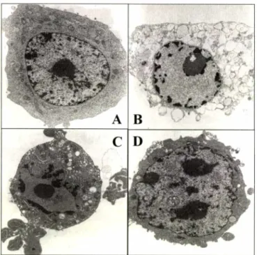
로죽은 세 포의 형태와도비교하기 위해 전자현미경 사진을 통해 분석해 보 았다.
그결과,약물을 처리하지 않은 정상세포는흑색종 세포의 특징인 커다란핵이 세포의 중심에 자리잡고 있었으며,
그 핵은 완전히 갖추어진 핵막으로둘러싸여 있었고,
비교적 매끄러운 세 포막을 가지고 있는 것으로 나타났다.
또한 골지체와 리소좀 둥 의세포 소기관들이 뚜렷이 보이는 반면, cisplatin
을 처리한 세포는
apoptotic body
라고할수 있는작은 입자들이 세포막주위에생겼으며
,
핵속의chrom atin
은농축되어 있었다. H eptaplatin
과sunpla
를처리한 세포에서도apoptotic body
라고 할 수 있는 입자들이 세포막 주위와 핵막 주위에 뭉쳐있음을 확인할 수 있 었고,
또한파괴되어진핵막을확인할수있었다.
특히,heptaplatin
D N A frag m e n tatio n
Apoptosis에의해죽게되는 세포의 가장 일반적인 특징 중 하 나는 agarose gel 전기영동상에서 나타나는 DNA fragmentation 이다
.
이것은 DNA가smear되는형태로나타나는 necrosis와확 실히 구분되는 장점을가지고있다.
따라서 cisplatin과 sunpla가 SK-MEL-28 세포의 DNA에미치는 영향을 알아보기 위해서 이 들각각의 약물을최종농도가0, 5, 10, 50 그리고 100 |oM이 되 게세포에 처리한후 agarose gel 전기영동을통해 DNA의변화 된모양을 확인해 보았다.
그 결과,
약물을 처리하지 않은 세포 의 DNA(control)은 절단된 형태로 나오지 않았고,
cisplatin과 sunpla를 처리한 세포의 DNA는 3시간이 지난후 처리한모든 농도에서 ladder 모양으로 절단된 형태를 띄고 있음을 확인하였 으며,그 절단된 모양이 모두 유사한형태인 것을 알수 있었다 (Fig. 2).T U N E L assay
사전에 보고되어진 cell cycle 분석 실험19〉과전자현미경에 의 한 분석 그리고 DNA fragmentation에 의한 분석들을 통해 cisplatin, heptaplatin 그리고 sunpla가 apoptosis^. SK-MEL-28
cisplatin
^ A
i i
_ _ _
heptaplatin
sunpla
cisplatin
■一 …. ,
heptaplatin
sunpla
Fluorescence Intensity (FL1)
- TUNEL
assay was performed to identify that apoptosis of SK-MEL-28 cells were induced by cisplatin, heptaplatin and sunpla. SK-MEL-28 cells were treated with concentrations of 0, 1, 5 and 10 fiM of each drugs and cultured for 24 hrs and 48 hrs. The black color is 0 |iM (control), the yellow color is 1 나M ,the green color is 5 (iM, the blue color is 10 uM.Fig. 1 -
Electronic photomicrographs present SK-MEL-28 cells which cultured with 100 (iM drug treatment for 24 hrs. A : control, B : cisplatin, C : heptaplatin, D : sunpla.cisplatin sunpla
M C 5 10 50 100 5 10 50 100 |
나M l
Fig. 2 -
DNA fragmentation of SK-MEL-28 cells by cisplatin and sunpla. SK-MEL 28 cells were treated concentrations of 0,5, 10,50 and 100 |iM of cisplatin and sunpla for 3 hrs. The markers indicate that M is a DNA marker, and C is a control which was cultured with media only.
을 처리한 세포에서는 세포막으로 둘러싸여져 가는 apoptotic body를 확인할 수 있었다
.
그리고 sunpla를 처리한 세포에서는 apoptosis의또다른특징인 chromatin을포함한 채다양한크기 로조각나 있는 핵을관찰할수 있었다(Fig. 1).세포를 게하는 것을 확인할 수 있었다
.
하지만,apoptosis와 necrosis를구분히는것에 대한이실험들이 가지고있는단점들24 hrs 48 hrs
SJaqEnN
II 9 CJ ig.
F
인체 흑색종 세포에서
Cisplatin, Heptaplatin, Sunpla에 의한
Apoptosis의 유도
1511
IOO^
iM
1 0 | iM
C
cisplatin
heptaplatin
unpla
]()(V!vf
l( U iM
1 y iM
■ᅵNippBwpy i
때문에
cisplatin, heptaplatin
그리고 선점:라이 세가지 약물이apoptosis
로세포를죽이는것을 더확실하게 확인하고,
또한 앞의결과들을더욱 확고하게 해줄수 있는
T U N EL assay
방법으■로실험을해보았다
. Cisplatin, heptaplatin
그리고sunpla
각 각의 최종농도를0, 1, 5,
그리고10 pM
이되게SK-MEL-28
세 포에 처리하고24
시간과48
시간동안배양히여flow cytometer
로분석하였다.
그 결과Fig. 3
에서 볼수 있듯이24
시간 배양 시cisplatin, heptaplatin
그리고sunpla
모두에서 농도가증가할 수록 형광강도가증가하고 있는것으로 나타났다.
이는이전 결 과들로부터 예상된 바와 같이cisplatin
에 의해 가장 크게apoptosis
가유도되어졌으며, sunpla
에의해서 가장 적게유도되었다
.
그리고48
시간배양시에는세가지 약물모두에 의해서24
시간 배양시보다형광강도가크게 증가되어졌는데
, sunpla
의경 우에는농도가증가함에 따라 형광강도가증가되는 것으로나타 났지만cisplatin
파heptaplatin
의경우에는비슷한잉상으로5 uM
Fluorescence Intensity (FL1)
Fig. 4 - Fas expression in SK-MEL-28 cells treated with 1,10 and
100 jiM of cisplatin, heptaplatin and sunpla, respectively, for 24 hrs. Fas expression experiment was performed as described in Materials and Methods.보다는 1 0
|iM
에서오히려 형광강도가감소했음을 알수 있었다.
이는D N A fragm entation
에서 보여진 결과와 상응하는 것으로sunpla
의 경우농도가중가할수록절단된D N A band
의 형광이 강해졌으며, cisplatin
의 경우는5
ᅣ싫을처리했을 때가lO ^ M
을처리했을 때보다절단된
DNA
의 양이 적음을알 수 있었다.
Fas expression
Fas
는세포의 표면에서 발현되어apoptosis
가일어나도록 유도한다고보고되어져있다
.^
따라서 세포에cisplatin, heptaplatin
그 리고sunpla
를각각 0,
1 , 1 0 그리고 1 0 0|iJVl
으로 처리하고24
시 간과48
시간 동안 배양한 후anti-CD95 F IT C
항 체(anti-Fas- FIT C
)를시용해 결합시킨후flow cytom etei
로 분석하였다. Fig.
4
에서 이세 가지 약물에 의해 농도와 시간에 의존적으로 형광강도가강해진 것을 볼수 있어
Fas
의발현이 약물의 농도가증가할수록그리고익물
i :
처리한후배양한 시간이 길어질수록증가한다는 것을 알수 있었다
.
따라서cisplatin, heptaplatin
그리 고sunpla
모두Fas
의 발현을증가시킴에 의해SK-MEL-28
세포를
apoptosis
로죽게하는기전임을 확인 할수 있었다.
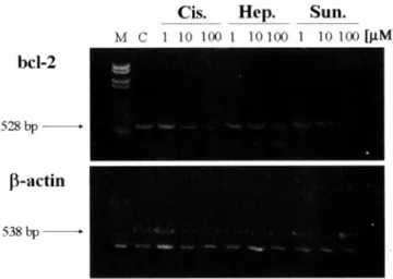
RT-PCR
에의한bcl-2
발현세포에서 일어나는 모든 현상은궁극적으로 어떠한 유전자가 발현또는 감소하느냐에 달려있다
.
따라서RT-PCR
벙법에 의해 서apoptosis
억제 유전자인bcl-
2의발현을 확인해 보았다.
이미 보고되어진 바에 의하면bcl-
2는apoptosis
억 제유 전 자(anti- apoptotic gene
)로써apoptosis
가증가할수록 세포내bcl-
2의 발 현은 감소하는 것으로 알려져 있다.
전술한 실험들에 의해Cis. H ep. Sun.
M C 1 10 100 1 10 100 1 10 100
[\lM]
bcl-2
528 bp ——
p -a c tin
538 bp ——
Fig. 5 - Bcl-2 gene expression in SK-MEL-28 cells treated with 1,
10 and 100 jiM of cisplatin, heptaplatin and sunpla, respectively, for 6hrs. Then, total RNA was isolated, and RT-PCR was perfomed and the DNAs were identified by 1% agarose gel electrophoresis. Lane M; protein size markers, C; control, 1; 1 fiM drug, 10; 10 (iM drug, 100;
100 liM drug, p-actin; house keeping gene.
001
s
s
3
s
0
cs oo
ᄋC
H S g s g o
81
80
95
8 0
£5 0 0
2acl£rlz10
cisplatin, heptaplatin 그리고 sunpla는 모두 농도가증가할수록 농도 의존적으로 apoptosis를중가시키는것을확인했으며
,
이를 바탕으로이들 약물의 농도가중가할수록b d -2의발현이 감소하 는지를 확인해 보았다.
세포에 cisplatin, heptaplatin, 그리고 sunpla를최종농도가0, 1,
10 그리고 100 nM 이되게 처리하여 배양한후
,세포내전체 RNA를문리하고cDNA를합성S]여 PCR 을수행하였다.
그 결과,
bcl-2의발현은약물처리 후 6시간을배 양한 세포에서 약물의농도가증가할수록그 발현이 감소하는것 을볼수있었다(Fig. 5). 즉 cisplatin, heptaplatin 그리고 sunpla 모두 1pM
보다는 1야iM
에서 발현이 더감소했으며 1 0nM
보 다는 100|oM에서더감소하였다.
따라서 이약물들은 SK-MEL- 28 cell에 대해 apotosis를유도하며,
그기전은 Fas의발현을증 가시킴에 의해 야기되는apoptosis 억제유전자인 bcl-2의발현감 소에 의한 것으로 확인되었다.
Fas
와bcl-2
유전자는수많은apoptosis
관련protein
들에의해 연관되어져 있음이 보고되어져 있는 바,
이 약물들에 의한caspase-1
또는caspase-3
등의 세포 내apoptosis
관련protein
들의 발현도 연구되어질 가치가있다고사료되어진다.
감사의 글
본 연구는 한국과학재단우수연구센터
(R-11-2002-100-03003)
의지원을받아수행되었기에 이에감사드립니다.
또한heptapaltin
과
sunpla
를 제공해주신 이화여자대학교 김대기 박사께 감사드립니다
.
문 헌
1) Trauth, B. C., Klas, C., Peters, A. M., Matzku, S., Moller, R, Falk, W, Debatin, K. M. and Krammer, E H .: Science 245,301 (1989).
2) McGahon, A. J., Martin, S. J., Bissonnette, R., Mahboubi, A., Shi, Y, Mogil, R., Nishioka, N. and Green, D. R. : The end of the cell line : methods for the study of apoptosis in vitro.
Method Cell Biol. 46,153 (1995).
3) Suda, T, Takahashi, T., Golstem, R and Nagata, S. : Molecular cloning and expression of the Fas ligand, a novel member of the tumor necrosis factor family. Cell 75, 1169 (1993).
4) Cho, Y. B.,Kim K. H. and Kim D. K. : Pharmacokinetucs, tissue distribution, and excretion of cis-malonato[(4R,5R)-4,5- bis(aminomethyl)-2-isopropyl-l,3-dioxolane]platinum( II) in dogs. Drug Metab. Dispos. 23,1280 (1995).
5) Owen-Schanb, L. B., Yonehara, S., Crump, W. D. and Grimm, E. A. : J. Cell Immunol.
140
,197 (1992).6) McGahon, A. J., Nishioka, W K.,Martin, S. J., Mahboubi, A., Cotter, T. G. and Green, D. R .: Regulation of the Fas Apoptotic cell Death Pathway by Abl. J. Biol. Chem. 270, 22625 (1995).
7) Orlinick, J. R., Vaishnaw, A., Elkon, K. B. and Chao. M. V : Requirement of cystein-rich receptors of the Fas Receptors for Binding by the Fas Ligand. J. Biol Chem. 272, 28889 (1997).
8) Itoh, N., Yonehara, S., Ishil, A., Yonehara, M.’ Mizushima, S., Sameshima, M., Hase, A., Seto, Y. and Negata, S. : The polypeptide encoded by the cDNA for human cell surface antigen Fas can mediate apoptosis. Cell 66,233 (1991).
9) Oehm, A., Behrmann, I., Falk, W, Pawlita, M., Maier, G., Klas, C.,Li-Weber, M., Richards, S., Dhein, J., Trauth, B. C., Ponstingl, H. and Krammer, R H. : Purification and molecular cloning of the APO-1 cell surface antigen, a member of the tumor necrosis factor/nerve growth factor receptor super
family, sequence identity with the Fasantigen. /. Biol. Chem.
267,10709 (1992).
10) Yonehara, S., Ishii, A. and Yonehara, M. : A cell-killing monoclonal antibidy (anti-Fas) to a cell surface antigene co
down regulated with the receptor of tumor necrosis factor. J.
Exp. Med. 169, 1747 (1989).
11) Trauth, B. C.,Klas, C., Peters, A. M. J., Matzku, S., Moller, R, Falk, W., Debatin, K. M. and Krammer, R H .: Science 245, 301 (1989).
12) Nagata, S. and Golstein, R : Science 267,1449 (1995).
13) Boise, L. H.,Gottschalk, A. R., Quintans, J. and Thombson, C.
B. : Cum Topics Microbiol. Immunol. 200, 107 (1995).
14) Hockenbery, D. M. : Bcl-2, a novel regulator of cell death.
BioEssays 17,631 (1995).
15) Korsmeyer S. J. : Bcl-2 initiate a new category of oncogenes;
regulators of cell death. Bolld 80,879 (1992).
16) Oltvai Z. N.,Milliman, C. L. and Korsmeyer, S. J. : Bcl-2 heterodimerizes in vivo with a conserved homolog box, that accelerates programmed cell death. Cell 369, 609 (1991).
17) Ym, X. M.,Oltvai, Z. N. and Korsmeyer, S. J. : BH1 and BH2 domains of bcl-2 are required for inhibition of apoptosis and heterodimerization with bax. Nature 369,321 (1994).
18)
서정선:
2 0세기말의 의•
생물학의 새로운비전,
아포프토시스 (Apoptosis). Korean Society of Medical Biochemistry and Molecular Biology News. 30.19) Choi, S. L. and Myung, R K .: Cell viability and flow cytometry analysis of a novel antitumor agent, Heptaplatin in human melanoma cell line, SK-MEL-28. Yakhak Hoeji 47(6),345 (2003).
20) Kerr, J. E R.,Wyllie, A. H. and Currie, A. R. : Apoptosis : a basic biological phenomenon with wide-ranging implications in tissue kinetics. Br. J. Cancer 26, 239 (1972).