Copyright ⓒ 2010, The Microbiological Society of Korea
갓김치에서 분리된 유산균의 활성산소종에 대한 저항성과 항산화 활성
임성미
*
동명대학교 식품공학과Resistance to Reactive Oxygen Species and Antioxidant Activities of Some Strains of Lactic Acid Bacteria from the
Mustard Leaf Kimchi
Sung-Mee Lim
Department of Food Science and Technology, Tongmyong University, Busan 608-735, Republic of Korea (Received November 10, 2010/Accepted December 21, 2010)
In present study, five strains of Lactobacillus acidophilus GK20, Lactobacillus brevis GK55, Lacto- bacillus paracasei GK74, Lactobacillus plantarum GK81, and Leuconostoc mesenteroides GK104 isolated from the mustard leaf kimchi were investigated for resistance to reactive oxygen species (ROS) and antioxidant activity. L. acidophilus GK20, L. brevis GK55, L. paracasei GK74, and L. plantarum GK81 were resistant to hydrogen peroxide (0.5 mM), showing a survival rate of 50% or more. In particular, L. acidophilus GK20 and L. paracasei GK74 were the most superoxide anions-resistant and L. paracasei GK74 and L. plantarum GK81 were most likely survive hydroxyl radicals. Meanwhile, the intracellular cell-free extract (ICFE) from L. plantarum GK81 exhibited significantly higher DPPH radical scavenging values (96.4±2.8%) than the intact cells (IC). The ICFE of L. plantarum GK81 showed the highest superoxide radical scavenging ability and chelating activity for Fe
2+ions among the 5 lactic acid bacteria (LAB) tested, and IC and ICFE from L. plantarum GK81 demonstrated excellent reducing activity, which was higher than those of BHA and vitamin C as a positive control.
Keywords: Fe
2+-chelating activity, Lactobacillus plantarum, radical scavenging ability, reactive oxygen species, reducing activity
산화적 스트레스(oxidative stress)는 살아있는 생명체에 유 해한 생화학 반응을 유발하고 죽상동맥경화증을 비롯한 각종 혈관계 질환, 돌연변이, 암, 신경퇴행성질환, 면역기능 장애 및 노화의 원인으로 알려져 있다(7). 생체의 에너지 생산을 위한 호흡 대사 과정 중 일정량의 산소는 반응성이 높은 활성산소종 (reactive oxygen species, ROS)이라는 유해 물질을 생성하게 되는데 그 종류로는 hydroxyl radical (OH·), superoxide anion radical(O2¯·), peroxyl radical (RO2·)과 같이 쌍을 이루지 못 한 전자를 가지는 free radical과 과산화수소(H2O2), 일중항산 소(singlet oxygen) 및 오존(O3) 등이 있다(30). Free radical에 의해 생성된 단백질 산화생성물은 세포 내 촉매 기능을 담당하 고 있는 효소의 기능을 상실케 하며(26), 또한 다가불포화 지 방산과 콜레스테롤의 과산화를 유발하여 2차 산물인 malondial-
* For correspondence. E-mail: [email protected]; Tel: +82-51-629- 1714; Fax: +82-51-629-1709
dehyde나 4-hydroxynonenal 등의 지질 과산화물을 축적하여 세포막 투과성의 변화와 단백질합성 능력을 감소시킨다(12).
또한 ROS는 세포의 DNA 가닥을 분리시키고 자매 염색분체 의 교환을 유도하여 악성 형질 전환을 유발하여 발암의 요인이 된다(29).
활성산소를 소거하는 기능이 있는 항산화 물질로는 vitamin C, glutathione, uric acid와 같은 수용성 항산화제, tocopherol, carotenoids, flavonoid/flavonoids 등의 지용성 항산화제, tert- butylhydroxytoluene (BHT), tert-butylhydroxyanisol (BHA) 등의 합성 항산화제, superoxide dismutase (SOD), catalase, glutathione peroxidase 등의 항산화효소가 있으며(7) 이들은 활성산소 중간물질을 제거하여 산화적 손상으로부터 세포를 보호하고 세포의 자연사(apoptosis)에 대하여 저항할 수 있다 (3, 35). 합성 항산화제는 산화 억제 효과는 뛰어나지만, 독성 에 의한 여러 부작용을 초래할 수 있으므로 최근 천연물을 이
용한 항산화력 측정에 관한 연구가 활발히 진행되고 있다. 가 자, 소목, 지유, 복분자 및 초두구 등 다양한 한약재들은 토코 페롤 보다 더 높은 항산화력이 있으며(20), lemon vervena, chamomile 등의 허브류 및 오레가노, 육두구, 계피 등의 향신 료들도 높은 항산화력이 있는 것으로 보고되고 있다(8, 33). 한 편 일부 미생물의 Mn-SOD, Cu-SOD는 활성산소를 비독성화 시키고(39), NADH, NADPH, glutathione 및 uric acid 같은 항산화제를 생산하여 hydroxyl radical을 분해할 수 있다고 알 려져 있다(1). Bifidobacterium infantis, Bifidobacterium breve, Bifidobacterium adolescentis 및 Bifidobacterium longum는 NADH peroxidase에 의해 hydrogen peroxide를 분해할 수 있 었다(32). 김치에서 분리된 Lactobacillus plantarum의 세포와 세포를 파쇄하여 얻은 세포추출물은 지질 과산화를 저해하였 는데 이는 SOD 활성에 기인하기 보다는 금속이온을 chelating 하는 활성이 있어 높은 항산화력을 나타내었는데 이들은 ROS 에 대한 저항력도 강한 것으로 밝혀졌다(16). 한편, Lacto- bacillus lactis 및 L. plantarum와 같이 SOD를 생산하는 유산 균은 대장의 염증 개선에도 효과가 있는 것으로 알려졌다(37).
본 연구는 probiotic bacteria의 선정 기준의 하나인 항산화 활성을 조사하기 위해 갓김치에서 분리된 유산균 5종을 대상 으로 hydrogen peroxide, superoxide anions 및 hydroxyl radical 등의 ROS에 대한 저항력과 DPPH radical, superoxide radical 소거능, 환원력 및 Fe2+ chelating 활성 등을 측정하여 산화 억제 능력을 실험한 결과이다.
재료 및 방법
사용배지 및 시약
유산균 분리 및 배양에 사용된 배지 Lactobacilli MRS agar 및 broth는 Difco (USA)사 제품, 분리된 유산균의 동정에 사 용된 API 50 CHL kit는 bioMériux (France)사 제품, ROS에 대한 저항성 측정과 radical 소거능, 환원력 및 Fe2+-chelating 활성 측정에 사용된 시약은 모두 Sigma Co. (USA)에서 구입 하여 사용하였다.
유산균 분리 및 동정
가정에서 담궈 3개월 가량 숙성시킨 갓김치(pH 3.8-5.2) 8 종을 수집하여 채취한 시료 50 g과 멸균생리식염수(0.85%
NaCl 용액) 450 ml을 혼합하여 2분간 균질화한 후 시료용액 1 ml를 1% CaCO3가 첨가된 Lactobacilli MRS agar 배지에 접종하여 37°C, 24-48시간 동안 미호기성 조건(10% O2, Anoxomat system, MART Co., Netherland)으로 배양한 후 투 명한 환을 생성하는 독립된 집락을 사면배지에 3회 계대 배양 하여 활성을 높인 후 사용하였다. 선발된 5종의 유산균을 대상 으로 Bergey’s Manual of Determinative Bacteriology (27)에 따라 배양학적 및 생화학적 특성과 API 50 CHL를 이용하여 당 발효능을 조사하여 추정되는 유산균명을 확인하였다.
ROS에 대한 저항성
Hydrogen peroxide에 대한 저항성 : H2O2에 대한 유산균 의 저항성은 Fu 등(11)의 방법을 일부 변형하여 측정하였다.
분리된 유산균은 Lactobacilli MRS broth 배지에 접종하여 37°C, 18시간 미호기적 조건에서 배양한 후 원심분리(7,000×g, 20분, 4°C)한 다음 phosphate buffered saline (PBS; 0.85%
NaCl, 2.68 mM KCl, 10 mM Na2HPO4 및 1.76 mM KH2PO4)으로 3회 세척하여 세포수 약 108 CFU/ml로 맞춰 동 일한 buffer에서 현탁시켰다. 0.5 mM H2O2 용액(35% H2O2
0.043 ml/DW 1,000 ml) 500 μl와 동량의 세포현탁액을 즉시 혼합하여 60분 간격으로 총 5시간 반응시킨 후 잔존하는 H2O2
을 제거하기 위해 다시 원심분리(20,000×g, 1분, 4°C)하여 pellet을 회수하였다. Pellet을 buffer에 재현탁 시킨 다음 단계 별로 희석하고 Lactobacilli MRS agar에 평판배양(37°C, 24시 간)하여 생성된 집락수를 측정하여 초기 균수에 대한 감소율을 나타내었다.
Superoxide anions에 대한 저항성 : Paraquat(1,1’-dimethyl- 4,4’-bipyridinium)에 의해 유도된 superoxide anions에 대한 유산균의 저항성은 Bauer 등(5)의 diffusion assay법에 따라 측 정하였다. 37°C에서 18시간 배양한 유산균은 원심분리(7,000×g, 20분, 4°C)하여 세포를 모으고 PBS로 3회 세척한 현탁 세포액 (약 108 CFU/ml) 100 μl를 Lactobacilli MRS agar 평판배지 위에 도말한 후 멸균수에 용해시킨 10 mM paraquat의 10 μl 를 균 접종한 평판배지 위에 spot하여 37°C에서 24시간 배양 하여 생성된 저해환을 측정하였다.
Hydroxyl radical에 대한 저항성 : Fenton 반응을 통해 발 생된 hydroxyl radical에 대한 유산균의 저항성은 Barreto 등 (4)에 따라 일부 변형하여 측정하였다. PBS에 용해시킨 10 mM terephthalic acid (1,4-benzenedicarboxylic acid), 유산균 세포(108 CFU/ml) 및 0.01 mM CuSO4·5H2O가 포함된 용액 내에서 hydroxyl radical을 발생시킨 다음 1 mM H2O2를 첨가 하여 반응시켜 15분 간격으로 용액을 채취한 다음 Lactobacilli MRS agar를 이용하여 생균수를 측정하여 저해율을 계산하였 다.
항산화 활성 측정
유산균의 항산화력 측정에 사용된 유산균의 세포는 Lin과 Chang (22)의 방법에 따라 준비하였다. 즉, Lactobacilli MRS broth 배지에 접종하여 37°C, 18시간 배양한 배양액을 원심분 리(7,000×g, 20분, 4°C)하여 회수한 pellet (intact cell, IC)은 PBS로 3회 세척 후 PBS에 재현탁하였다. 한편 intracellular cell-free extract (ICFE)는 IC를 탈이온수로 2회 세척한 후 ice- bath상에서 10분간 세포를 초음파(Qsonical, USA) 파쇄한 후 10,000×g, 10분간 원심분리하여 세포 파편을 제거하고 상등액 만을 회수하여 membrane filter (0.45 μm, Millipore Corp., USA)로 여과하여 얻었다. 유산균의 IC와 ICFE 항산화 활성은 DPPH (1,1-diphenyl-2-picryl hydrazyl), superoxide radical 소 거능 및 환원력을 측정하여 평가하였다. 양성 대조구로서 100 mg/L vitamin C 및 BHA을 사용하여 활성을 비교하였다.
Inhibition(%)
Incubation time with H2O2(h)
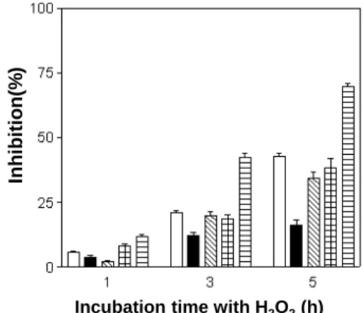
Fig. 1. Resistance to hydrogen peroxide (0.5 mM) of various lactic acid bacteria isolated from the mustard leaf Kimchi. Data represent the means of three experiments±standard deviation (error bars). (□), L. acidophilus GK20; (■), L. brevis GK55; (▧), L. paracasei GK74;
(▦), L. plantarum GK81; (▤), L. mesenteroides GK104.
DPPH radical 소거능 측정 : Brand-Williams 등(6)의 방 법을 일부 변형하여 DPPH radical 소거능을 측정하였다. 유산 균 IC 혹은 ICFE 100 μl에 에탄올에 녹인 4.0×10⁻⁴ M DPPH 용액 100 μl를 96 well plate에 loading한 다음 빛을 차단하여 20°C에서 30분간 반응시킨 후 517 nm에서 microplate reader (Spectrocount; Packard Instruments, USA)로 흡광도를 측정하 여 다음 식에 따라 DPPH radical 소거능을 계산하였다. DPPH radical 소거능 (%) = 1-(시험구 흡광도/ 대조구 흡광도) × 100
Superoxide radical 소거능 측정 : Superoxide radical 소 거능은 Kim 등(15) 방법에 따라 측정하였다. 즉, 96 well plate 에 sodium carbonate buffer (pH 10.0), xanthine, ethylenedia- minetetracetic acid (EDTA), bovine serum albumin 및 nitro blue tetrazolium 혼합용액 100 μl와 유산균의 IC 혹은 ICFE 100 μl를 가하고, xanthine oxidase를 최종적으로 12 mU가 되 도록 첨가하여 25°C에서 20분간 반응시킨 후 CuCl로 반응을 정지시킨 후 560 nm에서 흡광도를 측정하여 다음 식에 따라 superoxide radical 소거능을 계산하였다. Superoxide radical 소거능 (%) = 1-(시험구 흡광도/ 대조구 흡광도) × 100
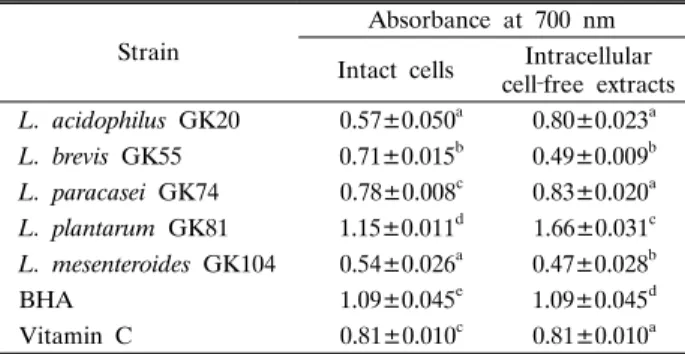
환원력 측정 : 유산균의 환원력은 Oyaizu (24)의 방법을 일 부 변형하여 측정하였다. 즉, 유산균 IC 혹은 ICFE (100 μl)은 1% potassium ferric cyanide (100 μl)와 sodium phosphate buffer (0.2 M, pH 7.0)(100 μl)를 혼합하여 50°C에서 20분간 반응시켰다. 반응 후 10% trichloroacetic acid를 첨가하여 3,000×g에서 5분간 원심분리하여 얻은 상등액은 0.1% ferric chloride 200 μl와 혼합하여 700 nm에서 흡광도를 측정하였다.
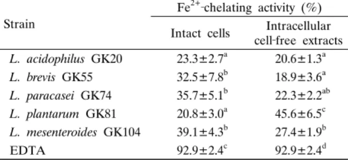
Fe2+-chelating 활성 측정 : 유산균의 Fe2+-chelating 능력 측정은 Yu 등(38) 방법에 따라 측정하였다. 유산균의 ICFE 0.2 ml, Tris-HCl buffer (pH 7.4) 0.8 ml, 1.8 mM FeSO4 0.1 ml, 10% hydroxylamine-HCl 0.32 ml 및 0.2 N HCl에 용해한 0.1% 2,2’-bipyridyl 0.8 ml을 혼합하여 522 nm에서 흡광도를 측정하여 Fe2+-chelating 활성을 측정하여 다음 식에 따라 계산 하였고, 이때 양성 대조구로서는 100 mg/L EDTA를 사용하였 다. Chelating 활성 (%) = 1-(시험구 흡광도/ 대조구 흡광도) × 100
통계처리
실험은 모두 3회 반복하여 실시하였으며, 유산균주 간의 차 이는 SPSS (version 12.0, USA) 프로그램의 분산분석(ANOVA) 을 이용하여 비교하였고, 평균간 유의성은 Duncan’s multiple range test를 사용하여 p<0.05 수준에서 시험구들과 대조구 간 의 유의적 차이를 검정하였다.
결과 및 고찰
갓김치로부터 유산균 분리
가정에서 담궈 숙성시킨 갓김치 8종을 수집하여 Lactobacilli MRS agar 평판배지에 배양하여 전형적인 유산균으로 추정되 는 균을 대상으로 생화학적 특성 및 당 발효능을 조사한 결과 는 Table 1과 같다. GK20은 L형 lactic acid 생산, catalase 음
성, lysine과 ornithine 가수분해 및 45°C 하에서 증식하며 arabinose, ribose, mannose 및 raffinose 등을 분해하고 동정 확률(ID) 99.9%를 나타내어 Lactobacillus acidophilus로 확인 되었다. GK55는 D형 lactic acid 생산, catalase 양성, arginine 가수분해하고 혐기적 조건하에서 증식하지 못하고 xylose, maltose, melibiose 및 gluconate 등을 이용하여 Lactobacillus brevis (ID=97.2%)로 추정된다. 또한 GK74는 glucose로부터 가스 생산, L형 lactic acid 생산, catalase 음성, 1-5% NaCl 하 에서 증식하며 sorbose, sorbitol, tagatose 및 arabitol 등을 발 효하는 능력이 있어 Lactobacillus paracasei (ID=99.2%)인 것 으로 추정된다. GK81은 L형 lactic acid 생산, catalase 양성, ornithine 가수분해, 호기적 조건하에서만 증식하며 mannitol, α -Methyl-D-mannoside, melezitose 및 gentiobiose 등의 당을 분해하는 능력이 있으므로 L. plantarum (ID=99.9%)으로 확인 되었다. GK104의 세포는 구형이며 D형 lactic acid 생산, catalase 음성, 45°C에서 증식할 수 없고, 혐기적 조건하에서만 증식하며 α-Methyl-D-glucoside, N-Acetyl glucosamine, salicine 및 turanose 등을 이용하는 능력이 있어 Leuconostoc mesenteroides (ID=99.8%)로 추정된다.
Park 등(25)에 따르면 갓김치 담금 직후 유산균수는 약 4.5 log cycle에서 숙성이 진행됨에 따라 서서히 증가하여 숙성 14 일만에 7 log cycle에 이르렀다고 보고하였으며, Shim과 Lee (31)에 따르면, recA 유전자를 특이적으로 증폭하는 PCR을 이 용하여 갓김치 내에 있는 유산균을 조사한 결과, L. plantarum 및 L. mesenteroides가 검출되었다고 하여 본 실험에서 분리된 유산균의 종류와 일부 일치하였다.
ROS에 대한 유산균의 저항성
Hydrogen peroxide, superoxide anions 및 hydroxyl radical 에 대한 분리 유산균의 저항성을 알아본 결과는 각각 Fig. 1, 2 및 3과 같다. 0.5 mM hydrogen peroxide에 대한 유산균의 저 항성 결과, 1시간 반응 후 대부분 균들의 저해율은 10% 내외 정도였으나, 반응 3시간 만에는 L. brevis GK55의 저해율이
Table 1. Differentiating characteristics and carbohydrate reactions of lactic acid bacteria from the mustard leaf Kimchi Contents GK 20 GK 55 GK 74 GK 81
GK 10Sugar 4
GK 20 GK 55 GK 74 GK 81 GK 104
Sugar
GK 20 GK 55 GK 74 GK 81 GK 104
Cell shapeRodsRodsRodsRodsCocciGlycerol‐‐‐‐‐Salicine+‐+++ Gram staining+++++Erythritol‐‐‐‐‐Cellobiose+‐++‐ Spores staining‐‐‐‐‐D‐Arabinose‐‐‐‐‐Maltose+++++ Acid‐fast staining‐‐‐‐‐L‐Arabinose++‐++Lactose+‐++‐ Motility‐‐‐‐‐Ribose+++++Melibiose++‐++ Gas from glucose‐++‐‐D‐Xylose‐+‐‐+Saccharose+‐+++ H2S production‐‐‐‐‐L‐Xylose‐‐‐‐‐Trehalose+‐+++ Lactic acidLDLLDAdonitol‐‐‐‐‐Inuline‐‐‐‐‐ Methyl red+++++β‐Methyl‐xyloside‐‐‐‐‐Melezitose‐‐++‐ Voges‐Proskauer‐‐‐‐‐Galactose+++++D‐Raffinose+‐‐‐+ Horse blood hemolysis‐‐‐‐‐D‐Glucose+++++Amidon‐‐‐‐‐ Sheep blood hemolysis‐‐‐‐‐D‐Fructose+++++Glycogene‐‐‐‐‐ Catalase‐+‐+‐D‐Mannose+‐+++Xylitol‐‐‐‐‐ Oxidase‐‐‐‐‐L‐Sorbose‐‐+‐‐β‐Gentiobiose‐‐++‐ Urease‐‐‐‐‐Rhamnose‐‐‐‐‐D‐Turanose‐‐+‐+ Arginine hydrolysis‐++‐‐Dulcitol‐‐‐‐‐D‐Lyxose‐‐‐‐‐ Lysine++‐‐+Inositol‐‐‐‐‐D‐Tagatose‐‐+‐‐ Ornithine+‐+++Mannitol‐‐+++D‐Fucose‐‐‐‐‐ Growth inaerobic condition++++‐Sorbitol‐‐++‐L‐Fucose‐‐‐‐‐ anaerobic+‐+‐+α‐Methyl‐D‐mannoside‐‐‐+‐D‐Arabitol‐‐+‐‐ Growth at 45℃++++‐α‐Methyl‐D‐glucoside‐‐‐‐+L‐Arabitol‐‐‐‐‐ Growth at pH 5.0‐9.0+++++N‐Acetyl glucosamine‐‐+++Gluconate‐++‐‐ Growth in1‐5% NaCl+++++Amygdaline+‐++‐2‐Ceto‐gluconate‐‐‐‐‐ 10% NaCl‐‐‐‐‐Arbutine‐‐++‐5‐Ceto‐gluconate‐‐‐‐‐ Esculine+‐+++
GK20 GK55 GK74 GK81 GK104 0
5 10 15 20
Strain
Clear zone (mm)
Fig. 2. Resistance to superoxide anions of various lactic acid bacteria isolated from the mustard leaf Kimchi. Data represent the means of three experiments±SD (error bars).
Inhibition(%)
Incubation time with hydroxyl radicals (min) Fig. 3. Resistance to hydroxyl radicals of various lactic acid bacteria isolated from the mustard leaf Kimchi. Data represent the means of three experiments±standard deviation (error bars). (□), L.
acidophilus GK20; (■), L. brevis GK55; (▧), L. paracasei GK74;
(▦), L. plantarum GK81; (▤), L. mesenteroides GK104.
12.2%로 가장 낮은 반면, L. mesenteroides GK104의 저해율 은 42.4%로 가장 높았다. 또한 5시간 후에도 L. brevis GK55 의 생존율은 83.6%로 가장 높았고, 그 다음으로 L. acidophilus GK20, L. paracasei GK74 및 L. plantarum GK81도 50% 이 상의 생존율을 보여 비교적 안정하였으나, L. mesenteroides GK104의 생존율은 약 30.1%에 불과하여 실험 균주들 중 가 장 낮은 저항성을 나타내었다. 일반적으로 유산균은 catalase 음성 반응을 나타내는 것으로 알려져 있는데, 본 실험에 사용 된 L. brevis GK55와 L. plantarum GK81는 특이하게 catalase 를 생성하는 것으로 확인되었으며, 이들이 H2O2에 대한 저항 성이 높은 것은 catalase 생성 능력에 기인하는 것으로 추정된다.
한편, 유산균을 접종한 평판배지에 superoxide anions을 유 도하는 paraquat을 spot하여 배양한 후 생성된 저해환의 크기 를 측정하였다. Paraquat는 확산을 통해 세포막을 통과하여 산 소와 반응한 결과 superoxide anions을 생성하여 세포에 유해 성분으로 작용한다(34). L. acidophilus GK20와 L. paracasei GK74의 저해 정도가 가장 낮았으며, 반면 L. mesenteroides GK104에 대한 저해환의 크기가 가장 크게 나타났으므로 superoxide anions에 대한 저항성도 균주의 종류에 따라 다소 차이가 있었다.
Hydroxyl radical에 대한 저항성을 조사한 결과, 반응 15분 만에 L. acidophilus GK20, L. plantarum GK81 및 L. mesenter- oides GK104들의 생균 감소율은 약 20-25% 정도로 이들 균 주간에는 유의적인 차이가 없었으며, 이는 L. brevis GK55에 비해 유의적으로 다소 낮았다. 모든 유산균들은 반응시간이 경 과함에 따라 저해율이 서서히 증가하는 경향을 나타내었는데, 반응 60분 후에는 L. acidophilus GK20, L. brevis GK55 및 L. mesenteroides GK104는 95% 이상의 높은 사멸율을 보인 반면, L. paracasei GK74는 약 24.4%, L. plantarum GK81는 약 33.7% 정도 생존한 것으로 나타났다.
Kullisaar 등(17)에 따르면, 항산화력이 없는 Lactobacillus fermentum E-338-1-1 균주는 0.4 mM hydrogen peroxide와 반응시켜 240분 동안에만 생존이 가능하였으나, 항산화력이 있 는 L. fermentum E-3과 E-18 균주는 각각 450과 390분 후에
도 생존하였으며, 1 mM 농도 하에서 E-3와 E-18 균주는 180 및 150분 동안 생존하였다고 보고하였다. 또한 E-338-1-1은 hydroxyl radicals 존재 하에서 약 15분 정도에만 생존하였으 나, E-3과 E-18은 34분 동안 생존 가능하였으며, 또한 이들 균 주는 superoxide anions에 대한 저항성도 있는 것으로 보고하 였다. Lee 등(19)은 김치에서 분리된 L. plantarum KCTC 3099 는 1 mM hydrogen peroxide와 0.4 mM hydroxyl radical의 존재 하에 8시간 반응시킨 후에도 생존하였고, paraquat에 의 해 발생된 superoxide anion에 의해 영향을 받지 않는 것으로 나타났다.
유산균의 항산화 활성
갓김치에서 분리된 유산균들의 IC와 ICFE에 대한 DPPH radical 소거 활성을 측정한 결과는 Table 2와 같다. IC에 의한 DPPH radical 소거능은 L. acidophilus GK20, L. brevis GK55 및 L. mesenteroides GK104들은 5% 유의수준에서 차 이가 없이 다른 2종의 유산균에 비해 낮은 활성을 보인 반면, L. paracasei GK74의 소거능은 47.6±6.3%에 이르렀고, L.
plantarum GK81은 실험 균주 중 가장 높은 소거능(70.8±
10.1%)을 나타내었다. 하지만 이들의 활성은 양성 대조구인 BHA나 vitamin C보다는 낮은 수준이었다. 한편, ICFE의 소거 능을 측정한 결과, 가장 낮은 소거능은 L. brevis GK55와 L.
mesenteroides GK104에서 나타났고, 가장 높은 소거능은 L.
plantarum GK81에서 나타났는데 이들의 소거 활성은 BHA와 vitamin C와 유의적인 차이가 없을 정도로 높게 나타났다. 또 한 동일한 균주의 IC와 ICFE들간의 활성을 비교한 결과, GK20, GK74 및 GK81은 ICFE에서 더 높은 활성을 보인 반 면, GK55와 GK104의 경우는 IC에서 더 높은 소거능을 나타 내었다.
Superoxide radical의 소거능을 측정한 결과는 Table 3과 같 다. IC의 superoxide radical 소거능은 L. mesenteroides GK104
Table 2. DPPH radical scavenging activity of various lactic acid bacteria isolated from the mustard leaf Kimchi
Strain
DPPH radical scavenging activity (%) Intact cells Intracellular
cell‐free extracts L. acidophilus GK20 31.1±8.6a 55.6±8.7a L. brevis GK55 42.2±7.4ab 26.0±3.9b L. paracasei GK74 47.6±6.3b 61.5±5.6a L. plantarum GK81 70.8±10.1c 96.4±2.8c L. mesenteroides GK104 30.7±1.8a 24.3±4.0b
BHA 89.1±2.9d 89.1±2.9c
Vitamin C 95.5±0.5d 95.5±0.5c
DPPH radical scavenging activity was defined as [1‐A517(sample)/
A517(control)] × 100%.
Data represent the means of three experiments±SD (error bars).
a‐d Means with different letters in a column are significantly different at p<0.05 by Duncan’s multiple range test.
Table 3. Superoxide radical scavenging activity of various lactic acid bacteria isolated from the mustard leaf Kimchi
Strain
Superoxide radical scavenging activity (%)
Intact cells Intracellular cell‐free extracts L. acidophilus GK20 42.1±7.9a 65.6±9.0a L. brevis GK55 66.2±2.4b 50.0±5.6b L. paracasei GK74 53.1±3.3c 69.8±0.2a L. plantarum GK81 62.1±8.8b 80.6±4.7c L. mesenteroides GK104 19.9±1.3d 18.0±0.5d
BHA 90.3±1.7e 90.3±1.7e
Vitamin C 97.9±0.8e 97.9±0.8e
Superoxide radical scavenging activity was defined as [1‐A560(sample)/
A560(control)]×100%.
Data represent the means of three experiments±SD (error bars).
a‐e Means with different letters in a column are significantly different at p<0.05 by Duncan’s multiple range test.
Table 4. Reducing activity of various lactic acid bacteria isolated from the mustard leaf Kimchi
Strain
Absorbance at 700 nm Intact cells Intracellular
cell‐free extracts L. acidophilus GK20 0.57±0.050a 0.80±0.023a L. brevis GK55 0.71±0.015b 0.49±0.009b L. paracasei GK74 0.78±0.008c 0.83±0.020a L. plantarum GK81 1.15±0.011d 1.66±0.031c L. mesenteroides GK104 0.54±0.026a 0.47±0.028b
BHA 1.09±0.045e 1.09±0.045d
Vitamin C 0.81±0.010c 0.81±0.010a Data represent the means of three experiments±SD (error bars).
a‐e Means with different letters in a column are significantly different at p<0.05 by Duncan’s multiple range test.
에서 가장 낮은(19.9±1.3%) 반면, L. brevis GK55와 L.
plantarum GK81은 각각 66.2±2.4%와 62.1±8.8%로 높은 활 성을 나타내었다. ICFE의 경우에도 L. mesenteroides GK104 에서 가장 낮은(18.0±0.5%) 반면, L. plantarum GK81의 소거 활성이 가장 높게 나타났으나(80.6±4.7%), 이는 BHA와 vitamin C의 활성과는 유의적인 차이가 있었다.
산화 과정에 생성된 free radical을 비롯한 각종 ROS 및 독 성 생성물들은 생물학적 분자를 공격하여 생체 내 세포를 손상 시킨다. DPPH radical은 화학적으로 유도되는 비교적 안정한 radical로서 다른 원자나 분자로부터 전자를 공여 받아 비가역 적으로 결합하면 보라색이 엷어지면서 흡광도가 감소하게 되 는 원리를 이용하여 radical 소거 활성을 측정할 수 있다.
DPPH radical 소거능이 높으면 free radical을 환원시키거나 상쇄시키는 능력이 높아 항산화 활성이 있다는 것을 의미한다 (2). Cho 등(9)은 김치에서 분리한 L. plantarum YS712 균주 가 95.8%로 가장 높은 radical 소거능을 나타내었으며, L.
plantarum ATCC 8014 (95.3%), L. plantarum L155 (95.2%), Enterococcus sp. 01 (94.9%), Lactococcus sp. KU107
(94.6%) 순으로 나타났다고 보고한 바 있다. Kaizu 등(14)의 보고에 따르면, 7종의 lactobacilli 균주는 70%의 산화 억제 효 과를 나타내었고, 이 중에서 헤테로발효형인 Lactobacillus sp.
SBT 2038의 활성이 가장 높았으며, 특히 Lactobacillus sp.
SBT 2028와 L. casei ssp. rhamnosus SBT 2257의 ICFE는 비타민 E가 결핍된 식이를 투여한 rat에게서도 항산화 효과가 뚜렷하게 나타났다고 보고하였다. Lee 등(18)은 γ-amino- butyric acid를 생산하는 L. brevis BJ20에 의해 발효된 다시마 는 양성 대조구 BHA보다 더 높은 DPPH, superoxide 소거 활 성을 나타내었고, xanthine oxidase 저해능도 뛰어났다고 보고 하였다. 한편, 결장암 유발 원인의 하나인 결장 내의 산화적 스 트레스 억제 효과를 조사한 결과, L. casei strain Shirota로 발 효시킨 탈지유에 의해 2,2-diphenyl-1-picrylhydrazyl radical 소 거 활성이 증가되었다고 하였고, hamster에게 14일간 발효 탈 지유를 투여했을 때 가열 처리한 탈지유에 비해 radical 소거능 이 높게 나타났다고 하였다(23). L. casei, L. acidophilus, L.
lactis는 강력한 2,2-diphenyl-1-picrylhydrazyl, malonaldialdehyde, hydrogen peroxide radical 소거능이 있고, 또한 linoeic acid 과산화 억제 효과는 L. casei 활성이 가장 높고, L. acidophilus 와 L. lactis 순으로 항산화 효과가 나타났다고 하였다(13). 또 한 L. mesenteroides ssp. cremoris, Lactobacillus jensenii ATCC 25258 및 L. acidophilus ATCC 4356도 높은 radical 소거능을 보였는데, 이들의 활성은 단백질 분해와 관련이 있으며, 높은 항산화 활성을 가지는 부분은 4-20 kDa의 분자량 범위 내에서 많은 펩타이드를 가지며, 특히 소수성 아미노산의 함량이 더 높게 나타났다고 알려져 있다(36).
유산균의 IC와 ICFE의 환원력 측정을 위해 700 nm에서 흡 광도를 측정한 결과는 Table 4와 같다. IC에 의한 환원력은 L.
acidophilus GK20와 L. mesenteroides GK104에 의해 가장 낮 게 나타난 반면, L. plantarum GK81의 환원력은 가장 높게 나 타났는데 이는 BHA와 vitamin C보다 유의적으로 더 높게 나 타났으며, L. paracasei GK74의 환원력도 vitamin C와 비슷한 수준인 것으로 확인되었다. 한편, L. plantarum GK81의 ICFE 환원력도 실험 균주 중 가장 높았으며, 양성 대조구 보다도 유 의적으로 높았다. 또한 L. acidophilus GK20, L. paracasei
Table 5. Fe2+‐chelating activity of various lactic acid bacteria isolated from the mustard leaf Kimchi
Strain
Fe2+‐chelating activity (%) Intact cells Intracellular
cell‐free extracts L. acidophilus GK20 23.3±2.7a 20.6±1.3a L. brevis GK55 32.5±7.8b 18.9±3.6a L. paracasei GK74 35.7±5.1b 22.3±2.2ab L. plantarum GK81 20.8±3.0a 45.6±6.5c L. mesenteroides GK104 39.1±4.3b 27.4±1.9b
EDTA 92.9±2.4c 92.9±2.4d
Fe2+‐chelating activity was defined as [1‐A522(sample)/A522(control)] × 100%.
Data represent the means of three experiments±SD (error bars).
a‐d Means with different letters in a column are significantly different at p<0.05 by Duncan’s multiple range test.
GK74 및 L. plantarum GK81들의 환원력은 IC보다 ICFE에서 더 높았으나, L. brevis GK55와 L. mesenteroides GK104는 IC에서 좀 더 높은 환원력을 나타내었다.
Saide와 Gilliland (28)에 따르면, Lactobacillus delbrueckii ssp. lactis, L. delbrueckii ssp. bulgaricus, L. acidophilus 및 L. casei의 항산화력은 유산균의 세포보다는 세포추출물에서 더 높게 나타났다고 보고하였고, 이들 세포추출물의 항산화력 과 환원력 사이에는 명백한 상관관계는 없는 것으로 보고하였 다. 하지만 Lee 등(19)이 보고한 L. plantarum KCTC 3099는 세포와 세포추출물 모두에서 높은 항산화력을 보였다고 하여 본 실험에서도 다른 균주에 비해 L. plantarum의 항산화력이 다소 높았다. 한편, L. acidophilus 606 세포에서 얻어진 수용 성 다당류로부터 강력한 항산화 활성을 나타내었다고 보고하 였다(10).
유산균 IC와 ICFE의 Fe2+-chelating 활성을 측정한 결과는 Table 5와 같다. L. acidophilus GK20과 L. plantarum GK81 IC의 Fe2+-chelating 활성은 약 20-23% 정도였으나, L. brevis GK55, L. paracasei GK74 및 L. mesenteroides GK104 IC는 30% 이상을 나타내었다. 한편 ICFE의 chelating 활성은 L.
acidophilus GK20, L. paracasei GK74 및 L. mesenteroides GK104는 모두 20% 이상이었으며, L. plantarum GK81은 45% 이상의 활성을 나타내었다. 하지만 실험 균주 모두 양성 대조구인 EDTA에 비해 Fe2+-chelating 활성은 유의적으로 낮 은 수준이었다.
Lin과 Yen (21)는 L. acidophilus, L. bulgaricus, S.
thermophilus 및 B. longum의 ICFE의 항산화 활성을 조사한 결과, S. thermophilus 821가 가장 높은 Fe2+ chelating 활성을 나타내었고, B. longum 15708은 가장 높은 Cu2+ chelating 활 성을 보였다. 또한 L. plantarum KCTC 3099는 SOD 활성을 거의 나타나지 않은 반면, Fe2+ (13.6 mg/L)와 Cu2+ (23.9 mg/L) 에 대해선 높은 chelating 활성을 나타내었다고 보고하였다 (19). Fe2+, Cu2+ 등의 금속이온은 생체 내 지질 과산화물이나 과산화수소 생성을 촉진하므로 이들에 대한 chelator는 Fe2+와 hydrogen peroxide 시스템으로부터 hydroxyl radical 생성을
감소시키므로 chelating 활성이 높을수록 지방 산화 반응의 촉 매 작용을 감소시킬 수 있다(33).
이상의 결과에서 미루어볼 때, 갓김치에서 분리된 5종의 유 산균 모두 radical 소거능, 환원력 및 금속이온 chelating 활성 을 가지고 있는 것으로 확인되었으나, 항산화력의 크기는 균주 의 종류에 따라 다소 차이가 있었으며, 실험 균주 중 L.
plantarum GK81은 다른 균주에 비해 ROS에 대한 저항성이 크고, radical 소거능 및 환원력이 크므로 이를 이용한 지방 산 화 방지제 혹은 노화 억제제로서의 개발 가치가 있는 것으로 판단된다.
적요
갓김치로부터 분리된 유산균 5종(Lactobacillus acidophilus GK20, Lactobacillus brevis GK55, Lactobacillus paracasei GK74, Lactobacillus plantarum GK81, 및 Leuconostoc mesen- teroides GK104)의 활성산소종에 대한 저항성 및 항산화 활성 을 조사하였다. Hydrogen peroxide (0.5 mM)와 반응 5시간 후 L. acidophilus GK20, L. brevis GK55, L. paracasei GK74 및 L. plantarum GK81들은 50% 이상의 생존율을 보였으며, superoxide anions에 의한 저해율은 L. acidophilus GK20와 L.
paracasei GK74가 가장 낮았고, hydroxyl radical과의 반응에 서는 L. paracasei GK74와 L. plantarum GK81이 가장 안정 하였다. 한편, L. plantarum GK81는 실험 균주 중 가장 높은 DPPH 소거능(70.8±10.1%)을 나타내었는데, 세포(IC)보다는 세포추출물(ICFE)의 활성이 더 높았다. 또한 L. plantarum GK81의 ICFE는 다른 균주들에 비해 높은 superoxide radical 소거능을 보였고, 이 균주의 IC와 ICFE에 의한 환원력은 BHA 와 vitamin C보다 유의적으로 더 높게 나타났으며, L.
paracasei GK74 IC의 환원력도 vitamin C와 비슷한 수준으로 나타났다. L. brevis GK55, L. paracasei GK74 및 L.
mesenteroides GK104 IC의 Fe2+-chelating 활성은 30% 이상 이었고, L. plantarum GK81의 ICFE는 45% 이상의 활성을 나 타내었다. 따라서 실험 균주 중 L. plantarum GK81은 다른 균 들에 비해 ROS에 대해 비교적 안정하고, 항산화 활성도 가장 높은 것으로 확인되었다.
참고문헌
1. Akaike, T., K. Sato, S. Ijiri, Y. Miyamoto, M. Kondo, M. Ando, and H. Maeda. 1992. Bactericidal activity of alkyl peroxyl radicals generated by heme-iron-catalyzed decomposition of organic peroxides. Arch. Biochem. Biophys. 294, 55-63.
2. Aoshima, H., H. Tsunoue, H. Koda, and Y. Kiso. 2004. Aging of whisky increased 1,1-diphenyl-2-picrylhydrazyl radical scavenging activity. J. Agr. Food Chem. 52, 5240-5244.
3. Bai, J., A.M. Rodriguez, J.A. Melendez, and A.I. Cederbaum.
1999. Over-expression of catalase in cytosolic or mitochondrial compartment protests Hep G2 cells against oxidative injury. J.
Bio. Chem. 274, 26217-26224.
4. Barreto, J.C., G.S. Smith, N.H.P. Strobel, P.A. McQuillin, and
T.A. Miller. 1995. Terephthalic acid: a dosimeter for the detection of hydroxyl radicals in vitro. Life Sci. 56, 89-96.
5. Bauer, A.W., W.M.M. Kirby, J.C. Sherris, and M. Turck. 1966.
Antibiotic susceptibility testing by a standardized single disk method. Am. J. Clin. Pathol. 45, 493-496.
6. Brand-Williams, W., M.E. Cuvelier, and G. Barnes. 1985. Use of a free radical method to evaluate antioxidant activity. Lebensm.
Wiss. Technol. 28, 25-30.
7. Castro, L. and B.A. Freeman. 2001. Reactive oxygen species in human health and disease. Nutrition 17, 161-165.
8. Chang, J.H., K.M. Yoo, and I.K. Hwang. 2006. Screening of natural herb methanol extracts for antioxidant activity in V79-4 cells. Kor. J. Food Cookery Sci. 22, 428-437.
9. Cho, Y.H., J.Y. Imm, H.Y. Kim, S.G. Hong, S.J. Hwang, D.J.
Park, and S.J. Oh. 2009. Isolation and partial characterization of isoflavone transforming Lactobacillus plantarum YS712 for potential probiotic use. Kor. J. Food Sci. Ani. Resour. 29, 640-646.
10. Choi, S.S., Y. Kim, K.S. Han, S. You, S. Oh, and S.H. Kim.
2006. Effects of Lactobacillus strains on cancer cell proliferation and oxidative stress in vitro. Lett. Appl. Microbiol. 42, 452-458.
11. Fu, R.Y., R.S. Bongers, I.I. Swam, J. Chen, D. Molenaar, M.
Kleerebezem, J. Hugenholtz, and Y. Li. 2006. Introducing glutathione biosynthetic capability into Lactococcus lactis subsp.
cremoris NZ9000 improves the oxidative-stress resistance of the host. Metab. Eng. 8, 662-671.
12. Gardner, H.W. 1975. Decomposition of linoleic acid hydro- peroxides. J. Agric. Food Chem. 23, 129-136.
13. Jain, S., H. Yadav, and P.R. Sinha. 2009. Antioxidant and cholesterol assimilation activities of selected lactobacilli and lactococci cultures. J. Dairy Res. 76, 385-391.
14. Kaizu, H., M. Sasaki, H. Nakajima, and Y. Suzuki. 1993. Effect of antioxidative lactic acid bacteria on rats fed a diet deficient in vitamin E. J. Dairy Sci. 76, 2493-2499.
15. Kim, E.Y., I.H. Baik, J.H. Kim, S.R. Kim, and M.R. Rhyu. 2004.
Screening of the antioxidant activity of some medicinal plants.
Kor. J. Food Sci. Technol. 36, 333-338.
16. Kim, H.S. and J.S. Ham. 2003. Antioxidative ability of lactic acid bacteria. Kor. J. Food Sci. Ani. Resour. 23, 186-192.
17. Kullisaar, T., M. Zilmer, M. Mikelsaar, T. Vihalemm, H. Annuk, C. Kairane, and A. Kilk. 2002. Two antioxidative lactobacilli strains as promising probiotics. Int. J. Food Microbiol. 72, 215- 224.
18. Lee, B.J., J.S. Kim, Y.M. Kang, J.H. Lim, Y.M. Kim, M.S. Lee, M.H. Jeong, C.B. Ahn, and J.Y. Je. 2010. Antioxidant activity and γ-aminobutyric acid (GABA) content in sea tangle fermented by Lactobacillus brevis BJ20 isolated from traditional fermented foods. Food Chem. 122, 271-276.
19. Lee, J., K.T. Hwang, M.S. Heo, J.H. Lee, and K.Y. Park. 2005.
Resistance of Lactobacillus plantarum KCTC 3099 from Kimchi to oxidative stress. J. Med. Food 8, 299-304.
20. Lee, S.E., N.S. Seong, C.G. Park, and J.S. Seong. 2002.
Screening for antioxidative activity of oriental medicinal plant materials. Kor. J. Med. Crop. Sci. 10, 171-176.
21. Lin, M.Y. and C.L. Yen. 1999. Antioxidative ability of lactic acid bacteria. J. Agric. Food Chem. 47, 1460-1466.
22. Lin, M.Y. and F.J. Chang. 2000. Antioxidative effect of intestinal bacteria Bifidobacterium longum ATCC 15708 and Lactobacillus acidophilus ATCC 4356. Digest. Dis. Sci. 45, 1617-1622.
23. Nishino, T., H.S. Sone, H.K. Hayakawa, and F. Ishikawa. 2000.
Transit of radical scavenging activity of milk products prepared by mailliard reaction and Lactobacillus casei strain Shirota fermentation through the hamster intestine. J. Dairy Sci. 83,
915-922.
24. Oyaizu, M. 1986. Antioxidative activities of browing reaction prepared from glucosamine. Jpn. J. Nutr. 44, 307-315.
25. Park, S.K., S.S. Chun, Y.S. Cho, J.S. Moon, J.S. Choi, and S.W.
Lee. 1995. Changes in mineral, pigment, texture, sensory score and microflora during fermentation of Gat(Leaf Mustard)-Kimchi.
Kor. J. Post-Harvest Sci. Technol. Agri. Products 2, 131-138.
26. Pelicano, H., D. Carney, and P. Huang. 2004. ROS stress in cancer cells and therapeutic implications. Drug Resist. Update 7, 97-110.
27. Rogosa, M. 1986. Bergey’s Manual of Determinative Bacteri- ology. In R.E. Buchanan and N.E. Gibbons (eds.). Williams &
Wilkins, Baltimore, MD, USA.
28. Saide, J.A.O. and S.E. Gilliland. 2005. Antioxidative activity of Lactobacilli measured by oxygen radical absorbance capacity. J.
Dairy Sci. 88, 1352-1357.
29. Sallmyr, A., J. Fan, and F.V. Rassool. 2008. Genomic instability in myeloid malignancies: Increased reactive oxygen species (ROS), DNA double strand breaks (DSBs) and error-prone repair.
Cancer Lett. 270, 1-9.
30. Scandalios, J.G. 2005. Oxidative stress: molecular perception and transduction of signals triggering antioxidant gene defenses. Braz.
J. Med. Biol. Res. 38, 995-1014.
31. Shim, S.M. and J.H. Lee. 2008. PCR-Based detection of lactic acid bacteria in Korean fermented vegetables with recA gene targeted species-specific primers. Kor. J. Microbiol. Biotechnol.
36, 96-100.
32. Shimamura, S., F. Abe, N. Ishibashi, H. Miyakawa, T. Yaeshima, T. araya, and M. Tomita. 1992. Relationship between oxygen sensitivity and oxygen metabolism of Bifidobacterium species. J.
Dairy Sci. 75, 3296-3306.
33. Su, L., J.J. Yin, D. Charles, K. Zhou, J. Moore, and L. Yu. 2007.
Total phenolic contents, chelating capacities, and radical- scavenging properties of black peppercorn, nutmeg, rosehip, cinnamon and oregano leaf. Food Chem. 100, 990-997.
34. Tampo, Y., M. Tsukamoto, and M. Yonaha. 1999. Superoxide production from paraquat evoke by exogenous NADPH in pulmonary endothelial cells. Free Radical Bio. Med. 27, 588-595.
35. Tome, M.E., A.F. Baker, G. Powis, C.M. Payne, and M.M.
Briehl. 2001. Catalase-overexpressing thymocytes are resistant to glucocorticoid-induced apoptosis and exhibit increased net tumor growth. Cancer Res. 61, 2766-2773.
36. Virtanen, T., A. Pihlanto, S. Akkanen, and H. Korhonen. 2007.
Development of antioxidant activity in milk whey during fermentation with lactic acid bacteria. J. Appl. Microbiol. 102, 106-115.
37. Wei Han, M.D., A. Mercenier, A.A. Belgnaoui, S. Pavan, F.
Lamine, I.I. Swam, M. Kleerebezem, C.S. Cartier, M. Hisbergues, L. Bueno, V. Theodorou, and J. Fioramonti. 2006. Improvement of an experimental colitis in rats by lactic acid bacteria producing superoxide dismutase. Inflamm. Bowel Dis. 12, 1044-1052.
38. Yu, L., J. Perret, D. Davy, J. Wilson, and C.L. Melby. 2002.
Antioxidant properties of cereal products. J. Food Sci. 67, 2600-2603.
39. Zitzelsberger, W., F. Gotz, and K.H. Schleifer. 1984. Distribution of superoxide dismutases oxides and NADH peroxides and various streptococci. FEMS Microbiol. Lett. 21, 243-246.