• 검색 결과가 없습니다.

Breast Tumor Cell Nuclei Segmentation in Histopathology Images using EfficientUnet++ and Multi-organ Transfer Learning

N/A
N/A
Protected

Academic year: 2021

Share "Breast Tumor Cell Nuclei Segmentation in Histopathology Images using EfficientUnet++ and Multi-organ Transfer Learning"

Copied!
12
0
0

로드 중.... (전체 텍스트 보기)

전체 글

(1)

https://doi.org/10.9717/kmms.2021.24.8.1000

Breast Tumor Cell Nuclei Segmentation in Histopathology Images using EfficientUnet++ and Multi-organ

Transfer Learning

Tuan Le Dinh

, Seong-Geun Kwon

††

, Suk-Hwan Lee

†††

, Ki-Ryong Kwon

††††

ABSTRACT

In recent years, using Deep Learning methods to apply for medical and biomedical image analysis has seen many advancements. In clinical, using Deep Learning-based approaches for cancer image analysis is one of the key applications for cancer detection and treatment. However, the scarcity and shortage of labeling images make the task of cancer detection and analysis difficult to reach high accuracy.

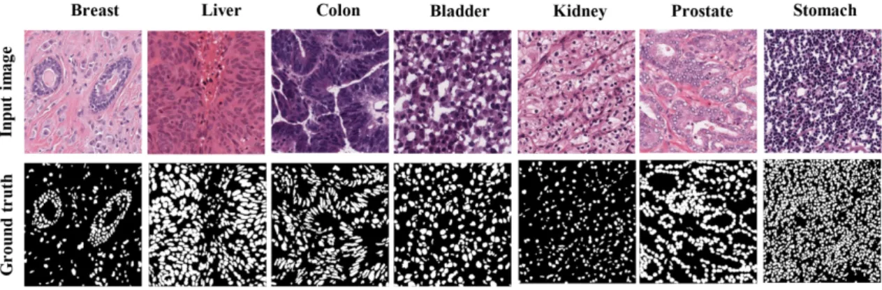
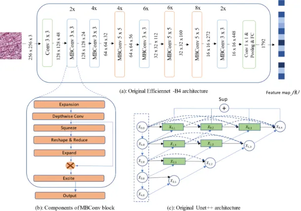
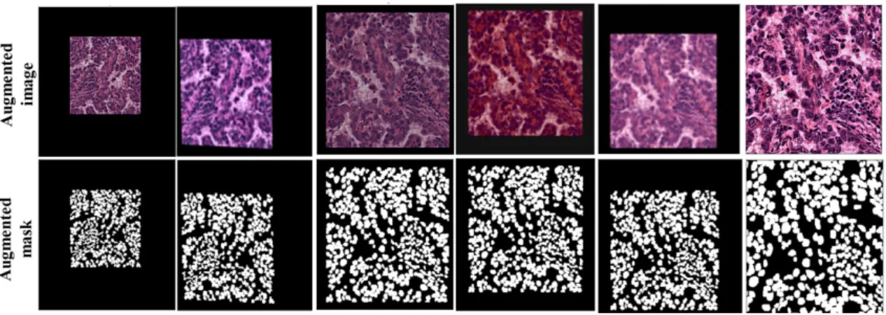
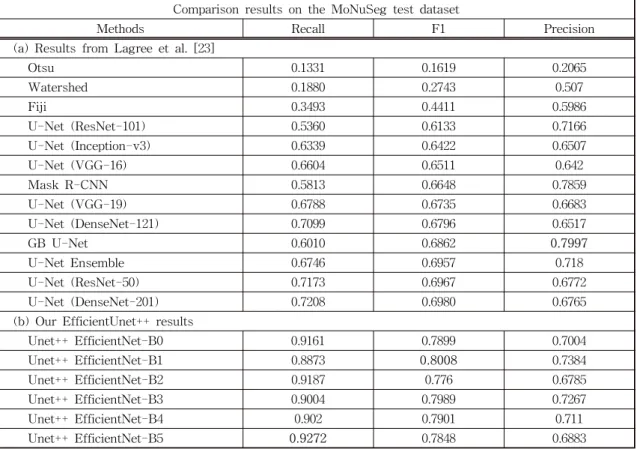
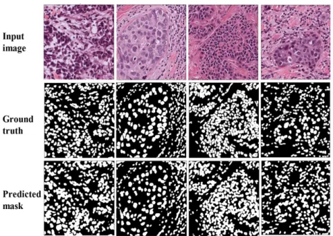
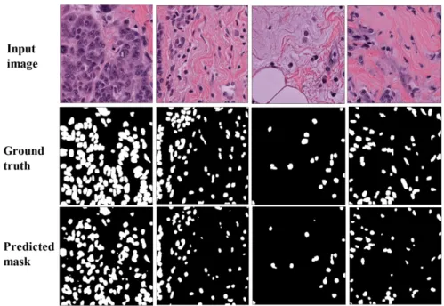
In 2015, the Unet model was introduced and gained much attention from researchers in the field. The success of Unet model is the ability to produce high accuracy with very few input images. Since the development of Unet, there are many variants and modifications of Unet related architecture. This paper proposes a new approach of using Unet++ with pretrained EfficientNet as backbone architecture for breast tumor cell nuclei segmentation and uses the multi-organ transfer learning approach to segment nuclei of breast tumor cells. We attempt to experiment and evaluate the performance of the network on the MonuSeg training dataset and Triple Negative Breast Cancer (TNBC) testing dataset, both are Hematoxylin and Eosin (H & E)-stained images. The results have shown that EfficientUnet++ architecture and the multi-organ transfer learning approach had outperformed other techniques and produced notable accuracy for breast tumor cell nuclei segmentation.

Key words: Deep Learning, Image Segmentation, Nuclei Segmentation, Unet, Unet++, EfficientNet, Hematoxylin and Eosin (H & E), Breast Cancer

※ Corresponding Author : Ki-Ryong Kwon, Address: 45 Yongso-ro, Nam-gu, Busan, Pukyong National University, Daeyon campus, TEL : +82-51-629-6257, FAX : +82-51- 629-6230, E-mail : [email protected]

Receipt date : Aug. 4, 2021, Approval date : Aug. 12, 2021

†††

Dept. of Artificial Intelligence Convergence, Pukyong National University (E-mail : [email protected])

†††

Dept. of Electronics Engineering, Kyungil University (E-mail : [email protected])

†††

Dept. of Computer Engineering, Dong A University (E-mail : [email protected])

††††

Dept. of Artificial Intelligence Convergence, Pukyong National University

※ This research was supported by the Basic Science Research Program through the National Research Foun- dation of Korea(NRF) funded by the Ministry of Educa- tion (2020R1I1A306659411, 2020R1F1A1069124), and the MSIT(Ministry of Science and ICT), Korea, under the ITRC (Information Technology Research Center) support program (IITP-2021-2020-0-01797) supervised by the IITP (Institute for Information & Communications Tech- nology Planning & Evaluation).

1. INTRODUCTION

Breast cancer is the most common type of wom- en's cancer and is the second cause of women's cancer death each year [1]. Early breast cancer di- agnosis is vital for cancer patients, the survival rate of 5-year for breast cancer patients can reach 90% with early diagnosis [1]. Currently, breast

cancer detection modalities have evolved with many types, and most of them use image-based approaches to screen and detect the tumor mass.

We can name them such as electrical impedance-

based imaging, breast ultrasound, scintimammog-

raphy, computed tomography (CT), positron emis-

sion tomography (PET), thermography, optical

imaging, mammography, and magnetic resonance

(2)

imaging (MRI) [2]. Breast cancer diagnosis often starts with mammography imaging analysis using Deep Learning to detect the existence of cancer tu- mors [3]. However, sometimes the method comes to an undetermined conclusion, and in situations like this, the breast cancer diagnosis needs to use biopsy and H & E-stained image analysis to have a more accurate diagnosis [4].

Hematoxylin and Eosin-stained is one of the primary techniques used for histological staining, because of its simplicity and the capability to illus- trate many different tissue structures [5]. The bi- opsy specimens are processed on slides and then the Hematoxylin material stains the nuclei with dark color, demonstrating clear details of the intra- nuclear, and the Eosin component stains the cell cytoplasm and tissue fibers with pink, red, or or- ange colors in many intensity levels. The slide im- ages were captured by whole slide digital scanners, stored for cancer tumor cell detection, or for the breast cancer staging process. Conventionally, digitized histopathology images are analyzed using laborious and manual techniques, which are time- consuming processes and limited by the experience of pathologists. Thus, we need an accurate and quantitative analysis approach for histopathology images. Computer-aid diagnosis (CAD) comes to the place and then follows with many state-of- the-art (SOTA) CAD methods for automated im- age analysis histopathology imagery [6].

Recently, the machine learning approach applied for histopathology image analysis has taken a leap, especially for Deep Learning-based methods that increase the efficiency and accuracy of histopatho- logical diagnosis [7]. The work of S. Bhattacharjee et al. [8] tried to predict the histological grade of prostate cancer biopsy using Convolutional Neural Network. P. Naylor et al. [9] had proposed a Deep Learning-based method for the segmentation of H

& E-stained nuclei images. The novelty of the method is that it considers the segmentation task similar to the regression task of distance map so

that the network can learn and tackle the problem of touching nuclei cells. Another work of Janowc- zyk et al. [10] had presented thoroughly Deep Learning techniques applied for seven Digital Pathology tasks and compared the results of Deep Learning-based methods with other state-of-the- art hand-crafted feature-based. Zhao Z et al. [11]

proposed PFA-ScanNet which stands for Pyrami- dal Feature Aggregation ScanNet, a neural net specific for breast cancer metastasis detection.

Graham et al. [12] proposed the Minimal infor- mation loss dilated network (MILD-Net), a Deep Neural network applied for the gland and lumen segmentation tasks. Ho et al. [13] introduced Deep Multi-Magnification, a Deep Neural Network with multi-encoder, multi-decoder, multi-concatenation specific for multi-class tissue segmentation from digital whole slide images.

2. RELATED WORKS

Most Deep Learning-based models applied for image segmentation tasks use some types of tech- niques related to encoder-decoder architecture.

There are two categories of encoder-decoder for image segmentation problems, the general seg- mentation, and the medical, biomedical segmenta- tion approaches [14]. The early work of Noh et al.

[15] had proposed a network that learns the decon- volution network and contains the deconvolution and unpooling layers. The network achieved out- standing results on the PASCAL VOC 2012 dataset and produced the best accuracy on the Microsoft COCO dataset. Another paper focused on the en- coder-decoder approach is the work of Badrinar- ayanan et al. [16] named Segnet. The novelty of the network is the upsampling layer in the decoder path, the pooling indices allow the network to re- duce the need for learning to upsample.

The scarcity of labeled training datasets and the

demand for high accuracy segmentation raises the

need to have a dedicated purpose neural net for

(3)

medical and biomedical images. In 2015, Olaf Ron- neberger et al. [17] published the Unet paper in which they proposed a U-shape encoder-decoder architecture that contains two symmetric paths, the contracting path, and the expanding path. The network can train end-to-end using a small num- ber of training images, and surpass the accuracy of other best methods on the ISBI dataset with the high performance of segmentation results.

Since the outstanding performance of Unet, there is a fast growth of the Unet related papers in medical and biomedical image segmentation tasks. Some of the Unet variant models such as 3D Unet, Attention Unet, Inception Unet, Residual U-Net, Dense Unet, Unet++, Adversarial Unet, etc had gained significant improvement on a specific type of Medical, Biomedical images [18]. The work of B. Baheti et al. [19] tried to combine pretrained EfficientNet and Unet as encoder and decoder of an architecture which had ranked in first place for image segmentation in the IDD lite challenge.

To enhance the accuracy of segmentation tasks for Medical, Biomedical images, Zhongwei Zhou et al. [20] had introduced the Unet++ model which re-designed the skip pathway of the original Unet.

They believed that when reducing the semantic gaps between encoder and decoder, the optimizer will carry out easier learning tasks and hence boost up the performance. In 2019, The EfficientNet pa- per was introduced by Mixing Tan and Quoc Le [21], and had become popular in the Machine Learning community. The basic idea of the paper is to scale the network in all dimensions, both the depth, the width of the neural net, and the image size. The new scaling method helps to create a novel EfficientNet model and achieve outstanding results compared to other ConvNets.

Using pretrained ConvNets as the backbone and then upsampling with Unet variant architecture as a decoder has become a novel approach and has achieved many improvements for segmentation tasks in recent years. The early evaluation of the

Unet++ with EfficientNet as a backbone for medi- cal image segmentation tasks can refer to the work of Le Duy Huỳnh et al. [22]. In the paper, the author had run experiments with datasets from the EndoCV2020 challenge and compared the result with other SOTAs. The Unet++ with pretrained EfficientNet as the backbone had outperformed other methods with high accuracy.

The problem facing when conducting experi- ments for breast tumor cell nuclei segmentation is the availability of labeled training datasets. To overcome the scarcity problem of training data, Lagree et al. [23] proposed the idea of multi-organ transfer learning, the neural net will be trained on the multi-organ H & E-stained cell datasets of liv- er, prostate, kidney, lung, colon, brain, bladder and stomach, then will be inferenced on the breast tu- mor cell dataset to evaluate the capability of trans- fer learning. This study follows the idea of the pre- viously mentioned paper. We will run experiments on Unet++ with pretrained EfficientNet models from B0 to B5 and then compare the results with other SOTAs. More details about the training da- tasets and the configurations to conduct experi- ments of multi-organs transfer learning will be discussed in the Methodology section.

3. METHODOLOGY

We use Unet++ with EfficientNet as the back- bone of the model. We conduct our experiments with pretrained EfficientNet from B0 to B5 which was trained on the ImageNet dataset with highly effective compound coefficients. The model pro- duced more efficiency and accuracy than previous ConvNets, while the parameter numbers are smaller.

3.1 Dataset

MonuSeg stands for Multi-organ Nucleus Seg-

mentation, the dataset was published in the official

satellite event of MICCAI 2018. The challenge was

opened aim to find the best methods to segment

(4)

Fig. 1. Some sample images from the MonuSeg dataset.

Fig. 2. Some sample images from the TNBC dataset.

the nuclei in H&E-stained cell images [24]. The original MonuSeg includes thirty training images, each with size 1000 × 1000, from seven organs and contains total 21,623 annotated nuclei boundaries.

The test set has 14 images that are the same size as the training set and have total 7,223 annotated nucleis. In our experiment, we moved 6 breast tu- mor cell images from the original MonuSeg to the test set, thus the total number of training images was 24 and was split into 80% train and 20%

validation.

The test set in our experiment includes 58 im- ages, we select 8 images from the MonuSeg data- sets and 50 images from the Triple Negative Breast Cancer (TNBC) dataset. The TNBC dataset is the work of P. Naylor et al. [9] which was generated at the Curie Institute, all slides are taken from Triple Negative Breast Cancer patients and then export histopathology images with Philips Ultra

Fast Scanner 1.6 RA. The TNBC consists of 50 images with a total of 4022 annotated cells, and with an average of 80 cells per sample image. The largest number of cells per image is 293 and the smallest number of cells is 5. One pathologist ex- pert and two trained research fellows carried out the annotation for the training and testing dataset.

The results were peer-checked then if there was disagreement, the team needed to discuss and come to the final decision.

3.2 EfficientNet Encoder

The EfficientNet family starts with Efficient

Net-B0 at 5.3 million parameters to the largest

model EfficientNet-B7 with 66 million. The ele-

mentary unit of EfficientNet is the mobile bottle-

neck (MBConv) using squeeze-and-excitation

components for optimization purposes [25]. Each

EfficientNet model differs in parameter size and its

(5)

Fig. 3. Original Efficienet-B4 and original Unet++ architecture.

number of modules, but the overall architecture re- mains the same from B0 to B7. The important con- tribution of the EfficientNet is the new compound scaling method, that uniformly scales the network in terms of depth, width, and resolution of input images. The scaling method is demonstrated in the following principle:

(1)

In which, α, β, γ are constant coefficients which can be calculated by a small grid search on the original small model. And φ is coefficient pre- defined by users that control resources for model scaling. If we scale the network depth α

N

, width β

N

, and resolution γ

N

then we get 2

N

times more computational resources.

3.3 Unet++ decoder

The decoder block of Unet++ connects with the encoder block of EfficientNet through a sequence of nested dense convolutional blocks. The skip pathway of the Unet++ is formulated as follow:

(2)

In which, the x

i,j

is the output of node X

i,j

. We

denote i as the indexes of downsampling layers

along the encoder and j as indexes of the convolu-

tional layer blocks along the skip pathway. We

have H (·) are the convolution operations flow by

an activation function, U (·) are the upsampling

layers and [] are concatenate layers. Overall, the

difference between Unet and Unet++ is the rede-

sign skip pathway, in which, Unet architecture just

has plain connections, features from decoders re-

ceive directly from encoders, and in Unet++ we

(6)

Fig. 4. Some sample of augmented images after applied Albumentations library.

have dense convolution blocks that reduce the se- mantic gap between the contracting path and the expansive path.

3.4 Data augmentation

We use Albumentations as the library for our training data augmentation pipeline. The library is a fast, robust, and easy-to-use tool for augmenting our training data [26]. Medical and biomedical im- age analysis confronts the problem of data scarcity due to the cost of labeling data and the availability of data sources. Using data augmentation techni- ques can help to solve the problem as well as im- prove the neural net performance against over- fitting during the training process. We use lots of data augmentation operations to enrich our data from 19 images to 5320 images for training and 5 images to 1400 images for validation. All the im- ages were resized into 256 × 256 then using aug- mentation techniques like HorizontalFlip, ShiftScale Rotate, RandomCrop, RandomBrightness, Random Gamma, etc to scale up the number of training images.

3.5 Training

We conduct experiments on a machine with Intel(R) Core (TM) i7-8700K CPU @ 3.70GHz in- stalled memory (RAM) 16GB, NVDIA GeForce RTX 2070 8GB.

We train our model using Sigmoid as the activa- tion function, Adam optimizer with a learning rate of 0.0001, and Dice Coefficient as the loss function.

The formula of Dice Loss function is defined as below:

(3) In which, p

i

denotes the predicted pixel and g

i

denotes the ground truth. In our nuclei segmenta- tion task, the p

i

are pixels that our trained models predict as nuclei, and the g

i

are nuclei pixels that were labeled by experts.

We execute image augmentation operations on- the-fly mode, simultaneously with the training process using the Albumenations library. We use StainTools (https://github.com/Peter554/StainTools) as stain normalization library for H & E-stained images. All the models and pretrained backbone were implemented on the Segmentation Models re- pository and available on GitHub (https://github.

com/qubvel/segmentation_models.pytorch).

4. EXPERIMENTAL RESULT AND DISCUSSION We evaluate our models based on three metrics Recall, F1 score, and Precision with the threshold value equal to 0.5. Our proposed EfficienetUnet++

results had outperformed others SOTAs on the nu-

clei segmentation task in terms of Recall and F1

(7)

Table 1. Quantitative results comparison between EfficientUnet++ and other segmentation methods on the MonuSeg dataset.

Comparison results on the MoNuSeg test dataset

Methods Recall F1 Precision

(a) Results from Lagree et al. [23]

Otsu 0.1331 0.1619 0.2065

Watershed 0.1880 0.2743 0.507

Fiji 0.3493 0.4411 0.5986

U-Net (ResNet-101) 0.5360 0.6133 0.7166

U-Net (Inception-v3) 0.6339 0.6422 0.6507

U-Net (VGG-16) 0.6604 0.6511 0.642

Mask R-CNN 0.5813 0.6648 0.7859

U-Net (VGG-19) 0.6788 0.6735 0.6683

U-Net (DenseNet-121) 0.7099 0.6796 0.6517

GB U-Net 0.6010 0.6862 0.7997

U-Net Ensemble 0.6746 0.6957 0.718

U-Net (ResNet-50) 0.7173 0.6967 0.6772

U-Net (DenseNet-201) 0.7208 0.6980 0.6765

(b) Our EfficientUnet++ results

Unet++ EfficientNet-B0 0.9161 0.7899 0.7004

Unet++ EfficientNet-B1 0.8873 0.8008 0.7384

Unet++ EfficientNet-B2 0.9187 0.776 0.6785

Unet++ EfficientNet-B3 0.9004 0.7989 0.7267

Unet++ EfficientNet-B4 0.902 0.7901 0.711

Unet++ EfficientNet-B5 0.9272 0.7848 0.6883

metrics, except for Precision, the GB U-net pro- posed by Lagree et al. [23] had produced higher result compared to the EfficientNet family.

On the MonuSeg dataset, Unet++ with Efficient Net-B5 and EfficientNet-B1 produced the best re- sult for Recall metric (0.9272) and F1 metric (0.8008). EfficientUnet++ performance is superior with a wide margin on Recall metric compared with traditional segmentation methods like Otsu, Watershed, and Fiji. Similar to the Recall metric on the F1 metric, EfficientUnet++ results exceed a large margin compared with traditional segmen- tation methods and produced higher accuracy than Deep Learning-based methods. However, results on Precision remain to the same degree as other Deep Learning-based methods. Fig. 5 shows you the qualitative output results of Unet++ with EfficientNet-B1 as the backbone when inferencing

on MonuSeg dataset.

On the TNBC dataset, Unet++ with Efficient Net-B5 and B1 exceeds other methods in terms of Recall (0.9343) and F1 (0.6785) metrics, the Effi- cientUnet++ do not pass the GB-Net (0.8102) on Precision metric evaluation. Similar to the Monu Seg dataset, EfficientUnet++ also outperformed other Deep Learning-based segmentation methods on Recall and F1, however, in Precision metric EfficientUnet++ produced not as good results as other Deep Learning-based methods. Fig. 6 shows you the qualitative output results of Unet++ with EfficientNet-B1 as the backbone when inferencing on the TNBC dataset.

5. CONCLUSION

In this work, we show you the effectiveness of

(8)

Fig. 5. Qualitative output results of Unet++ with EfficientNet-B1 as the backbone on the MonuSeg dataset.

Table 2. Quantitative results comparison between EfficientUnet++ and other segmentation methods on the TNBC dataset.

Comparison results on the TNBC test dataset

Methods Recall F1 Precision

(a) Results from Lagree et al. [23]

U-Net (VGG-19) 0.4585 0.5099 0.5741

U-Net (Inception-v3) 0.3925 0.4703 0.5864

U-Net (VGG-16) 0.4940 0.5042 0.5149

U-Net (ResNet-101) 0.4139 0.5427 0.7882

U-Net (ResNet-50) 0.5281 0.5638 0.6048

U-Net Ensemble 0.5176 0.6068 0.7331

U-Net (DenseNet-121) 0.5745 0.6216 0.6772

U-Net (DenseNet-201) 0.5745 0.6298 0.697

GB U-Net 0.5541 0.6581 0.8102

Mask R-CNN 0.6172 0.6732 0.7403

(b) Our EfficientUnet++ results

Unet++ EfficientNet-B0 0.921 0.5986 0.4999

Unet++ EfficientNet-B1 0.8942 0.6785 0.5931

Unet++ EfficientNet-B2 0.9209 0.5877 0.4875

Unet++ EfficientNet-B3 0.9156 0.6024 0.5105

Unet++ EfficientNet-B4 0.9033 0.6183 0.5278

Unet++ EfficientNet-B5 0.9343 0.5612 0.4571

multi-organ transfer learning applied for breast cancer tumor cells. The Unet++ with pretrained EfficientNet from B0 to B5 was selected to run the

experiment on seven organ cell images. Our ex-

periments exhibited the best result in terms of

Recall and F1 score compared with previous works

(9)

Fig. 6. Qualitative output results of Unet++ with EfficientNet-B1 as the backbone on the TNBC dataset.

that run on the Monuseg datasets and inference on the TNBC datasets. The results demonstrate that Deep Learning models trained on different organ cell images can produce a good performance for breast cancer tumor cells on segment nuclei tasks.

Compared with traditional methods, the Efficient Unet++ outperformed with a large accuracy margin.

Compared with other Deep Learning methods, our proposed models produced more excellent results than Mask RCNN, Unet Ensemble, Unet with Resnet-50, Resnet-101, VGG-16, VGG-19, Dense Net-121, Densenet-201, Inception-v3 as the back- bone, and GB U-net proposed in the paper Lagree et al. [23].

To enhance the network accuracy, we need more training data feeding to our network. More training data help the network to improve the generalization and prevent from being overfitting. Recently, many techniques to enrich the labeled training dataset had been investigated, two approaches that were popular in medical and biomedical communities are scalable crowd-sourcing annotation [27] and GAN for generating new medical and biomedical images [28]. The scalable crowd-sourcing annotation ap-

proach focus to utilize the crowd workforce to pro- duce more labeling data with the supervision of ex- pert domains. While the GAN approach mainly lev- erages the family of Generative Adversarial Net- work to augment original data. In future work, we will try to exploit those two methods to increase the dataset for our models and will take more nu- merical evaluation on the benefit of those two data enrichment methods.

REFERENCE

[ 1 ] R.L. Siegel, K.D. Miller, H.E. Fuchs, and A.

Jemal, “Cancer statistics,” A Cancer Journal for Clinicians 71, Vol. 71, pp. 7-33, 2021.

[ 2 ] S.V. Sree, E.Y. Ng, R.U. Acharya, and O.

Faust, “Breast imaging: A survey,” World Journal of Clinical Oncology, Vol. 2, pp. 171- 178, 2011.

[ 3 ] S.Y. Kwon, Y.J. Kim, and G.G. Kim, “A deep learning-based automatic segmentation of breast mass in breast imaging,” Journal of the Korean Society for Multimedia, Vol. 21, No.

12, pp. 1363-1369, Dec. 2018.

(10)

[ 4 ] M. Lekka, “Atomic force microscopy: A tip for diagnosing cancer,” Nature Nanotechnology, Vol. 7, pp. 691-692, 2012.

[ 5 ] J.D. Bancroft and C. Layton, “The hematox- ylins and eosin,” in Bancroft's Theory and Practice of Histological Techniques (Eighth Edition), Elsevier, 2019, pp. 126-138.

[ 6 ] M.N. Gurcan, L.E. Boucheron, A. Can, A.

Madabhushi, N.M. Rajpoot, and B. Yener,

“Histopathological image analysis: A Review,”

IEEE Reviews in Biomedical Engineering, Vol. 2, pp. 147-171, 2009.

[ 7 ] G. Litjens, C. Sánchez, N. Timofeeva et al.,

“Deep Learning as a Tool for Increased Accuracy and Efficiency of Histopathological Diagnosis,” Scientific Reports, Vol. 6, No.

26286, 2016.

[ 8 ] S. Bhattacharjee, D. Prakash, C.H. Kim, and H.K. Choi, “Multichannel Convolution Neural Network Classification for the Detection of Histological Pattern in Prostate Biopsy Images,” Journal of the Korean Society for Multimedia, Vol. 23, No. 12, pp. 1486-1495, Dec. 2020.

[ 9 ] P. Naylor, M. Laé, F. Reyal, and T. Walter,

“Segmentation of Nuclei in Histopathology Images by Deep Regression of the Distance Map,” IEEE Transactions on Medical Imag- ing, Vol. 38, No. 2, pp. 448-459, Feb. 2018.

[10] A. Janowczyk and A. Madabhushi, “Deep learning for digital pathology image analysis:

A comprehensive tutorial with selected use cases,” Journal of Pathology Informatics, Vol.

7, pp. 29, Jul. 2016.

[11] Z. Zhao, H. Lin, H. Chen, and P.A. Heng,

“PFA-ScanNet: Pyramidal Feature Aggrega- Tion with Synergistic Learning for Breast Cancer Metastasis Analysis,” Medical Image Computing and Computer Assisted Inter- vention–MICCAI 2019, pp. 586-594, 2019.

[12] S. Graham et al., “MILD-Net: Minimal Infor- mation Loss Dilated Network for Gland

Instance Segmentation in Colon Histology Images,” Medical Image Analysis, Vol. 52, pp.

199-211, 2019.

[13] D.J. Ho et al., “Deep Multi-Magnification Networks for Multi-Class Breast Cancer Image Segmentation,” Computerized Medical Ima- ging and Graphics, Vol. 88, No. 101866, 2021.

[14] S. Minaee, Y.Y. Boykov, F. Porikli, A.J. Plaza, N. Kehtarnavaz, and D. Terzopoulos, “Image Segmentation Using Deep Learning: A Sur- vey,” arXiv preprint, arXiv:2001.05566, 2020.

[15] H. Noh, S. Hong, and B. Han, “Learning Decon- volution Network for Semantic Segmentation,”

2015 IEEE International Conference on Computer Vision (ICCV), pp. 1520-1528, 2015.

[16] V. Badrinarayanan, A. Kendall, and R. Cipolla,

“SegNet: A Deep Convolutional Encoder- Decoder Architecture for Image Segmentation,”

IEEE Transactions on Pattern Analysis and Machine Intelligence, Vol. 39, No. 12, pp. 2481- 2495, 2017.

[17] O. Ronneberger, P. Fischer, and T. Brox, “U- Net: Convolutional Networks for Biomedical Image Segmentation,” Medical Image Com- putting and Computer-Assisted Intervention- MICCAI 2015, vol. 9351, 2015.

[18] N. Siddique, S. Paheding, C. P. Elkin, and V.

Devabhaktuni, “U-Net and its Variants for Medical Image Segmentation: A Review of Theory and Applications,” IEEE Access, Vol.

9, pp. 82031-82057, 2021.

[19] B. Baheti, S. Innani, S. Gajre, and S. Talbar,

“Eff-UNet: A Novel Architecture for Semantic Segmentation in Unstructured Environment,”

in IEEE/CVF Conference on Computer Vision and Pattern Recognition Workshops (CVPRW), 2020.

[20] Z. Zhou, M.M.R Siddiquee, N. Tajbakhsh, and

J. Liang, “UNet++: A Nested U-Net Architec-

ture for Medical Image Segmentation,” Deep

Learning in Medical Image Analysis and

Multimodal Learning for Clinical Decision

(11)

Support. DLMIA 2018, ML-CDS 2018. Lec- ture Notes in Computer Science, Vol. 11045, pp. 3-11, 2018.

[21] M. Tan and Q.V. Le, “EfficientNet: Rethinking Model Scaling for Convolutional Neural Networks,” in Proceedings of International Conference on Machine Learning, 2019.

[22] L.D. Huynh and N. Boutry, “A U-Net++ with Pre-trained EfficientNet Backbone for Seg- mentation of Diseases and Artifacts in Endoscopy Images and Videos,” in EndoCV

@ISBI, 2020.

[23] A. Lagree, M. Mohebpour, N. Meti et al., “A Review and Comparison of Breast Tumor Cell Nuclei Segmentation Performances Using Deep Convolutional Neural Networks,” Scien- tific Reports, Vol. 11, No. 8025, 2021.

[24] N. Kumar et al., “A Multi-Organ Nucleus Segmentation Challenge,” IEEE Transactions on Medical Imaging, Vol. 39, No. 5, pp. 1380- 1391, May 2020.

[25] M. Sandler, A. Howard, M. Zhu, A. Zhmogi-

nov, and L. Chen, “MobileNetV2: Inverted Residuals and Linear Bottlenecks," in 2018 IEEE/CVF Conference on Computer Vision and Pattern Recognition, 2018.

[26] A. Buslaev, V.I. Iglovikov, E. Khvedchenya, A. Parinov, M. Druzhinin, and A.A. Kalinin,

“Albumentations: Fast and Flexible Image Augmentations,” Information, Vol. 11, No. 2, pp. 125, 2020.

[27] M. Amgad, L.A. Atteya, H. Hussein, K.H.

Mohammed, E. Hafiz, M.A. Elsebaie, A.M.

Alhusseiny, M.A. AlMoslemany, A.M. Elmatb- oly, P.A. Pappalardo et al., “NuCLS: A Scalable Crowdsourcing, Deep Learning Approach and Dataset for Nucleus Classification, Localization and Segmentation,” arXiv preprint, arXiv:2102.

09099, 2021.

[28] X. Yi, E. Walia and P. Babyn, “Generative

Adversarial Network in Medical Imaging: A

Review,” Medical Image Analysis, Vol. 58,

No. 101552, 2019.

(12)

Tuan Le Dinh

received the B.S. degree in Com- puter Science from University of Engineering and Technology, Vietnam National University, in 2018. He is currently studying a Master course in Artificial In- telligence Convergence at Pu- kyong National University, Korea. His research inter- ests include medical and biomedical image Processing, bioinformatics and deep learning.

Seong-Geun Kwon

received a B.S., a M.S., and a Ph.D. degree in Electrical Eng- ineering from Kyungpook Na- tional University, Korea in 1996, 1998, and 2002 respectively. He worked at Samsung Electronics Company from 2002-2011. He is currently a professor in the Department of Electronics Engineering at Kyungil University. His research inter- ests include multimedia security and watermarking, mobile broadcasting, and digital image processing.

Suk-Hwan Lee

received a B.S., a M.S., and a Ph.

D. degree in Electrical Engin- eering from Kyungpook Natio- nal University, Korea in 1999, 2001, and 2004 respectively. He is currently a professor in the Department of Computer Engin- eering at Donga University. His research interests in- clude multimedia security, digital image processing, and computer graphics.

Ki-Ryong Kwon

received the B.S., M.S., and Ph.

D. degrees in electronics engin- eering from Kyungpook Natio- nal University in 1986, 1990, and 1994 respectively. He worked at Hyundai Motor Company from 1986-1988 and at Pusan Univer- sity of Foreign Language from 1996-2006. He is cur- rently a professor in Dept. of IT Convergence & Appli- cation Engineering at the Pukyong National University.

He has researched University of Minnesota in USA

from 2000-2002 with Post-Doc, and Colorado State

University from 2011-2012 with visiting professor. He

was the General President of Korea Multimedia

Society from 2015-2016. He was also a director of

IEEE R10 Changwon section. He is currently chairman

of Global FinTech Industry Promotion Center. His re-

search interests are in the area of digital signal and

image processing, multimedia security and deepfake

image, bioinformatics, visual information processing.

수치

Fig. 1. Some sample images from the MonuSeg dataset.
Fig. 3. Original Efficienet-B4 and original Unet++ architecture.
Fig. 4. Some sample of augmented images after applied Albumentations library.
Table 1. Quantitative results comparison between EfficientUnet++ and other segmentation methods on the MonuSeg  dataset.
+3

참조

관련 문서

Intraoperative photograph shows grossly total removal of tumor using the navigation system

In this study, using concept mapping of a variety of learning techniques in the area of science, especially biology, has a positive effect on learning

A retrospective analysis of invasive tumor percentages of GSS tumor samples showed that samples with high tumor content correlated well with FNAB samples

Beichel,“Automated 3-D segmentation of lungs with lung cancer in CT data using a novel robust active shape model approach,” IEEE Trans.. 저작물의

paper proposes proposes proposes proposes a a a a new new new new contrast contrast contrast contrast enhancement enhancement enhancement enhancement method

 Students needing additional information about grading policies and procedures should meet with their faculty advisor, Executive Director/Chairperson or a

Hypoxic cell cytotoxins Complementary cytotoxicity with radiation on euoxic and hypoxic tumor cells.. Antimetabolites Nucleotide pool perturbation Lowering

Patients with breast cancer; ovarian cancer; renal cell carcinoma; pancreatic neuroendocrine cancer; colorectal cancer; head and neck cancer; non-small cell lung