Mammography시 Thyroid에 미치는 산란선량에 관한 연구
이미화1,2, 동경래3,4,a, 박서주1, 황선광1
1 경희대학교 동서신의학병원 영상의학과
2 연세대학교 역학건강증진학과
3 광주보건대학 방사선과
4 조선대학교 원자력공학과
The Effect of Scattering Dose on the Thyroid During Mammography
Mi-Hwa Lee
1,2, Kyung-Rae Dong
3,4,a, Seo-Joo Park
1, and Sun-Kwang Whang
11 Department of Diagnostic Radiology, East-West Neo Medical Center, Seoul 134-727, Korea
2 Department of Epidemiology and Health Promotion, Yonsei university, Seoul 120-752, Korea
3 Department of Radiological Technology, Gwangju Health College University, 506-701 Gwangju, Korea
4 Department of Nuclear Engineering, Chosun University, 501-759 Gwangju, Korea
(Received September 2, 2010; Revised September 13, 2010; Accepted September 20, 2010)
Abstract: This study examined the effect of the scattering dose on the thyroid during a mammography examination. One hundred subjects for a mammography examination were enrolled in this study. The average glandular dose (AGD) and thyroid scattering dose (TSD) were measured. Statistical analysis was carried out using the percentage, t-test and co-variance. The mean radiation exposure to the breast and thyroid was 1.08 ± 0.16 and 0.14 ± 0.04 mGy, respectively. The percentage TSD to the AGD was 31.19%.
There was no difference between the Rt. and Lt., and CC to MLO, and radiation dose to the TSD was 13.78% of the breast. Therefore, the volume of radiation exposure to the thyroid was 54.12% in a single routine mammography examination. These results suggest that the TSD was increased by increasing radiation dose to the breast. A thyroid protector is considered necessary to decrease the level of radiation exposure.
Keywords: Scattering dose, Mammography, Thyroid, breast
1. INTRODUCTION
1)The number of people with breast cancer has been increasing at a rate of 10% a year. In particular, the incidence in patients aged twenty to forty years has increased rapidly due to lifestyle changes, increasing levels of obesity, low birth rate and breast-feeding, late marriage, and early
a. Corresponding author; [email protected]menarche or late menopause [1]. Routine check-ups and self examination are considered essential for early detection. The National Health Insurance Corporation in South Korea recommends women aged 40 years and over undergo a screening test for breast cancer every second year [2]. However, this would increase the level of radiation exposure by mammography, which is a major concern considering its relationship with breast cancer [3].
The thyroid gland is the first organ to be
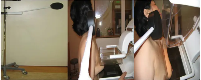
Fig. 1. The method of the scattering dose of thyroid.
damaged by radiation because of its proximity to the breast and that it is affected by estrogen.
There are many reports showing that breast cancer is related to thyroid gland diseases [4-7], nodular hyperplasia, hyperthyroidism and thyroid cancer. In other words, women with breast cancer generally have a disease of the thyroid gland [8,9]. Radiation from a mammography examination can damage the thyroid gland. The aim of this study was to determine the effect of an average glandular dose by mammography on the thyroid gland.
2. EXPERIMENTS
One hundred adult subjects undergoing a routine mammography examination at an East-West Neo Medical Center in South Korea between June 1, 2009 and August 26, 2009 were selected. The instruments used to check the volume of radiation were digital radiography (DR), Senography DS (General Electric Co., USA), Dosimeter (Victoreen Nero Max Model 8000, Japan) and Ionization Chamber (6000-523B, volume 400 cm, Japan). As shown in Fig. 1, the ionization chamber was adjusted for height by positioning it to the area of the thyroid gland.
When the cranio-cauudal (CC) view was taken, the chamber arm paralleled the shoulder line. The
posterior chamber contacted the neck and did not lean to one side but was placed against the chamber arm. The thyroid scattering dose (TSD) was measured under this configuration. The method of the medio-lateral oblique (MLO) view was the same as the CC view but the inclination of the chamber was equal to that of the neck in order maintain the position between the thyroid gland and chamber. The thyroid scattering dose was measured using a Dosimeter and Ionization chamber for Mammography. The average glandular dose (AGD) was confirmed from the Digital Image and communication in medicine (DICOM) header information. The AGD and TSD measurements were converted to mGy using the formula (1 Gy=100 rad, 1 R=0.95 rad).
Statistical analysis was carried out using the Statistical package for the Social Sciences for window (v.15.0). The percentage, t-test and Co-Variance were used.
3. RESULTS AND DISCUSSION
3.1 Mean value of radiation exposure to breast and thyroid
Each level of radiation exposure to the breast
and thyroid of the subjects is given in the
appendix. Table 1 shows the mean and standard
AGDc 1.07 ± 0.17 1.07 ± 0.20 1.10 ± 0.18 1.08 ± 0.20 1.08 ± 0.16
TSDd 0.14 ± 0.05 0.15 ± 0.05 0.14 ± 0.05 0.15 ± 0.05 0.14 ± 0.04
Note) aCC= Cranio-Caudal; bMLO= Medio-Lateral Oblique; cAGD= Average Glandular Dose; dTSD= Thyroid Scattering Dose
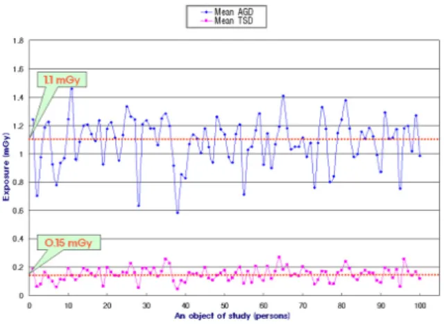
Fig. 2. The values of AGD and TSD per one subject.
Table 2. The percentage of TSD to AGD in total 100 populations.
AGDa TSDb Percentage
1.08 0.14 13.78
Note)
aAGD=Glandular Scattering dose;
bTSD=
Thyroid Scattering Dose
deviation to identify the AGD and TSD according to each position. The AGE value and TSD in a total of 100 people was 1.084 ± 0.169 mGy and 0.149 ± 0.047 mGy, respectively. Figure 2 shows the sampling components concerning the differences in the AGD to TSD. The mean AGD and TSD were 1.1 mGy and 0.15 mGy, respectively.
3.2 The percentage of TSD to AGD in the 100 subjects
Table 2 shows the percentage of TSD to AGD.
The level of exposure to the thyroid was estimated to be 13.78% using the formula, (TSD Mean/AGD Mean)x100, assuming the mean level of radiation exposure to the breast is 100%.
3.3 The relationship between Rt. and Lt., and CC to MLO
There was no difference between the Rt. and Lt., and CC to MLO in the AGD and TSD (Table 3).
3.4 The difference in radiation exposure between the AGD and TSD
The percentage radiation exposure to the AGD and TSD compared to the breast was 15.54% and 31.19%, respectively (Table 4). The percentage was calculated using the formula, (SD/Mean)x100.
3.5 Discussion
Radiation exposure during a medical treatment
has been given a great deal of weight when
determining the level of artificial radiation exposure
[10,11]. The ICRP (ICRP publication 60 in 1991)
reported that there was no lower limit of the
radiation dose in a medical treatment for optimal
treatment and the protection of patients. The risk
increases with the increasing level of radiation
exposure, and it is believed that there is no
threshold dose of risk [12,13]. The ICRP (2007),
which is an international organization of radiation
protection, reported that < 3 mGy was the average
glandular dose from mammography in the direction
of the breast (50% lipid, 50% genetic) pressed as
much as 4.2 cm in a Mo target+Mo filter,
film/screen system, and imaged in the up and
down directions [14]. They recommended that the
glandular dose be < 2 mGy or 1.5 mGy. Therefore,
Table 3. The relationship between Rt. and Lt., and CC to MLO (unit: Mean ± SD) Rt. CC-Lt. CCa Lt. CC-Lt. MLOb Rt. CC-Rt. MLO Lt. MLO-Lt. MOL
AGDc 1.07 ± 0.17 1.07 ± 0.20 1.10 ± 0.18 1.08 ± 0.20
TSDd 0.14 ± 0.05 0.15 ± 0.05 0.14 ± 0.05 0.15 ± 0.05
Note) aCC=Cranio-Caudal; bMLO=Medio-Lateral Oblique; cAGD=Average Glandular Dose; dTSD=Thyroid Scattering Dose
Table 4. The difference of radiation exposure between AGD and TSD.
Mean ± SD Co-Variance
AGDa 1.08 ± 0.16 15.54
TSDb 0.14 ± 0.04 31.19
Note) aAGD=Average Glandular Dose; bTSD=Thyroid Scattering Dose
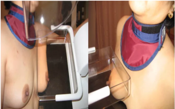
Fig. 3. An example of the thyroid protector.
measures need to be taken to decrease the level of radiation exposure to patients [15].
The thyroid is near the breast and is closely related to the organ controlling hormones. There is supporting evidence showing a great deal of radiation exposure to the thyroid and effect the scattering dose on the thyroid during mammography. This study found that the thyroid was exposed to 13.78% of the average glandular dose. Patients change their posture 4 times when they undergo a mammography examination.
Therefore, the thyroid receives 55.12% of the AGD.
A difference of 31.19% in TSD between patients suggests the possibility that patient A can receive 1 mGy but patient B receives 31 mGy, even though they were both exposed to a dose of 100 mGy. This means that the scattering dose increases with increasing radiation exposure depending on the condition of the breast (a thickness of compression, density, mass, and type and existence or nonexistence of the prosthesis).
The amount of the radiation cannot be ignored.
The ICRP publication (2007) reported that the level of the radiation exposure to the thyroid should not exceed 3 rem in a single year in a nonprofessional situation.
Some people believe that the level of the scattering dose from the breast is very small but the total volume is increased under the following conditions: when a follow up study is performed every 3-6 months, when magnification or a spot test and stereotactic biopsy is carried out after mammography, and taking the mammography many times at a single part of the breast. Although mammography is taken once a year, CT or angiography using radiation, which is performed continuously, does not affect the level of radiation exposure to the thyroid.
There is a 30-fold difference in scattering dose
to the thyroid between old and new models of
mammography equipment. Indeed, the management
of radiation must be completely controlled. It is
recommended that steps be taken to protect the
thyroid from the scattering dose. In addition, there
is a need for government regulation to limit the
level of radiation exposure from medical centers or
health care providers. Primarily, the radiology
technologist will play a key role in limiting the
level of radiation exposure. These results highlight
4. CONCLUSIONS
The thyroid was exposed to 13.78% of the AGD.
The results suggest that the thyroid is exposed to 55.12% of the scattering dose during a single routine mammography examination. The scattering dose to the thyroid increases with increasing radiation exposure to the breast. It is suggested that a thyroid protector be used to decrease the level of the radiation exposure.
REFERENCES
[1] S. S. Kim, J. Korean Breast Image Technol. 1, 52 (2005).
[2] M. S. Ryu, M. W. Choo, and C. S. Kim, J. Korean Breast Image Technol. 1, 61 (2005).
[3] I. J. Lee, K. Y. Park, and S. S. Kim, J. Radiol. Sci.
Techol. 29, 21 (2006).
[4] M. B. Goldman, Epidermiol. Rev. 12, 28 (1990).
[6] C, Giani, P. Fierabracci and R, Bonacci, J. Endocr.
Metab. 81, 990 (1986).
[7] P. P. A. Smyth, J .Endocrinol. Invest. 23, 42 (2000).
[8] A. A. Turken, Breast Cancer Res. 5, 110 (2003).
[9] Y. H. Kim, J. H. Choi, and S. S. Kim, J. Radiol.
Sci. Techol. 28, 241 (2005).
[10] K. S. Shin, J. H. Choi, and Y. H. Kim, J. Radiol.
Sci. Techol. 28, 293 (2005).
[11] Y. H. Kim, J. Radiol. Sci. Techol. 28, 173 (2005).
[12] International Atomic Energy Agency. Org [homepage on the Internet]. Vienna: IAEA Resource, Inc.;
c2003-08 [cited 2008 Agu 15]. Available from:
http://www-pub.iaea.org/MTCD/publications/PDF/SS -115-Web/Start.pdf (2008).
[13] Korea Food and Drug Administration [homepage on the Internet]. Seoul: The Association; c2006 [cited 2008 Nov 13]. A series of Management of the safety radiation (14): Guideline for checking the radiation dose. Available from: http://www.kfda.go.kr/open_
content/data/publication_view.php?seq=1521&av_pg=1
&textfield=환자&keyfield=v_all (2008).
[14] H. C. Kim, P. K. Cho, and S. S. Kim, J. Radiol. Sci.
Techol. 27, 55 (2004).
[15] W. G. Woo, J. Korean Breast Image Technol. 1, 22 (2005).