- 1 -
서 론
역사적으로 바늘을 이용한 조직검사는 1847년 Kun 등이 시행한 것으로 알려져 있으나 임상적으로 이용이 활발하게 된 것은 1970년대에 들어서이다.1) 여러 연구를 통하여 절개 생검 보다는 미세침흡인세포검사가 가격 성능 면에서도 뛰어 나며, 정확한 것을 확인하였다.2) 1980년대 들면서 미세침 흡 인세포검사의 여러가지 장점이 부각되면서 갑상선 결절의 조 직검사를 주도해 왔고 현재까지 표준 조직검사방법으로 사 용되고 있다.3) 미세침흡인세포검사의 갑상선암 진단정확도는 높지만, 진단특이도는 평균 83%로 매우 높지는 않으며 위음성 율이 2~18%로 무시할 수 없다.4-6) 특히 임상적으로 문제가 되 는 것은 현재 Bethesda 시스템의 atypia of undetermined singnificance의 해석이며, 10%에 달하는 inadequate specimen 도 환자와의 관계에 악영향을 미치는 중요한 단점이 된다. 또
한 미세침흡인세포검사는 여포성병변(follicular lesion)의 진 단 정확도가 상대적으로 낮다. 따라서 보다 적절한 치료 결정 을 위해서는 세침흡인세포검사의 한계를 극복할 수 있는 다른 수단이 필요하며, 실제 조직의 구성을 볼 수 있게 하는 초음파 유도하중심생검은 이런 약점을 보완할 수 있는 좋은 대안으 로 떠오르고 있다.
1. 초음파유도하중심생검
갑상선 결절의 조직검사 방법으로는 미세침흡인세포검사, 큰바늘흡인생검과 큰바늘생검 등 다양한 방법들이 사용되어 왔다.7,8) 갑상선결절의 진단에 초음파가 도입되고 1990년부터 스프링의 힘을 이용한 생검바늘이 개발되어 이의 유용성이 보 고되기 시작했고 이를 이용한 조직검사 방법을 이전과 구분하 여 초음파유도하중심생검이라고 부른다.9) 이는 기본적으로 미 세침흡인세포검사에 비해 비진단결과가 적고 조직의 구조를 유지한 상태로 비교적 많은 양의 조직을 얻을 수 있으며, 다양 한 면역화학 염색이 가능하다는 장점이 있다.
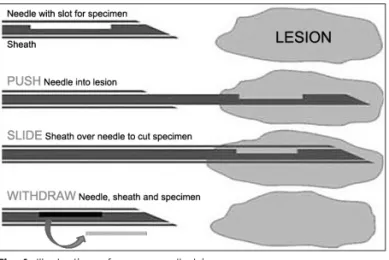
초음파유도하중심생검은 여러 회사에서 나오는 일회용 제 품을 이용하여 다음과 같은 방법으로 시행되며(Fig. 1) 현재까 지 16~21 gauge 바늘을 사용한 연구들이 보고되어 있고 그 중 에서 18~21 gauge가 많이 사용되고 있지만 국내에서는 주로 대한 두경부 종양 학회지
제 31 권 제 1 호 2015
갑상선 결절에서 초음파 유도하 중심생검의 역할
서울대학교 의과대학 분당서울대학교병원 이비인후과학교실
유 윤 종·안 순 현
=
Abstract
=The Role of Ultrasound Guided Core Needle Biopsy in Thyroid Nodule
Yoon-Jong Ryu, MD, Soon-Hyun Ahn, MD, PhD
Department of Otorhinolaryngology-Head and Neck Surgery, Seoul National University Bundang Hospital, Seoul National University College of Medicine, Seoul, Korea
Fine needle aspiration cytology(FNAC) holds a main role in assessing thyroid nodules. But nonnegligible rate of thyroid cytology is reported as uncertain, indeterminate or inadequate for diagnosis. Recently, the microhistologic evaluation by core needle biopsy(CNB) under ultrasound sonographical guidance has been reported to show high accuracy for the diagnose of thyroid nodules. Aim of this review was to furnish the state of the art of this topic by summarizing previous published data about indication, diagnostic performance, and complication of CNB in thy- roid lesions compared with FNAC
KEY WORDS
:Thyroid noduleㆍCore needle biopsyㆍFine needle aspiration cytologyㆍIndicationㆍComplication.Received : March 8, 2015 Accepted : April 29, 2015
교신저자 : 안순현, 463-707 경기도 성남시 분당구 구미로173번길 82 서울대학교 의과대학 분당서울대학교병원 이비인후과학교실 전화 : (031) 787-2034 ・ 전송 : (031) 787-4025
E-mail : [email protected]
online©MLComm
- 2 -
도는 1.5~2.3%로19-21) 미세침흡인세포검사에서 보고된 1~6.4%와22) 큰 차이를 보이지 않으며 통증 및 불편감에 있어 서도 미세침 흡인세포검사와 유의한 차이는 없는 것으로 나타
났다.23,24) 그러나 여러 번 검사를 시행하는 경우, 결절의 위치
가 갑상선 깊은 부위에 있어서 정상 갑상선을 많이 거쳐야 하 는 경우, 혈관이 풍부한 결절 등에 대해서는 출혈 및 통증의 빈도가 더 증가할 수 있는 점이 고려되어야 하겠다.25)
3. 적응증
초음파유도하중심생검의 구체적 적응증은 아직 확립되어 있지 않지만 현재까지의 모든 권고안들은 미세침흡인세포검사 의 보조적인 수단으로의 이용을 추천하고 있다. National Cancer Instute conference에서는 미세침흡인세포검사에서 비 진단결과를 보일 경우에 초음파유도하중심생검이 도움이 될 수 있다고 언급하였다.7) 2010년에 발표된 AACE/AME/ETA 권 고안5)에서는 미세침흡인세포검사로 정확한 진단이 어려운 림 프종 또는 역형성암종이 의심되는 경우나 이전 미세침흡인세 포검사에서 반복적으로 비진단결과가 나온 결절들에 한해서 제한적으로 초음파유도하중심생검을 사용할 것을 권고하고 있다. 2014년 공개된 American Thyroid Association 권고안 초 안에서는 미세침흡인세포검사에서 비진단결과 또는 불확정결 과로 진단될 경우에 추가적인 정보를 얻을 수 있다는 정도로 기술하고 있다.
현재 분당서울대병원에서는 첫 검사로 대개 미세침흡인세포 검사를 시행하며, 일부 매우 견고하게 보이는 경우에 한해서 영상의학과에서 필요에 따라 중심생검을 일차로 시행하기도 한다. 또 비진단결과 또는 불확정결과가 나올 경우 환자와의 관 계에서 문제가 생길 가능성이 있는 일차의료기관에서는 미세 침흡인세포검사와 중심생검을 함께 시행하는 경우도 흔한 것 으로 보인다. 저자의 경우 첫 흡인세포검사 결과에서 적절한 정 보를 얻지 못했을 경우 2차 검사로 중심생검을 적용하고 있다.
결 과
초음파유도하중심생검의 진단정확도에 대한 체계적인 연구 결과는 아직 부족한 편이데 미세침흡인세포검사에 비해 대체 로 진단예민도가 높고 위음성율이 매우 낮은 편이다.26) 그러나 Renshaw 등은 초음파유도하중심생검이 유두암종에 대한 낮 은 민감도를 보이는 것으로 상반된 결과를 보고하였는데27) 이 는 초음파유도하중심생검을 시행하는 검사자의 숙련도가 많 은 영향을 미친다고 해석할 수 있겠다.
최근의 연구들은 대게 특정 임상상황에서 검사방법에 따른 비교가 주류를 이루고 있다. 1차 미세침흡입세포검사에서 비진
단결과19,21) 또는 불확정결과18,20)를 얻은 경우에 초음파유도하
중심생검의 시행이 높은 진단율을 보이는 것으로 알려져 있으 18 gauge를 사용하고 있다.
2. 안정성 1) 종양 파종
바늘을 이용한 조직검사 방법이 소개되고 우선 논란이 된 것은 바늘이 통과한 경로에 암세포가 퍼질 수 있는 가능성 여부에 대한 것이다. 여기에 대해서 동물을 이용한 실험이 많 은데 Eriksson 등은 synergic mice에서 다양한 종양을 다리 에 만들고 조직검사의 방법에 따른 종양의 재발률을 보았다.
일반적으로 시행 되는 미세침흡입세포검사, 절제생검 등은 생존에 큰 영향을 주지 못하였다. 다만 미세침흡인세포검사 를 복강을 통해서 고의적으로 긴 경로로 하였을 경우는 유의 하게 높은 전이와 사망률을 보여 미세침흡인으로 인해 종양 세포가 퍼질 가능성을 보여 주었다.10) 이런 바늘을 이용한 검 사에서 종양세포가 퍼지는 것은 바늘의 크기와 관련이 있는 것으로 알려져 있다. 18~22 gauge의 얇은 바늘을 사용한 경 우에는 종양세포가 바늘구멍을 통해서 밖으로 나올 수 있음 은 보고가 되어 있으나11,12) 실제로 주사경로에 종양이 발생한 보고는 적다. 이에 비해 굵은 바늘을 이용한 경우는 종양의 발생이 드물지 않게 보고된 바 있다.13,14) 현재의 초음파유도하 중심생검은 과거와 달리 잘 고안된 기계를 사용하므로 시술에 따른 종양의 전이 가능성은 적다고 하지만 여전히 주의를 기 울여야 할 문제이며, 가능하면 작은 크기의 바늘을 이용하고 있다.
2) 합병증
초음파유도하중심생검은 실시간으로 중심부 바늘의 움직 임을 관찰하면서 조절하는 정교한 시술이 가능하기 때문에 숙련된 시술자에 의해 시행되는 경우 안전한 검사법으로 알려 져 있다. 여러 기관에서 보고된 합병증의 빈도는 2.5% 정도이
며15-21) 보고된 합병증으로는 혈종, 객혈, 감염, 갑상선 부종 및
일시적인 목소리이상 등이다. 일반적으로 미세침흡인세포검사 와 비교하여 통증이 더 많고 혈종의 발생률이 높다고 생각되 나 최근의 연구에 의하면 초음파유도하중심생검의 출혈의 빈
Fig. 1. Illustration of core needle biopsy.
- 3 -
며, 초음파소견상 악성이 강력하게 의심되는 경우28) 및 석회화 로 단단해진 결절29)에서도 유용하게 적용될 수 있음이 보고되 고 있다.결 론
역사적으로 볼 때 바늘을 이용한 생검이 먼저 시도되었다 가 질병의 파급 가능성과 생검을 위해 특수하게 제작된 바늘 이 일반적으로 구하기가 어려운 문제 등으로 미세침흡인세포 검사로 발전하였다. 고해상도 초음파 및 상품화된 중심생검을 위한 기구들이 개발 보급됨으로 인해 여러 분야에서 초음파 유도하중심생검이 적용되고 있다. 갑상선결절의 경우에도 대 부분의 경우 세침흡입세포검사에서 초음파 유도가 필요하고 상대적으로 높은 진단정확도로 인해 초음파유도하중심생검 의 장점이 분명한 것으로 보인다. 현재까지로는 미세침흡인세 포검사에서 불확실한 결과가 나온 경우 2차검사에 주로 이용 되지만 점차 적응증을 넓혀갈 것으로 생각된다.
중심 단어 : 갑상선결절 ・중심생검 ・미세침흡인세포검사 ・적 응증 ・합병증.
References
1) Linsk JA. Aspiration cytology in Sweden: The Karolinska Group.
Diagnostic Cytopathology. 1985;1:332-335.
2) Layfield LJ, Chrischilles EA, Cohen MB, Bottles K. The palpa- ble breast nodule. A cost-effectiveness analysis of alternate di- agnostic approaches. Cancer. 1993;72:1642-1651.
3) Wang C, Vickery AL, Jr, Maloof F. Needle biopsy of the thyroid.
Surgery, Gynecology & Obstetrics. 1976;143:365-368.
4) Tee YY, Lowe AJ, Brand CA, Judson RT. Fine-needle aspira- tion may miss a third of all malignancy in palpable thyroid nod- ules: A comprehensive literature review. Annals of Surgery. 2007;
246:714-720.
5) Gharib H, Papini E, Paschke R, Duick DS, Valcavi R, Hegedus L, et al. American Association of Clinical Endocrinologists, As- sociazione Medici Endocrinologi, and EuropeanThyroid Asso- ciation Medical Guidelines for Clinical Practice for the Diag- nosis and Management of Thyroid Nodules. Endocrine practice:
Official journal of the American College of Endocrinology and the American Association of Clinical Endocrinologists. 2010;16 (1):1-43.
6) Wang CC, Friedman L, Kennedy GC, Wang H, Kebebew E, Steward DL, et al. A large multicenter correlation study of thy- roid nodule cytopathology and histopathology. Thyroid: Official journal of the American Thyroid Association. 2011;21:243-251.
7) Pitman MB, Abele J, Ali SZ, Duick D, Elsheikh TM, Jeffrey RB, et al. Techniques for thyroid FNA: A synopsis of the Nation- al Cancer Institute Thyroid Fine-Needle Aspiration State of the Science Conference. Diagnostic Cytopathology. 2008;36:407-424.
8) Silverman JF, West RL, Finley JL, Larkin EW, Park HK, Swanson MS, et al. Fine-needle aspiration versus large-needle biopsy or cutting biopsy in evaluation of thyroid nodules. Diag- nostic Cytopathology. 1986;2:25-30.
9) Novoa E, Gurtler N, Arnoux A, Kraft M. Role of ultrasound- guided core-needle biopsy in the assessment of head and neck lesions: A meta-analysis and systematic review of the literature.
Head & Neck. 2012;34:1497-1503.
10) Eriksson O, Hagmar B, Ryd W. Effects of fine-needle aspiration and other biopsy procedures on tumor dissemination in mice. Can- cer. 1984;54:73-78.
11) Engzell U, Esposti PL, Rubio C, Sigurdson A, Zajicek J. Inves- tigation on tumour spread in connection with aspiration biopsy.
Acta radiologica: Therapy, Physics, Biology. 1971;10: 385-398.
12) Struve-Christensen E. Iatrogenic dissemination of tumour cells.
Dissemination of tumour cells along the needle track after percu- taneous, transthoracic lung biopsy. Danish Medical Bulletin. 1978;
25:82-87.
13) Clarke BG, Leadbetter WF, Campbell JS. Implantation of cancer of the prostate in site of perineal needle biopsy: report of a case.
The Journal of Urology. 1953;70:937-939.
14) Burkholder GV, Kaufman JJ. Local implantation of carcinoma of the prostate with percutaneous needle biopsy. The Journal of Urology. 1966;95:801-804.
15) Quinn SF, Nelson HA, Demlow TA. Thyroid biopsies: Fine-nee- dle aspiration biopsy versus spring-activated core biopsy needle in 102 patients. Journal of Vascular and Interventional Radiolo- gy: JVIR. 1994;5:619-623.
16) Taki S, Kakuda K, Kakuma K, Annen Y, Katada S, Yamashita R, et al. Thyroid nodules: Evaluation with US-guided core biop- sy with an automated biopsy gun. Radiology. 1997;202:874-877.
17) Screaton NJ, Berman LH, Grant JW. US-guided core-needle bi- opsy of the thyroid gland. Radiology. 2003;226:827-832.
18) Park KT, Ahn SH, Mo JH, Park YJ, Park do J, Choi SI, et al.
Role of core needle biopsy and ultrasonographic finding in man- agement of indeterminate thyroid nodules. Head & Neck. 2011;33:
160-165.
19) Na DG, Kim JH, Sung JY, Baek JH, Jung KC, Lee H, et al. Core- needle biopsy is more useful than repeat fine-needle aspiration in thyroid nodules read as nondiagnostic or atypia of undetermined significance by the Bethesda system for reporting thyroid cytopa- thology. Thyroid: Official journal of the American Thyroid Asso- ciation. 2012;22:468-475.
20) Sung JY, Na DG, Kim KS, Yoo H, Lee H, Kim JH, et al. Diag- nostic accuracy of fine-needle aspiration versus core-needle bi- opsy for the diagnosis of thyroid malignancy in a clinical cohort.
European Radiology. 2012;22:1564-1572.
21) Samir AE, Vij A, Seale MK, Desai G, Halpern E, Faquin WC, et al. Ultrasoundguided percutaneous thyroid nodule core biopsy:
Clinical utility in patients with prior nondiagnostic fine-needle aspirate. Thyroid: Official journal of the American Thyroid As- sociation. 2012;22:461-467.
22) Polyzos SA, Anastasilakis AD. Clinical complications following
- 4 -
thyroid fine-needle biopsy: A Systematic review. Clinical endo- crinology. 2009;71:157-165.
23) Carpi A, Rossi G, Nicolini A, Iervasi G, Russo M, Mechanick J.
Does large needle aspiration biopsy add pain to the thyroid nodule evaluation? PloS one. 2013;8:e58016.
24) Nasrollah N, Trimboli P, Rossi F, Amendola S, Guidobaldi L, Ventura C, et al. Patient’s comfort with and tolerability of thyroid core needle biopsy. Endocrine. 2014;45:79-83.
25) Baek J. Core needle biopsy of thyroid nodules: Consensus state- ment and recommendations. J Korean Soc Ultrasound Med.
2013;32:95-102.
26) Li L, Chen BD, Zhu HF, Wu S, Wei D, Zhang JQ, et al. Compar- ison of pre-operation diagnosis of thyroid cancer with fine needle
aspiration and core-needle biopsy: A meta-analysis. Asian Pacif- ic journal of cancer prevention: APJCP. 2014;15:7187-7193.
27) Renshaw AA, Pinnar N. Comparison of thyroid fine-needle as- piration and core needle biopsy. American Journal of Clinical Pa- thology. 2007;128:370-374.
28) Ha EJ, Baek JH, Lee JH, Song DE, Kim JK, Shong YK, et al. So- nographically suspicious thyroid nodules with initially benign cy- tologic results: The role of a core needle biopsy. Thyroid: Official Journal of the American Thyroid Association. 2013;23:703-708.
29) Ha EJ, Baek JH, Lee JH, Kim JK, Kim JK, Lim HK, et al. Core needle biopsy can minimise the non-diagnostic results and need for diagnostic surgery in patients with calcified thyroid nodules.
European radiology. 2014;24:1403-1409.