Corresponding Author: Kwang Soon Song, M.D., Department of Orthopedic Surgery, Keimyung University School of Medicine, 56 Dalseong-ro, Jung-gu, Daegu 700-712, Korea
Tel : +82-53-250-7729 E-mail : [email protected]
Abstract
The purpose of this thesis was to evaluate effectiveness of minimal open reduction and circular ring external fixation for treating of tibial pilon fractures with serious soft-tissue injuries or open wounds. The retrospective outcome study was performed as one of the methodology to conduct this research. The study was conducted on fifteen patients with tibial pilon fractures associated with serious soft-tissue injuries or open wounds. Patients were treated with minimal open reduction and circular ring external fixation and all of them were monitored for more than one year. Out of fifteen patients, thirteen of them were males and two were females, and their mean age was 45.3 years old (ranging from 29 to 62). Reduction status was evaluated with radiology based on the Burwell and Charnley’s criteria. Ankle function was graded using the criteria suggested by Mast and Teipner. The length of time that the circular ring external fixation device remained in place varied for each patient according to time until bony union. Any other complications were recorded throughout the study. Bony union was achieved within average 17.2 weeks in 13 out 15 fracture (86.6%). For 2 fractures complicated by infection, bony union was achieved at an average of 37 weeks after eradication of the infection. During the final follow-up examination, radiographs revealed that 7 patients had anatomic reduction, 6 had fair reduction, and 2 had poor reduction. Clinically, 7 patients had good function, 4 had fair function, and 4 had poor function. Range of motion of ankle joint at last follow up.
Showed average 17 degree of dorsiflexion and 13 degree of plantarflexion. There were 3 types of complications found during the study: pin-tract infection (1 patient), osteomyelitis (1 patient), and posttraumatic arthritis (1 patient). In conclusion, the combination of minimal open reduction and circular ring external fixation could be considered as an alternative treatment for tibial pilon fracture associated with serious soft-tissue injuries or open wounds.
Key Words :
Circular ring external fixation, Minimal open reduction, Pilon fracture, Tibia Department of Orthopedic Surgery, Keimyung University School of Medicine,Daegu, Korea
Ki Cheor Bae, M.D., Kwang Soon Song, M.D.
Treatment of Tibial Pilon Fractures with Minimal Open Reduction
and Circular Ring External Fixation
Introduction
Tibial pilon fractures, or fractures of the articular surface of the distal tibia, account for 5%
to 7% [1,2] of all tibial fractures. Pilon fractures are high-energy, explosive intra-articular fractures produced by axial loading force. They are very difficult to reduce anatomically, because of severe comminution. About 10% to 30% of such fractures are related to serious soft-tissue injuries or open wounds [3-5].
In cases of closed tibial pilon fracture, open reduction and internal fixation is preferred generally. When open wound with extensive soft tissue injury or severe comminution with displacement are noted, external fixation is preferred [6,7]. If one try open reduction and internal fixation in cased of high energy trauma with open wound, there are increased risk of infection, skin necrosis or non-union by further soft tissue injury during open reduction, which may cause poor result requiring other unnecessary procedures.
We conducted this study to evaluate the results and complications of using a combination of minimal open reduction and circular ring external fixation to treat tibial pilon fractures with serious soft-tissue injuries or open wounds.
Materials and Methods
We evaluated all patients’ medical records and radiographs retrospectively. We recalled all patients and performed physical examinations in 15 patients with pilon fractures who had open or severe soft- tissue damage and who were treated with minimal open reduction and circular ring external fixation between December 1995 and September 2006 (Table 1).
The mean age of the patients was 45.3 years (range, 29 to 62 years); there were 13 men and 2 women. The mean follow-up period was 17.8 months (range, 13 to 29 months).
Causes of injury were as follows: falling from a height, 9 patients (60%); motor vehicle accident, 5 patients (33.3%); falling, 1 patient (6.7%). Of the motor vehicle accidents, 4 (26.6%) involved a car and 1 (6.7%) involved a motorcycle. Eleven patients had closed fractures and 4 had open fractures.
We used the Rüedi-Allgöwer classification system, which stratifies fractures into 3 types according to the degree of comminution and displacement of the articular surface. This study was composed of tibial pilon fractures associated w i t h s e r i o u s s o f t-t i s s u e i n j u r i e s s u c h a s s u b c u t a n e o u s h e m o r r h a g e, b l u i s h s k i n discolorization and bullae formation or open wounds, and were treated with minimal open reduction and circular ring external fixation. Of the 15 patients, 4 (26.6%) had a type II fracture and 11 (73.3%) had a type III. To determine the degree of damage to soft tissue in closed fractures, we used the Tscherne classification [8]. Of 11 cases of closed fractures, 8 were grade 2 and 3 were grade 3.
The time elapsed from injury to surgery was an average of 8.4 days. We attempted reduction in all patients at the earliest point after their general condition had been stabilized, performing surgery generally within 2 weeks of injury.
All cases had combined fibular fractures, fibular fracture was reduced and fixed with AO plate &
screws at first. To maintain the correct length of bone, via a longitudinal skin incision along the posterior border of fractured fibula. Then we applied external ring fixator. K-wires were inserted into tibia shaft above the fracture site and talus, calcaneus below fracture site, from medial side. After application of ring fixator, we applied distraction forces for
ligamentotaxis and confirmed the reduction status by image intensifier. When articular incongruence is noted, we made a minimal skin incision for limited open reduction and followed by internal fixation using a lag screw or K-wires.
We confirmed reduction status using an image intensifier. Circular ring external fixation devices were used in all patients. To determine the reduction state (anatomic, fair, or poor) of the fractures, we evaluated trimalleolar displacement, angulation, and displacement of the talus according
to the strict radiographic criteria of Burwell and Charnley [9]. We used the time until removal of the circular ring external fixation device to calculate the time to bony union.
Time interval for bone union was calculated from the day of injury to the day of removal of ring external fixator. All patients were followed more than 1 year, and we analyzed clinical result by range of motion, presence of swelling or pain, using the Mast and Teipner’s functional evaluation method [10], and the results were recorded as Table 1. Patients’ data
Type
Case Age/Sex Mechanism of Injury R-A
Class
a
Closed/Open
Union Time (weeks)
Follow- (months)Up
Radiologic Results
b
Functional
Results
c
Complications1 48/M Fall
e
III Open 26 13 Fair Good2 35/M Fall
e
III Open 30 27 Poor Poor Valgusalignment
3 62/M TA
d
II Closed 15 13 Anatomic Good4 58/M TA
d
II Open 15 13 Anatomic Good5 29/M Fall
e
III Closed 21 13 Fair Good6 46/F Fall
e
III Closed 10 29 Anatomic Poor7 30/F TA
d
III Closed 13 23 Fair Fair8 46/M Fall down III Closed 17 27 Poor Poor
9 46/M TA
d
II Closed (34) 18 Fair Fair Pin tractinfection
10 51/M Fall
e
III Closed (40) 24 Anatomic Poor Infection11 62/M TA
d
II Open 24 13 Anatomic Good12 42/M Fall
e
III Closed 15 15 Fair Fair13 33/M Fall
e
III Closed 12 13 Anatomic Good14 51/M Fall
e
III Closed 13 13 Anatomic Fair15 41/M Fall
e
III Closed 10 13 Fair Gooda
Classification system of Rüedi and Allgöwer (23).b
Radiographic criteria of reduction by Burwell and Charnley (8).c
Function criteria by Mast and Teipner (16).d
Traffic accident.e
Fall from a height.good, fair or poor. We also noted postoperative complications.
Results
The average elapsed time until union was 17.2 weeks (range, 10 to 30 weeks) for 13 patients (86.6%); the other 2 (13.3%) had infective nonunion. Fracture reduction was anatomic in 7 patients (46.6%), fair in 6 (40%), and poor in 2 (13.3%), according to the Burwell and Charnley method [9]. There were 4 cases of type II fracture and 11 cases of type III fractures, by Rüedi- Allgöwer classification system. Of these 4 patients of type II fractures, 3 patients had anatomical reduction was achieved in 3 patients (75%) and fair reduction in 1 (25%). Of the 11 cases of type III, anatomic reduction was achieved in 4 patients (36.4%), fair in 5 (45.5%), and poor in 2 (18.2%).
Treatment results were determined to be good in 7 patients (46.6%), fair in 4 (26.6%), and poor in 4 (26.6%) according Mast and Teipner’s clinical function evaluation system. Of the 4 cases of type II fractures, result was good in 3 patients (75%), fair in 1 patients (25%). Of the 11 cases of type III fractures, result was in 4 patients (36.4%), fair in 3 (27.2%), and poor in 4 (36.4%).
We experienced 3 cases of complication; one patient with a Rüedi-Allgöwer type III and Tschene grade 2 fracture had soft-tissue necrosis and osteomyelitis. We performed bacterial cultures, intravenous administration of suitable antibiotics, curettage and wound irrigation. The other patient with a Rüedi-Allgöwer type II and Tschene grade 2 fracture had a pin-tract infection. But we achieved union at an average of 37 weeks. One patient with Rüedi-Allgöwer type III and open fracture resulted valgus deformity and osteoarthritis, so we performed ankle arthrodesis with success.
Discussion
Tibial pilon fracture is fracture of the region constituting the top of the ankle joint and is caused mainly by high-energy axial compression forces. In these fractures, severe comminution is caused by crush rather than by rotation force.
According to the degree of damage, these fractures can be accompanied by malleolar fractures, displacement, and bone loss of the metaphysis such that widespread soft-tissue damage may occur.
Several studies have reported that main causes for pilon fractures are falls from a height or traffic accidents [11,12]; this was the case with our patients. Several authors have attempted to classify pilon fractures, but this is difficult because of the complicated properties of the fractures themselves. In 1953, Lauge-Hansen [13]
described pilon fractures as pronation-dorsiflexion fractures, and in 1986, Ovadia and Beals divided these same fracture types into 5 groups on the basis of amount of displacement and comminution [14]. In 1969, Rüedi and Allgöwer classified fractures into 3 types, according to the degree of comminution and displacement of the articular sur face, which was useful in determining prognosis; their now-universal classification system is the one that we used [15].
Because we targeted patients with tibial pilon fractures accompanied by severe soft-tissue damage or open wounds whose fractures were treated with minimal open reduction and external fixation using the circular ring external fixation device, all had fractures classified as type II or III under the Rüedi-Allgöwer system.
Treating tibial pilon fractures is difficult, because they combine severe swelling and damage to soft tissue (Fig. 1), especially when the displacement of an articular fragment is severe or is
accompanied by metaphysial bone defect or when the degree of comminution is severe. Tibial pilon fractures have long been treated with nonsurgical methods, with open reduction and internal fixation, with a restrictive open reduction and external fixation, and with a manual reduction and external fixation [10,16,17]. Ayeni [18] reported poor results in 50% of patients treated with nonsurgical methods such as a plaster splint or cast. Bone [1] used a cast and calcaneus skeletal traction after closed reduction. However, plaster casts can neither reduce a fractured articular surface nor secure and maintain the normal length of the fracture site, and when plaster casts are used in the presence of severe soft-tissue damage and an open wound, it is impossible to monitor the state of the soft tissue.
At present, the most popular treatment is open reduction and firm internal fixation using a plate and screw [14]. Rüedi and Allgöwer reported good results in 1973 after deciding on a treatment
protocol [3]. They restored the normal length of the fibula by reducing it [18]. Then they recovered the distal articular surface of the tibia [4]. Next, they did bone grafting to repair the metaphysial bone defect [5]. Finally, using a buttress plate, they attempted rigid internal fixation on the medial side of the distal tibia. They had these patients perform range-of-motion exercises early after surgery and noted good results in 73% [19,20]. However, most of those patients had sustained fractures caused by a low-energy impact and had only slight soft-tissue damage.
Kellam and Waddell [12] reported that 84% of their patients had good results from repairs to their type I fractures but that only 53% of those with type II or III fractures. In 1989, Bourne et al. [16]
reported that 80% of patients had satisfactory results in type I and II fractures, but only 32% had good results in type III fractures. Also, there were difficulties with open reduction and internal Fig. 1. A Rüedi-Allgöwer type III pilon fracture of the right ankle, a 51-year-old man who had a motor vehicle accident. Ankle swelling and multiple bullae developed.
fixation; Kang et al. [21] reported that rigid internal fixation with a buttress plate was possible in only 6 of 17 cases.
When open reduction and internal fixation is used as the first-line treatment for open fractures or severely damaged pilon fractures, complications such as skin necrosis, osteomyelitis, septic arthritis, nonunion, malunion, and superficial infection occur, requiring flap surgery for skin coverage, ankle arthrodesis, or amputation.
Posttraumatic arthritis has been reported to occur in 50% of patients with tibia pilon fracture [1,2,18,22]. Also, several studies have reported that when open reduction and internal fixation has been performed for Gustilo-Anderson type II and III open pilon fractures, the number of complications was very high [14,23,24] and that when open reduction and internal fixation is performed for pilon fractures accompanied by serious soft-tissue injury, the frequency of delayed complications and revision surgeries is likely to increase [25]. Teeny and Wiss [26] emphasized that if anatomic reduction cannot be conducted without soft-tissue complications before surgery, another type of treatment should be considered; they reported poor results for performing open reduction and internal
fixation in this situation. Primary open reduction and internal fixation produced unsatisfactory results in Rüedi-Allgöwer type III fractures and open fractures caused by high-energy trauma.
Because the complication rate for open reduction and internal fixation is high in high energy trauma patients, an alternative external fixation is attracting attention. Kellam and Waddell [12] and Mast et al. [27] reported that when an external fixation device is used in treating tibia pilon fracture, it is useful in securing the normal length of the fracture site and reconstructing and stabilizing the articular surface, and Tornetta et al.
[7] reported that 81% of patients had excellent results and low rate of complication when internal fixation and external fixation were integrated.
Bone et al. [28] reported excellent or good results in 6 cases, fair in 9, and poor in 5 by using a delta- frame external fixation device, including the ankle joint, in 20 cases of open comminuted pilon fracture.
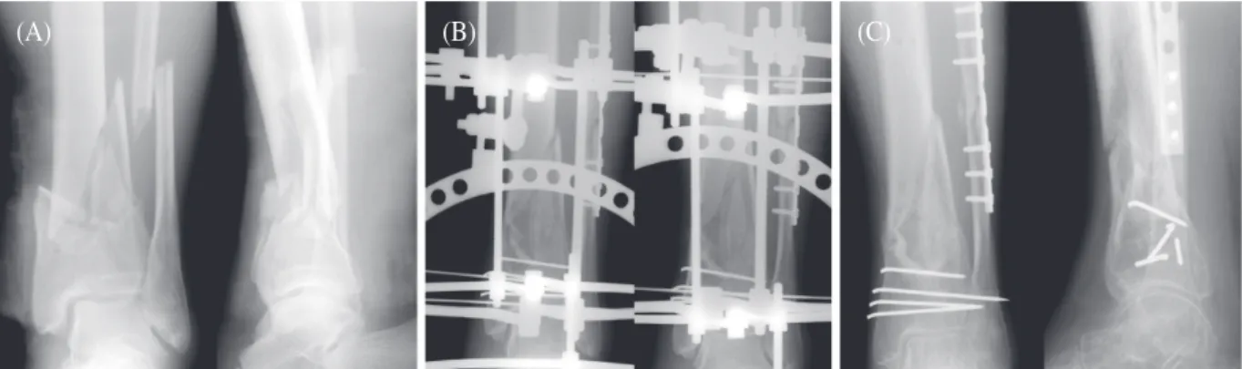
We used minimal open reduction and circular ring external fixation to treat tibia pilon fractures with by serious soft-tissue injuries or open wounds (Fig. 2) and obtained union in 13 (86.6%) of 15 patients by an average of 17.2 weeks (range, 10 to
Fig. 2. A: A 62-year-old man had a motor vehicle accident and sustained a Rüedi-Allgöwer type II pilon fracture of left ankle. B: The patient underwent minimal open reduction and circular ring external fixation with percutaneous pinning of the tibial pilon fracture and open reduction and internal fixation of the fibular shaft fracture. C: Follow-up radiographs show complete bone union. There were no complications.
(A) (B) (C)
30 weeks). We obtained good results in 7 patients (46.6%), fair in 4 (26.6%), and poor in 4 (26.6%), as evaluated by Mast and Teipner’s clinical function method. We did not encounter widespread skin necrosis, which is frequently a problem with internal fixation, in any of our patients. Three of our patients developed complications (soft-tissue necrosis and osteomyelitis, pin-site infection, and valgus deformity); thus, in comparison with studies in which other treatments were used, our results were at least as good, if not better.
We were able to obtain good results and had only a small number of complications because primary reduction was obtainable, along with restoration of normal bone length, through the use o f c i r c u l a r r i n g e x t e r n a l f i x a t i o n a n d ligamentotaxis.
Minimal open reduction in combination with circular ring external fixation leads to fewer complications and good union in tibial pilon fractures accompanied by serious soft-tissue injury and open wounds. Despite the brevity of our follow-up period, we believe that longer-term studies will confirm that this combination of treatment methods is a viable alternative for treating such fractures.
References
1. Bone LB. Fractures of the tibial plafond. The pilon fracture. Orthop Clin North Am 1987;18:95-104.
2. Mast J, Seligson D. Complex ankle fractures. In: Mayer MH, editor. The Multiply Injured Patient with Complex Fractures. Philadelphia: Lea & Febiger; 1984. P. 304.
3. Abelseth G, Buckley PE, Pineo GE, Hull R, Rose MS.
Incidence of deep-vein thrombosis in patients with fractures of the lower extremity distal to the hip. J Orthop Trauma 1996;10:230-5.
4. Barbieri R, Schenk RS, Koval K. Hybrid external
fixation in the treatment of tibial plafond fracture. Clin Orthop Relat Res 1996;332:16-22.
5. Blauth M, Bastian L, Krettek C, Knop C, Evans S.
Surgical options for the treatment of severe tibial pilon fracture: a study of the three techniques. J Orthop Trauma 2001;15:153-60.
6. French B, Tornetta P 3rd. Hybrid external fixation of tibia pilon fractures. Foot Ankle Clin 2000;5:853–71.
7. Tornetta P 3rd, Weiner L, Bergman M, Watnik N, Steuer J, Kelley M, et al. Pilon fractures: treatment with combined internal and external fixation. J Orthop Trauma 1993;7:489–96.
8. Tscherne H, Gotzen L. External articular transfixation of joint injuries with severe soft tissue damage. In:
Tscherne H, Gotzen L, edtors. Fractures with Soft- Tissue Injuries, Berlin: Springer-Verlag; 1984. 103-17.
9. Burwell HN, Charnley AD. The treatment of displaced fractures of the ankle by rigid internal fixation and early joint movement. J Bone Joint Surg Br 1965;47:634-60.
10. Mast JW, Teipner WA. A reproducible approach to the internal fixation of adult ankle fractures: rationale, technique, and early results. Orthop Clin North Am 1980;11:661-79.
11. Hwang SK, Park JS, Park HJ. Fractures of the tibial pilon. J Korean Orthop Assoc 1993;28:1747-57.
12. Kellam JF, Waddell JP. Fractures of the distal tibial metaphysis with intra-articular extension—the distal tibial explosion fracture. J Trauma 1979;19:593-601.
13. Lauge-Hansen N. Fractures of the ankle. V. Pronation- dorsiflexion fracture. AMA Arch Surg 1953;67:813-20.
14. Ovadia DN, Beals RK. Fractures of the tibial plafond.
J Bone Joint Surg Am 1986;68:543-51.
15. Rüedi TP, Matter P, Allgöwer M. Intra-articular fractures of the distal tibial end [German]. Helv Chir Acta 1968;35:556-82.
16. Bourne RB, Rorabec CH, Macnab J. Intra-articular fractures of the distal tibia: the pilon fracture. J Trauma 1983;23:591-5.
17. Griend RA, Savoie FH, Hughes JL. Fractures of the Ankle. In: Rockwood Jr CA, Green DP, Bucholz ,
edtors. Rockwood and Green’s Fractures in Adults.
Philadelphia: JB Lippincott; 1991. 1983-2039.
18. Ayeni JP. Pilon fractures of the tibia: a study based on 19 cases. Injury 1988;19:109-14.
19. Rüedi TP. Fractures of the lower end of the tibia into the ankle joint: results 9 years after open reduction and internal fixation. Injury 1973;5:130-4.
20. Rüedi TP, Allgöwer M. The operative treatment of intra-articular fractures of the lower end of the tibia.
Clin Orthop Relat Res 1979;138:105-10.
21. Kang CS, Pyun YS, Sohn SW, Song KS, Kang CH, Min BW, et al. A clinical study of the surgical treatment of pilon fracture. J Korean Orthop Assoc 1993;28:276-82.
22. Taylor JC. Fractures of the lower extremity. In:
Crenshaw AH, edtor. Campbell’s Operative Orthopaedics. St. Louis: Mosby Year Book; 1992. P.
795-893.
23. McNamara MG, Heckman JD, Corley FG. Severe open fractures of the lower extremity: a retrospective
evaluation of Mangled Extremity Severity Score (MESS). J Orthop Trauma 1994;8:81-7.
24. McFerran MA, Smith SW, Boulas HJ, Schwartz HS.
Complications encountered in the treatment of pilon fractures. J Orthop Trauma 1992;6:195-200.
25. Rommens PM, Claes P, De Boodt P, Stappaerts KH, Broos PL. Therapeutic procedure and long-term results in tibial pilon fracture in relation to primary soft tissue damage [German]. Unfallchirurg 1994;97:39-46.
26. Teeny SM, Wiss DA. Open reduction and internal fixation of tibial plafond fractures. Variables contribution to poor results and complications. Clin Orthop Relat Res 1993;292:108-17.
27. Mast JW, Spiegel PG, Pappas JN. Fractures of the tibial pilon. Clin Orthop Relat Res 1988;230:68-82.
28. Bone L, Stegemann P, McNamara K, Seibel R.
External fixation of severely comminuted and open tibial pilon fractures. Clin Orthop Relat Res 1993;292:101-7.