- 817 -
In vitro Antioxidant Activity of the Aqueous of Angelicae gigas Nakai Leaves Sung-Jin Park1, Jung-Han Yoon, Young-Eon Kim
3, Won-Byong Yoon
4 and Jong-Dai Kim
4†
12
Department of Tourism Food Service Cuisine, Hallym College, Chuncheon 200-711, Korea Department of Food Science and Nutrition, Hallym University, Chuncheon 200-702, Korea
3Korea Food Research Institute, Seongnam 463-746, Korea
4
Division of Food Biotechnology, Medical & Bio-material Research Center, Kangwon National University, Chuncheon 200-701, Korea
당귀 잎의 항산화 활성
박성진
1․윤정한
2․김영언
3․윤원병
4․김종대
4†1
한림성심대학교 관광외식조리과,
2한림대학교 식품영양학과,
3한국식품연구원,
4강원대학교 식품생명공학과
Abstract
Angelicae gigas Nakai has been used as a traditional medicine as well as an edible vegetable in South Korea.
In this study, the total phenolic and flavonoid content and antioxidants of A. gigas Nakai leaves were examined in vitro via hydroxyl-radical-scavenging activity, reducing power activity, metal chelating assay, and DPPH-free -radical-scavenging assay. Among all the extracts from A. gigas Nakai leaves, the ethanol extract showed the strongest effects in all of the assays. The EC
50values for the DPPH-radical-scavenging activities of ethanol, methanol, and water extracts were 31.47, 42.14, and 58.47 μg/mL, respectively. Among the extracts from A. gigas Kakai leaves, the ethanol extract had the highest levels of total phenolics (7.84 ± 1.46 mg TAN/g) and total flavonoids (4.23
± 0.03 mg QE/g), which correlated strongly with the individual phenolic-compound (p-hydroxybenzoic acid, vanillin, and trans-ferulic acid) contents. The ethanol extract also showed stronger antioxidant activity than tocopherol in hydroxyl- radical-scavenging activity assay. These results indicate that the ethanol extract of A. gigas Kakai leaves possesses significant antioxidant properties, which suggests its great potential as a functional-food ingredient in the food industry.
Key words :Angelicae gigas Nakai leaf, antioxidant, total phenolics, total flavonoids, phenolic acids
INTRODUCTION
1)
It is commonly recognized that reactive oxygen species (ROS) are involved in a variety of physiological and pathological processed, including cellular signal transduction, cell proliferation, differentitation and apotosis, as well as ischemia–reperfusion, inflammation, and many neurodegenerative disorders (1). In healthy individuals, ROS production is continuously balanced by natural antioxidative defence systems. Oxidative stress is a process where the physiological balance between pro-oxidants and antioxidants is disrupted
†
Corresponding author. E-mail:[email protected] Phone:82-33-250-6456, Fax:82-33-241-0508
in favour of the former, ensuing in potential damage for the organism (2). ROS production can induce DNA damages, protein carbonylation, and lipid peroxidation, leading to a variety of chronic health problems, such as cancer, aging, Parkinson’s disease, Alzheimer’s disease and amyotrophic lateral sclerosis (3).
There is increasing evidence that consumption of a variety
of phenolic compounds present in natural foods may lower
the risk of serious health disorders because of the antioxidants
activity of these compounds (4). When added to foods,
antioxidants minimize rancidity, retard the formation of toxic
oxidation products, maintain nutritional quality, and increase
shelf life (5). The antioxidant activity of extract of several
plants, including their leaves, bark, roots (6), fruits, seeds
(7) and seedcake (8) has been extensively studied.
Tocopherol, tertiary-butylhydroquinone (TBHQ), butylated hydroxytoluene (BHT) and butylated hydroxyanisol (BHA) are the most commonly used primary antioxidants in oils.
However, many researchers reported the adverse effects of synthetic antioxidants such as toxicity and carcinogenicity (9). Due to safety and limitation of synthetic antioxidant usage, natural antioxidants obtained from edible materials, edible by‐products and residual sources have become alternately interesting (10).
Antioxidants have diverse functions in biological systems, including defense against oxidative damage and participation in major signaling pathways in cells (11). One of the main functions of antioxidants is to prevent damage caused by the action of ROS (12). Antioxidants posses the ability to protect the cellular organelles from damages caused by free radicals induced oxidative stress. Free radicals include hydroxyl radical, superoxide anion radical and hydrogen peroxide.
Highly reactive free radicals which are formed by exogenous chemicals, stress or in the food system are capable of oxidizing biomolecules, resulting in cancer, coronary heart disease and hypertension (13).
The pharmaceutically active ingredient in Angelica gigas is a substance called coumarin. In addition, Angelica gigas contains decursinol, umbelliferone, and β-sitosterol. Coumarin derivatives have been used as anticoagulant to prevent acute myocardial infection bleeding (14). In addition, several kinds of Angelica exist including Angelica gigas (Korea), Angelica acutiloba Kotagawa (Korea), Angelica acutilova Kit. var.
(Japan) sugiyamae Hikino (Japan) and Angelica sinensis Diels (China). South Korean Angelica has shown to contain coumarin, decursin, decursinol, etc., and Chinese and Japanese Angelica have shown to contain phthalide ligustilide, butylidene phthalide, and butylphthalide (15). The components of Korean Angelica giga were studied by Ryu (16). In these studies, coumarin, decursin, decursinol and decursinol angelate were successfully identified and separated out.
Studies of this plant have primarily been conducted using root samples; thus, little information is available regarding the leaves of Angelicae gigas , which are consumed as a vegetable in Korea (17). To investigate the potential antioxidant properties of Angelicae gigas leaf, we utilized a number of in vitro assay systems, including hydroxyl radical scavenging assay, metal chelating assay, DPPH free radical scavenging assay, phenolic acids, total phenolic and total flavonoid measurement.
MATERIALS AND METHODS
Chemicals and Reagents
1,1-Diphenyl-2-picryhydrazyl (DPPH), α-tocopherol, 3-(2- pyridyl)-5,6-bis (4-phenyl-sulfonic acid)-1,2,4-triazine (ferrozine), trichloroacetic acid (TCA), sulfanilamide, naphthy-lethlenediamine dihydrochloride, tannic acid, Folin-Ciocalteu’s, BHT, and LPS ( E. coli 0111:B4) were purchased from Sigma (St. Louis, MO, USA). 2-Thiobarbituric acid (TBA) was purchased from Alfa Aesar (Karlsruhe, Germany). RPMI medium 1620 and FBS were acquired from Gibco BRL (Grand Island, NY, USA). The culture supplies (e.g., 96-well plates) were obtained from SPL Brand Products (SPL, Suwon, Korea).
All other chemicals were of analytical grade.
Preparation of the Extracts
Leaves of Angelicae gigas collected in September 2008 in PyeongChang, Korea, were dried in the shade at room temperature and then powdered. Two hundred grams of the leaf powder were extracted separately with 80% methanol, 80% ethanol at 70℃ and with hot water for 8 hr. The methanol (ME), ethanol extracts (EE), and hot water (WE) were filtered (100-mm Whatman filter paper; Maidstone, UK) and evaporated (CCA-1110; Eyela, Tokyo, Japan). The freeze- dried samples were weighed and kept in a refrigerator until further analysis.
Total Phenolics and Total Flavonoid Contents
Total phenolic compounds of the extracts were determined
using Folin-Ciocalteu reagent described by Singleton (18)
with slight modifications. Each extract (0.1 g) was diluted
to 1 mL with distilled water. The diluted samples were mixed
with 1 mL of diluted (1 : 10) Folin-Ciocalteu reagent and
incubated at 22℃ for 5 min. The mixtures were reacted with
1 mL of 10% sodium carbonate solution and allowed to stand
at 22℃ for 1 h. The absorbance was measured using a
microplate reader at 760 nm. Tannic acid (Tan) was used
as the standard to prepare a calibration curve. Total phenolic
concentrations were expressed as mg of tannic acid equivalent
(TAE) per g of extract. Total flavonoid content was
determined using a spectrometric method (19). Each extract
(0.5 mL) was serially mixed with 0.1 mL of 10% aluminum
nitrate, 0.1 mL of 1 M aqueous potassium acetate, and 4.3
mL of ethanol. The mixture was allowed to stand at room
temperature for 40 min, and then the absorbance was
measured spectrophotometrically at 415 nm. A standard curve
was prepared at 0, 2, 4, 6, 8, and 10 mg/mL of quercetin
as described above. Flavonoid contents were expressed as mg of quercetin equivalent (QE) per g of extract.
DPPH Radical Scavenging Activity
The antioxidant activity of the samples and control was determined by 2,2-diphenyl-1-picrylhydrazyl (DPPH) radical scavenging assay (20). Two mL of the extracted sample (1%, w/v) was mixed with 1 mL of 0.2 mM DPPH radical solution in 95% ethanol. The mixture was incubated at 25℃ for 30 min and measured the absorbance at 517 nm. The scavenging activity of DPPH radical was calculated by the following equation:
DPPH scavenging activity (%)=[1−(At/Ac)]×100, where Ac is the absorbance of the control reaction and At is the absorbance of the extract.
The efficient concentration (EC
50) was estimated from the percentage of DPPH scavenging activity plotted as function of the concentration of Angelicae gigas leaf extract, which was expressed in terms of the concentration (mg/mL) required for 50% reduction of DPPH.
Reducing Power Assay
A spectrometric method of Oyaizu (21) with slight modification was used to determine the reducing power of extracts. Each extract (1 mL) was mixed with 0.2 M sodium phosphate buffer (1 mL, pH 6.6) and 1% aqueous potassium ferricyanide (1 mL). The mixture was placed in a water bath at 50℃ for 20 min, immediately cooled to room temperature, and mixed with 1 mL of 15% trichloroacetic acid, which was centrifuged at 1,500 × g for 15 min. The collected supernatant (1 mL) was diluted with 1 mL of distilled water and then mixed with 1 mL of 0.1% ferric chloride to reduce K
3Fe(CN)
6to K
4Fe(CN)
6. The absorbance was measured spectrophotometrically at 700 nm. The efficient concentration (EC
50) was estimated from the absorbance plotted as function of the concentration of Angelicae gigas leaves extract, denoting the concentration (mg/mL) required to achieve absorbance of 0.5.
Fenton Reaction (Degradation of Deoxyribose)
The ability of leaf extract to prevent Fe
2+/H
2O
2‐induced decomposition of deoxyribose was assessed using the method of Halliwell and Gutteeridge (22). In brief, freshly prepared aqueous extract (0-100 μL) was added to a reaction mixture containing 120 μL of 20 mM deoxyribose, 400 μL of 0.1
M phosphate buffer, 40 μL of 20 mM hydrogen peroxide, and 40 μL of 500 μL FeSO
4, and the volume was made up to 800 μL with distilled water. The reaction mixture was incubated at 37℃ for 30 min, and the reaction was then stopped by addition of 0.5 mL of 2.8% trichloroacetic acid.
This was followed by addition of 0.4 mL of 0.6%
thiobarbituric acid solution. The tubes were subsequently incubated in boiling water for 20 min. The absorbance was measured at 532 nm in a spectrophotometer (UV 1601 PC, Shimadzu Co, Kyoto, Japan).
Metal Chelating Activity
The chelation of ferrous ions by the extracts was estimated as described previously (23). In brief, 1 mL of extract at various concentrations was mixed with 3.7 mL of absolute methanol and 0.1 mL of 1 mM FeCl
2. The reaction was initiated by the addition of 0.2 mL of 5 mM ferrozine followed by vigorous shaking; the mixture was then left to react at room temperature for 10 min. The absorbance was measured at 562 nm.
HPLC Analysis of Phenolic Acids
Phenolic monomers and dimers were analyzed using the high-performance liquid chromatography (HPLC, Waters M600E; Milfold, MA, USA) system equipped with a UV absorbance detector (280 nm, 0.05 AUFS). The separations of free phenolic acids were achieved in the reverse phase mode using Waters Spherisorb ODS2 column (250 mm × 4.6 mm i.d., 5 μm). The mobile phases were solvent A, 1 mM trifluoroacetic acid (TFA) in 10% (v/v) acetonitrile;
solvent B, 1 mM TFA in 40% (v/v) methanol and 40% (v/v) acetonitrile. The gradient profile was used as follows (24):A 90%, B 10% (initial); A 90%, B 10% (0-10 min); A 60%, B 40% (10-15 min); A 60%, B 40% (15-24 min); A 0%, B 5%, C 100%, (24-40 min); A 90%, B 10%, C 100%, (40‐45 min); and A 90%, B 10%, C 100%, (45-50 min) at a flow rate of 1 mL/min. Peaks were identified by retention times established from standard solutions. Pure phenolic acid standards (p-hydroxybenzoic acid, p-hydroxybenzaldehyde, vanillic acid, vanillin, coumaric acid, trans -ferulic acid, and trans -cinnamic acid) were dissolved in methanol and serially diluted to 10, 50, 100 mg/mL. Dilutions and peak areas were used to establish standard curves.
Statistical Analysis
The experimental results are expressed as means ± standard
deviation (SD) of three measurements at least. Statistical
Package for Social Science (version 12.0 for window) was used to analyze the variance (AVONA), and the significant level for all measurements was p < 0.05.
RESULTS AND DISCUSSION
Total Phenolics And Flavonoids Contents
The total phenolic and total flavonoid contents in each A. gigas leaf extracts are presented in Table 1. As shown in Table 1, the total phenolics and total flavonoids from the ethanol extraction showed highest values. It is reported that intense heat from ethanol was able to release cell wall phenolics or bound phenolics due to the breakdown of celluar consistuents, thus causing polyphenols to be extracted (25).
Table 1. Total phenols (mg TAN/g) and flavonoids (mg QE/g) in A. gigas leaf extracts
Extracts Yield (%)
aTotal phenols Flavonoids EE
c35.47 7.87 ± 1.46
b4.23 ± 0.03
ME
d32.87 7.47 ± 0.58 2.87 ± 0.26
WE
e31.25 7.43 ± 0.35 1.97 ± 0.79
aExtraction yield (%) is expressed as:
(freeze dried sample extract weight/ dried sample weight)X100.
bEach value is the mean ± S.D. of triplicate measurements.
c80% ethanol extract
d80% methanol extract
ehot water extract
DPPH Radical Scavenging Activity
Free radicals are known as the major cause of oxidative damage of biological molecules in human body, including coronary heart disease, aging, cancer and dementia (26). The effect of antioxidants on DPPH radical scavenging was
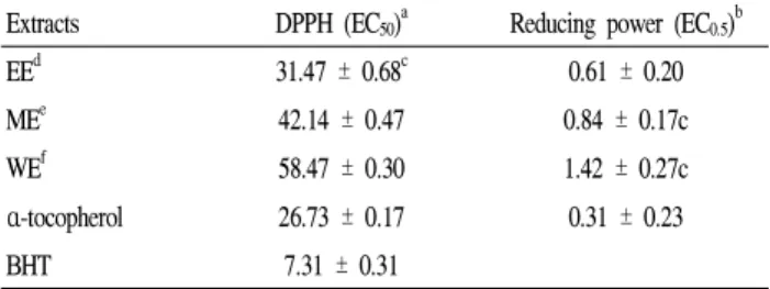
Table 2. Effective concentrations (mg/mL) of A. gigas leaf extracts in DPPH scavenging and reducing power assays
Extracts DPPH (EC
50)
aReducing power (EC
0.5)
bEE
d31.47 ± 0.68
c0.61 ± 0.20
ME
e42.14 ± 0.47 0.84 ± 0.17c
WE
f58.47 ± 0.30 1.42 ± 0.27c
α -tocopherol 26.73 ± 0.17 0.31 ± 0.23
BHT 7.31 ± 0.31 ‐
aEC50represents the effective concentration required to scavenge DPPH radicals by 50%.bEC0.5indicates the effective concentration required to achieve absorbance of 0.5.
cEach value is the mean ± S.D. of triplicate measurements.
d80% ethanol extract
e80% methanol extract
fhot water extract
α-tocopherol and BHT were used as positive control.
thought to be due to their hydrogen-donating ability. DPPH has been used extensively as a free radical to evaluate reducing substances (27).
Table 2 shows the DPPH radical scavenging activities of the various extracts. The EC
50values for the DPPH radical scavenging activity of EE, ME, and WE were 31.47, 42.14, and 58.47 μg/mL, respectively. Based on the EC
50results, EE showed the highest DPPH scavenging activity. α -Tocopherol and BHT, positive controls, had EC
50values of 26.73 and 7.31 μg/mL, respectively. Such observation agreed with several previous findings (28).
Reducing Power Assay
The reducing power of the extract of A. gigas leaf was determined and presented in Table 2. The reducing power of a compound is associated with antioxidant capability (29).
For the measurement of the reductive activity, we investigated the Fe
3+-Fe
2+transformation in presence of each extracts activity, which is an important mechanism of phenolic antioxidant action that is strongly correlated with other antioxidant properties (30). As shown in the Table 2, the EC
50value for EE was higher than ME and WE. The EC
50value obtained for α-tocopherol was 0.31 ± 0.07 μg/mL.
The effective reducing power was inversely correlated with the EC
50value as follows: α-tocopherol > EE > ME > WE.
The higher reducing ability of EE may be due to the high phenolic content which may act as reductones by donating electors to free radicals.
Fenton Reaction (Degradation of Deoxyribose)
·OH, the most reactive of all free radicals, is formed from O2
-and H
2O
2in the presence of metal ions (31). ·OH has the capacity to bond with the nucleotides in DNA, causing strand breakage that ultimately results in carcinogenesis, mutagenesis, and cytotoxicity (32). The ability of the extracts to prevent Fe
2+/H
2O
2-induced decomposition of deoxyribose was determined as an index of ·OH scavenging ability, and this is presented in Fig. 1. EE exhibited stronger antioxidant activity (IC
50= 43.22 ± 1.67 μg/mL) against ·OH using Fenton system than ME (IC
50= 53.21 ± 2.54 μg/mL) and WE (IC
50= 61.24 ± 1.89 μg/mL), whereas α-tocopherol at 68.64 ± 5.47 μ g/mL showed 50% hydroxyl radical scavenging activity.
The result implies that EE was more effective as a hydroxyl
radical scavenging than the other extracts or a positive control
(α-tocopherol).
Fig. 1. Hydroxyl radical scavenging activity of Angelicae gigas leaf extracts.
Each value is expressed as the mean ± S.D. (n = 3). EE: 80% ethanol extract, 80%
methanol extract, and WE: hot water extract
Metal Chelating Activity
It is reported that transition metal is involved in both initiation and propagation of oxygen free radicals in the organisms. Transition elements, such as iron and copper, are powerful catalysts of oxidation reactions because they contain one or more unpaired electrons that can participate in electron transfer reactions (33). The metal chelating ability of the A.
gigas leaf extracts was tested against that of EDTA by evaluating their capacity to complex with Fe
2+. As shown in Fig. 2, the ferrous ion chelating activity of the extracts and standard was: EDTA > EE > ME > WE. These data indicate a marked capacity for iron binding in the EE.
Fig. 2. Metal chelating activity of Angelicae gigas leaf extracts.
Each value is expressed as the mean ± S.D. (n = 3). EE: 80% ethanol extract, 80%
methanol extract, and WE: hot water extract
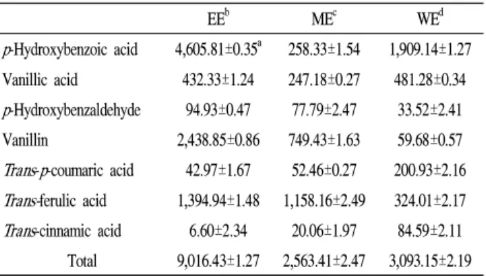
HPLC Analysis of Phenolic Acids
Vanillic acid, p -hydroxybenzoic, p -hydroxybenzaldehyde, vanillin, trans - p -coumaric acid, trans -ferulic acid, and trans -cinnamic acid were used for phenolic acid quantification.
The representative HPLC profile of selected phenolic standards is shown in Fig. 3. The contents of phenolic acids in A. gigas leaf extracts are shown in Table 3. The total amounts of phenolic acids from ethanol extraction showed three to four times higher than those of methanol and water extraction. Phenolic acids are classified as hydroxybenzoic acids (salicylic, gallic, and vanillic acids) and hydroxycinnamic acids (caffeic, chlorogenic, sinapic, fertaric, coumaric, and ferulic acids), which are responsible for sensory quality, antioxidant activity, and other physiological benefits (34). The hydroxycinnamic acids are known to be more effective antioxidants than the hydroxybenzoic acids.
Fig. 3. Typical HPLC chromatogram of phenolic acid standards.
Peak identification:(1) p-hydroxybenzoic acid; (2) vanillic acid; (3) p-hydroxybenzaldehyde;
(4) vanillin; (5) trans-p-coumaric acid; (6) trans-ferulic acid; (7) trans-cinnamic acid.
Table 3. Phenolic acids (μg/g) in the extracts of Angelicae gigas leaves
EE
bME
cWE
dp -Hydroxybenzoic acid 4,605.81±0.35
a258.33±1.54 1,909.14±1.27 Vanillic acid 432.33±1.24 247.18±0.27 481.28±0.34 p -Hydroxybenzaldehyde 94.93±0.47 77.79±2.47 33.52±2.41 Vanillin 2,438.85±0.86 749.43±1.63 59.68±0.57 Trans - p -coumaric acid 42.97±1.67 52.46±0.27 200.93±2.16 Trans- ferulic acid 1,394.94±1.48 1,158.16±2.49 324.01±2.17 Trans -cinnamic acid 6.60±2.34 20.06±1.97 84.59±2.11 Total 9,016.43±1.27 2,563.41±2.47 3,093.15±2.19
aEach value is the mean ± S.D. of triplicate measurements.
b80% ethanol extract
c80% methanol extract
dhot water extract