IIM. IIM-associated ILD (IIM-ILD) contributes to nearly 80%
of the mortality in IIM with a reported prevalence of 65% of newly diagnosed IIM cases
1-3. The antisynthetase syndrome is characterized by the presence of any one of several antisyn- thetase autoantibodies (perhaps occurring in up to one-third of DM and PM patients) in combination with fever, Raynaud’s phenomenon, polyarthritis, myositis, ILD, and “mechanic hands”
4,5. ILD often dominates the clinical picture and may be the presenting feature of the syndrome, as the acute onset of fever and respiratory distress occurs in nearly 50% of pa- tients
3,6,7. Acute respiratory distress syndrome (ARDS) is the most acute form of presentation, with rapid progressive respi- ratory failure; however, very few cases have been reported in association with IIM or the antisynthetase syndrome
8,9.
We report a case with antisynthetase syndrome, presenting with ARDS.
Case Report
A 60-year-old woman was admitted to the Emergency De- partment (ED) of our hospital for cough, shortness of breath, chills, and myalgia. There was no sputum. She was a tourist from Japan and had come to Korea three days earlier. The patient had been in good health until the past week, when
Introduction
The idiopathic inflammatory myopathies (IIM) are a rare group of heterogeneous connective tissue diseases affecting skeletal muscle and other organ systems. Subsets of IIM are clinically and pathologically assigned to one of three general subcategories: polymyositis (PM), dermatomyositis (DM), and inclusion body myositis. The lung is one of the most com- mon extramuscular targets in IIM, and interstitial lung disease (ILD) is a prevalent and often devastating manifestation of
Acute Respiratory Distress Syndrome as the Initial Clinical Manifestation of an Antisynthetase Syndrome
Seo-Hyun Kim, M.D. and I-Nae Park, M.D.
Department of Internal Medicine, Inje University Seoul Paik Hospital, Seoul, Korea
Antisynthetase syndrome has been recognized as an important cause of autoimmune inflammatory myopathy in a subset of patients with polymyositis and dermatomyositis. It is associated with serum antibody to aminoacyl-transfer RNA synthetases and is characterized by a constellation of manifestations, including fever, myositis, interstitial lung disease, mechanic’s hand-like cutaneous involvement, Raynaud phenomenon, and polyarthritis. Lung disease is the presenting feature in 50% of the cases. We report a case of a 60-year-old female with acute respiratory distress syndrome (ARDS), which later proved to be an unexpected and initial manifestation of anti-Jo-1 antibody–positive antisynthetase syndrome. The present case showed resolution of ARDS after treatment with high-dose corticosteroids. Given that steroids are not greatly beneficial in the treatment of ARDS, it is likely that the improvement of the respiratory symptoms in this patient also resulted from the prompt suppression of the inflammatory systemic response by corticosteroids.
Keywords: Respiratory Distress Syndrome, Adult; Immunoglobulins; Antisynthetase Syndrome
Copyright © 2016
The Korean Academy of Tuberculosis and Respiratory Diseases.
All rights reserved.
Address for correspondence: I-Nae Park, M.D.
Department of Internal Medicine, Inje University Seoul Paik Hospital, 9 Mareunnae-ro, Jung-gu, Seoul 04551, Korea
Phone: 82-2-2270-0004, Fax: 82-2-2285-2286 E-mail: [email protected]
Received: Jun. 1, 2015 Revised: Jun. 24, 2015 Accepted: Sep. 19, 2015
cc
It is identical to the Creative Commons Attribution Non-Commercial
License (http://creativecommons.org/licenses/by-nc/4.0/).
she developed a fever and headache. In response, she took acetaminophen for 3 days. She had no history of smoking and was not on any continuous therapy. On arrival to the ED, the patient’s body temperature was 37.8
oC, blood pressure 130/74 mm Hg, pulse rate 82 beats per minute, respiratory rate 24 breaths per minute, and oxygen saturation 86% while breath- ing ambient air. Crackles were heard at the base of lungs. Re- spiratory failure was confirmed by the arterial blood gas: PO
251 mm Hg, PCO
232 mm Hg, and pH 7.52. The white blood cell count was 7,500/μL (neutrophils, 80%; lymphocytes, 12%;
monocytes, 5%) and a hemoglobin value of 13.0 g/dL. C-reac- tive protein was increased to 12.7 mg/dL (normal, 0–0.8 mg/
dL). Procalcitonin was <0.05 ng/mL (normal, <0.05 ng/mL).
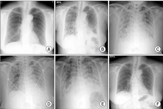
Chest radiography (Figure 1B) showed bilateral infiltrates in both lower fields. The patient was admitted to the Respiratory Department, where she received antibiotics (intravenous le- vofloxacin plus piperacillin-tazobactam plus oral clarithromy- cin) and oxygen. Further blood tests were conducted, which showed elevations of creatinine kinase (448 IU/L), lactate de- hydrogenase (596 IU/L), aspartate aminotransferase (144 IU/
L), and alanine aminotransferase (128 IU/L). On the second day of hospitalization, the patient’s respiratory failure wors- ened, and she was transferred to the intensive care unit, where endotracheal intubation and mechanical ventilation were performed. As she progressed to respiratory failure (Figure 1C), we thought she had severe community-acquired pneu- monia, and we administered hydrocortisone intravenously (100 mg then 50 mg every 6 hours) for 3 days. After 4 days on the ventilator (Figure 1D), we removed the endotracheal tube.
However, two days later, the patient’s body temperature was 39
oC, her respiratory rate was 35 breaths per minute, and she
complained of general weakness and dyspnea. Due to the de- velopment of ARDS (Figure 1E), we re-intubated the patient and began mechanical ventilation again. We thought her con- dition may be ventilator-associated pneumonia, and changed antibiotics from levofloxacin plus piperacillin-tazobactam to meropenem plus vancomycin. However, the patient’s high fever persisted and her chest radiography worsened. On the eighth day of hospitalization, there was still no improvement and the microbiological evaluation provided no evidence for bacterial, fungal, or viral infection under repeated endotrache- al aspiration cultures and protected bronchial washing cul- ture. We suspected inflammatory myopathy with ILD due to the increased muscle and liver enzyme values. Especially, cre- atinine kinase was elevated to >2,000 IU/L. Thus, we checked specific markers for connective tissue diseases. In addition, we reviewed her past medical history from her daughter. Before admission, the patient had suffered from frequent oral ulcers and eye dryness for several weeks. Two months ago, she un- derwent chest radiography (Figure 1A) at a Japanese hospital and the results were normal. One week before she arrived in Korea, she underwent a blood test due to multiple arthral- gias, which showed a high value of lactate dehydrogenase (422 IU/L). While we screened for the specific markers for inflammatory myopathy, an empirical therapy with high-dose corticosteroids was started (intravenous pulses of 125 mg every 6 hours for one day tapered 60 mg/day). The therapy led to a significant improvement of PaO
2/FIO
2ratio, and en- abled to wean the patient from mechanical ventilation on the 11th day after intubation. On the 13th day of hospitalization, laboratory results revealed positive anti–Jo-1 antibody with 6.4 index (normal range, <1.0 index), aldolase with 22.9 U/L
D E F
A B C
Figure 1. Chest X-ray film at 2 months
prior to admission (A), on admission
(B), before the first intubation (C), after
the first extubation (D), after the second
intubation (E), and at discharge (F).
(normal range, 0–7.6 U/L), and anti-Ro (SSA) antibody with
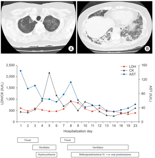
>240 U/mL (normal range, <10 U/mL). Rheumatoid factor, antinuclear antibody, anti-La (SSB) antibody, anti-centromere antibody, anti–Scl-70 antibody, anti–ds-DNA antibody, anti- ribonucleoprotein antibody, and anti-Sm antibody were all negative. On high-resolution computed tomography (HRCT) (Figure 2) after extubation, there were still irregular diffuse ground-glass opacity in both upper lungs and irregular patchy consolidation on both lower lungs without honeycombing. Al- though a histologic diagnosis was not made, we provisionally diagnosed the patient with ARDS as the initial clinical mani- festation of an anti–Jo-1 antibody–positive antisynthetase syn- drome. After 25 days of hospitalization (Figure 1F), the patient was discharged without oxygen. Figure 3 shows the whole course of laboratory values, clinical course, and treatments.
Discussion
Aminoacyl-tRNA synthetase (ARS) is a family of cytoplas- mic enzymes that catalyze the formation of aminoacyl-tRNA
from a specific amino acid and its cognate tRNA and play a crucial role in protein synthesis. Eight anti-ARS autoantibodies have been identified to date. These include antihistidyl (anti–
Jo-1), anti-threonyl (anti–PL-7), anti-alanyl (anti–PL-12), anti-isoleucyl (anti-OJ), anti-glycyl (anti-EJ), anti-asparaginyl (anti-KS), anti-phenylalanyl (anti-Zo), and anti–tyrosyl-tRNA synthetases
7. Based on a unique combination of clinical fea- tures commonly observed in patients with anti-ARS auto- antibodies, Targoff
4proposed a disease entity termed anti- synthetase syndrome. Antihistidyl tRNA synthetase antibody, often referred to as anti–Jo-1 antibody, is the most frequently detected antisynthetase autoantibody and is strongly associat- ed with the presence of ILD in both PM and DM. In one large cohort study, ILD was identified in 86% of anti–Jo-1 antibody–
positive myositis patients
10.
The breadth of presentation in IIM-ILD ranges from asymp- tomatic basilar lung fibrosis to an acute, rapidly progressive condition associated with ARDS and respiratory failure
3,6. The most common presenting symptoms are cough and dyspnea;
however, IIM-ILD may be associated with asymptomatic lung disease, as evidenced by the high frequency of radiographi-
1 2 3 4 5 6 7 8 9
2,500
2,000
1,500
1,000
500
LDH/CK(IU/L)
0
160
120
80
40
AST(IU/L)
0 10 11 12 13 14 16 19 23 Hospitalization day
LDH CK AST
Fever
Ventilator Hydrocortisone
Fever
Ventilator
Methylprednisolone IV oral prednisolone