Early Growth Response 1 Induces Epithelial-to-mesenchymal Transition via Snail
Hyun Min Jeon
1, Su Yeon Lee
1, Min Kyung Ju
1, Hye Gyeong Park
2and Ho Sung Kang
1*
1
Department of Molecular Biology, College of Natural Sciences,
2Nanobiotechnology Center, Pusan National University, Busan 609-735, Korea
Received July 25, 2013 /Revised August 8, 2013 /Accepted August 8, 2013The epithelial-to-mesenchymal transition (EMT) plays an essential role in embryogenesis and is in- volved in tumor metastasis and invasion; it significantly contributes to tumor progression and aggressiveness. The EMT is characterized by a loss of epithelial cell polarity as a result of the reduced expression of epithelial E-cadherin, a hallmark of the EMT, and the acquisition of mesenchymal-like cell morphology. Reactive oxygen species (ROS) such as O
2-, H
2O
2,and OH
-have been demonstrated to induce the EMT; although Snail is involved in ROS-induced EMT by transcriptionally repressing E-cadherin, its mechanism is not fully understood. In this study, we examined the effects of early growth response 1 (Egr-1) overexpression in noninvasive breast tumor cell line MCF-7 cells. Upon Egr-1 overexpression, MCF-7 cells lost epithelial cell polarity and became more spindle-shaped, in- dicating that Egr-1 may induce EMT. We found that Snail is implicated in Egr-1 induced EMT. We further demonstrate that the Egr-1-Snail axis is activated by ROS and plays a critical role(s) in ROS-in- duced EMT.
Key words : Egr-1, epithelial-to-mesenchymal transition (EMT), Snail, E-cadherin, reactive oxygen species (ROS)
*Corresponding author
*Tel : +82-51-510-2275, Fax : +82-51-513-9258
*E-mail : [email protected]
This is an Open-Access article distributed under the terms of the Creative Commons Attribution Non-Commercial License (http://creativecommons.org/licenses/by-nc/3.0) which permits unrestricted non-commercial use, distribution, and reproduction in any medium, provided the original work is properly cited.
Journal of Life Science 2013 Vol. 23. No. 8. 970~977 DOI : http://dx.doi.org/10.5352/JLS.2013.23.8.970
Introduction
EMT plays an essential role in embryogenesis and is in- volved in tumor metastasis, invasion, and angiogenesis [20, 25, 31]. During EMT, epithelial cells lose their polarity by reducing the expression of epithelial E-cadherin, a hallmark of EMT, and acquire mesenchymal-like cell morphology [20, 25, 31]. EMT can be induced by several tumor-stimulating cytokines, such as transforming growth factor (TGF)-β, Notch, hedgehog, and Gli in many invasive human carcino- mas [31]. Several transcription factors have been implicated in the transcriptional repression of E-cadherin, including zinc finger proteins of the Snail/Slug family, Twist1, Twist2, δ EF1/ZEB1, ZEB2/Sip1, and the basic helix-loop-helix factor E12/E47 [9, 23]. Snail is a zinc finger transcription factor that induces the EMT by directly repressing E-cadherin ex- pression [25]. Snail-expressing epithelial cells exhibit mi- gratory and invasive properties during tumor progression [20, 25, 37]. Snail is highly expressed in the invasive regions
of many different types of carcinomas [5, 29, 35]. RNA inter- ference-mediated Snail silencing results in a reduced tumor growth in vivo [22]. In addition, Snail exerts mammary tumor recurrence-promoting activities [18]. Recently, ROS such as O
2-, H
2O
2and OH
-have been demonstrated to induce EMT [6, 7, 14, 19, 23, 24, 26]. By ROS-mediated EMT, invasion, and metastasis, cancer cells could escape from oxida- tive-damaged tumors tissues [14]. ROS are produced by tu- mor microenvironments such as GD and hypoxia [31].
Although Snail is involved in ROS-induced EMT by tran- scriptionally repressing E-cadherin [28], its mechanism is not fully understood.
Early growth response-1 (Egr-1) is a Cys2-His2-type zinc- finger transcription factor [1, 30]. This zinc-finger protein is an immediate early growth response gene known to be in- duced by a broad range of extracellular stimuli such as an- ti-cancer drugs, growth factors, irradiation, hypoxia, and oxi- dative stress. It is involved in the regulation of cell pro- liferation, apoptosis, growth arrest, differentiation, trans- formation, senescence, and cancer development. Egr-1 regu- lates positively or negatively tumor growth [1, 2, 17, 30, 32-34, 36]. It acts as a tumor suppressor; however, many new evidences revealed that Egr-1 promotes cancer progression.
Egr-1 can induce apoptosis by directly regulating tumor sup-
pressors such as p53 [3]. In addition, Egr-1 plays a critical
role(s) in hypoxia-induced tumor progression, survival, and
angiogenesis [13, 21, 27, 38]. Although sustained Egr-1 ex-
Table 1. shRNA target sequences used in this study Genes Target sequence 5’ to 3’
Con shRNA Egr-1 shRNA Snail shRNA
AATTCTCCGAACGTGTCACGT AAGTTACTACCTCTTATCCAT GCGAGCTGCAGGACTCTAA
Table 2. Primer sequences used in this study
Sequence 5’ to 3’ Annealing °C
Real time PCR β-actin Egr-1 Snail
E-cadherin #1 E-cadherin #2
NM_001101.3 NM_001964.2 NM_005985 NM_004360
sense antisense sense antisense sense antisense sense antisense sense antisense
ACTCTTCCAGCCTTCCTTCC TGTTGGCGTACAGGTCTTTG AGGACAGGAGGAGGAGATGG GGAAGTGGGCAGAAAGGATTG ATCGGAAGCCTAACTACAGC CAGAGTCCCAGATGAGCATT GATTTTGAGGCCAAGCAGCA AGATGGGGGCTTCATTCACA AGCTGGACAGGGAGGATTTT TTCGAGGTTCTGGTATGGGG
62 55 55 55
ChIP assay
Egr-1 binding site #1 Egr-1 binding site #2 Egr-1 binding site #3 E-cadherin E4
sense antisense sense antisense sense antisense sense antisense
GTGCGTTTCCCTCGTCAATG GACACCTGACCTTCCGACG CCAGGGGGCGTCAGAAG GACGTCGAGCGAAGCGAG GGGCGTGGCAGATAAGG AGAAGAACCACTCGCTAGGC TCCATTTCTTGGTCTACGCC CACCTTCAGCCAACCTGTTT
62 62 62 55
pression induces anti-angiogenesis, growth arrest, and apop-
tosis, transient induction of Egr-1 is known to activate angio- genesis [16]. In addition, Egr-1 has been implicated in GD-in- duced necrosis and tumor progression [10].
In this study, we show that Egr-1 is implicated in ROS-in- duced EMT via Snail activation. In addition, we show that Egr-1-Snail axis plays a critical role(s) in ROS-induced tumor progression through regulating EMT.
Materials and Methods Cell culture and ROS treatment
MCF-7 was obtained from the American Type Culture Collection (ATCC, Manassas, VA, USA; authenticated by short tandem repeat profiling), and maintained in EMEM supplemented with 10% (v/v) heat-inactivated fetal bovine serum (FBS, Hyclone, Logan, UT, USA) and 1% pen- icillin-streptomycin (PS, Hyclone, Logan, UT, USA) in a 37℃
humidified incubator with 5% CO
2. The cells were treated
with reactive oxygen species [ROS, including 200 μM hydro- gen peroxide (H
2O
2; Sigma, MO, USA) and 10 μM mena- dione (an O
2-generator; Sigma, MO, USA)].
Transfection and short hairpin RNA (shRNA) interference
The expression vectors pcDNA3.1-Egr-1 (provided by Dr.
Thomas E. Eling, Laboratory of Molecular Carcinogenesis, National Institute of Environmental Health Sciences, USA) were transfected into MCF-7 cells using jetPEI (Polyplus- transfection, SA, USA) according to the manufacturer’s instructions. pSUPER vectors expressing shRNA for control, Egr-1, and Snail were generated from target-specific an- nealed oligonucleotides inserted into the HindIII and BglII sites of pSUPER.gfp/neo (Oligoengine Platform, Seattle, WA) (Table 1). All target sequences were designed and verified as specific for Egr-1, and Snail by Blast search against the human genome and transfected using jetPEI.
Western blotting and real-time PCR
Sodium dodecyl sulfate-polyacrylamide gel electro-
phoresis (SDS-PAGE) and Western blotting with antibodies
to Egr-1, Snail, and E-cadherin (Santa Cruz, CA, USA), and
α -tubulin (Biogenex, CA) was performed as described pre-
viously [10-12]. Transcript levels were assessed with re-
al-time quantitative PCR with the primers for Egr-1, Snail,
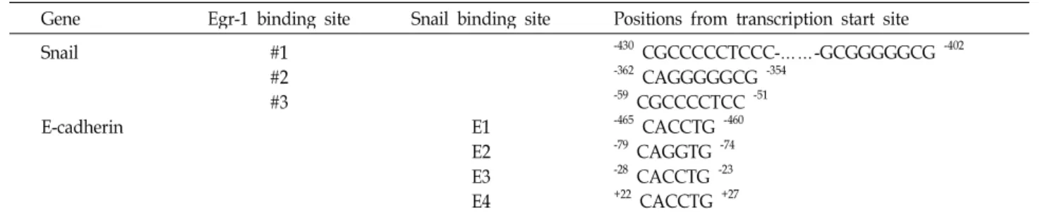
Table 3. Putative Egr-1 binding site and Snail binding site (E-box) in promoter region
Gene Egr-1 binding site Snail binding site Positions from transcription start site Snail
E-cadherin
#1
#2
#3 E1
E2E3 E4
-430 CGCCCCCTCCC-……-GCGGGGGCG -402
-362 CAGGGGGCG -354
-59 CGCCCCTCC -51
-465 CACCTG -460
-79 CAGGTG -74
-28 CACCTG -23
+22 CACCTG +27
E-cadherin, and β-actin (Table 2). Quantitative real-time PCR was conducted in a LightCycler (Roche Diagnostics, Mann- heim, Germany) using a SYBR Green kit (Roche Diagnostics).
Chromatin immunoprecipitation (ChIP) assay ChIP assays were performed using a ChIP assay kit (Millipore, Billerica, MA, USA). IgG or anti-Egr-1, anti-Snail (Santa Cruz, CA, USA) was used to immunoprecipitate DNA-containing complexes. ChIP-enriched DNA was ana- lyzed by PCR using primers (Table 2) complementary to the promoter regions containing Egr-1 and Snail binding site, respectively (Table 3).
Measurement of circularity
Circularity was measured with the Axiovision LE soft- ware (Release 4.8 version)’s Measure command that calcu- lates object circularity using the formula circularity = 4π (area/perimeter
2). Circularity value of 1.0 indicates a perfect circle. As the value approaches 0.0, it indicates an increas- ingly elongated polygon.
Statistical analysis
Data were analyzed by the Student’s t-test and p<0.05 was considered statistically significant.
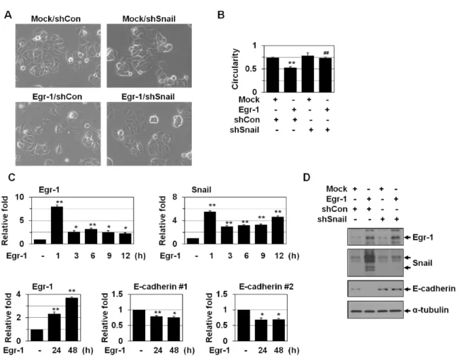
Results and Discussion Egr-1 induces EMT via Snail activation
Egr-1 is involved in tumor cell proliferation and apoptosis.
We examined the effects of Egr-1 overexpression in non- invasive breast tumor cell line MCF-7 cells. Upon Egr-1 over- expression, MCF-7 cells lost epithelial cell polarity and be- came more spindle-shaped, indicating that Egr-1 may induce EMT (Fig. 1A). Spindle quantification also showed Egr-1 in- duction of EMT (Fig. 1B). Egr-1 overexpression also de- creased the levels of E-cadherin, one of the hallmarks of EMT, as revealed by real-time PCR (Fig. 1C) and Western
analysis (Fig. 1D). We examined if Egr-1 directly regulates
the E-cadherin expression. However, Egr-1 binding sites
were not found in the promoter of E-cadherin. Thus, we
postulated that Egr-1 may reduce E-cadherin indirectly by
activating other transcriptional factors. Several transcription
factors such as Snail/Slug, Twist1, Twist2, δEF1/ZEB1,
ZEB2/Sip1, and E12/E47 have been implicated in EMT and
the transcriptional repression of E-cadherin [9, 23]. Among
these transcriptional factors, Snail is a typical transcription
factor for EMT and E-cadherin downregulation [25]. Thus,
we examined whether Snail is involved in Egr-1-induced
EMT. Snail shRNA prevented the EMT (Fig. 1A and B), in-
dicating that Egr-1 may induce EMT via Snail activation. In
addition, Egr-1 overexpression increased Snail levels (Fig. 1C
and D), indicating that Egr-1 acts as an upstream mediator
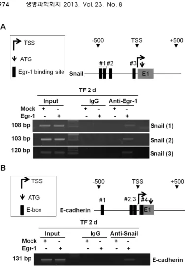
of Snail activation. Next, we checked whether Egr-1 regu-
lates Snail expression through directly binding to the pro-
moter of Snail. Four Egr-1 binding sites were found in the
human Snail promoter (Fig. 2A). Egr-1 overexpression en-
hanced the binding of Egr-1 to the sites of the Snail promoter
(Fig. 2A) indicating expression of Snail by Egr-1. And then
we performed ChIP assay to examine in vivo binding of
Snail to the promoter of E-cadherin. There are 4 E-boxes in
human E-cadherin promoter that can be recognized by Snail
[4, 15]; -465/-460, -79/-74, -28/-23, +22/+27 from tran-
scription start site (Table 3). Snail has been shown to bind
to the E-boxes 1, 3, 4, but not to E-box 2 and suppressed
E-cadherin gene activity; E-box 4 had the most repression
ability compared with other E-boxes on human E-cadherin
gene expression [4, 15]. Therefore, ChIP analysis of Egr-1
and Snail on the E-cadherin promoter was conducted with
E-box 4 region. Egr-1 overexpression enhanced the binding
of Snail to the E-box sites of the E-cadherin (Fig. 2B), indicat-
ing that Egr-1-induced Snail protein interacts directly with
E-box 4 in the E-cadherin promoter. Our results showed that
Egr-1 induces EMT by activating Snail.
Fig. 1. Egr-1 induces EMT via Snail activation. (A) MCF-7 cells were cotransfected with Egr-1 and Snail shRNA for 2 days. The cell morphology was observed under a phase-contrast microscope (X200). (B) Images were then analyzed by circularity calculation. Circularity value of 1.0 indicates a perfect circle. As the value approaches 0.0, it indicates an increasingly elongated polygon. The results (77-126 cells in each group) are mean±SE. **
p
<0.01 versus control; ##p
<0.01 versus Egr-1. (C) MCF-7 cells were transfected with Egr-1 for the indicated times and then analyzed by real-time PCR using primers for Egr-1, Snail, E-cadherin (#1, #2) and β-actin. *p
<0.05, **p
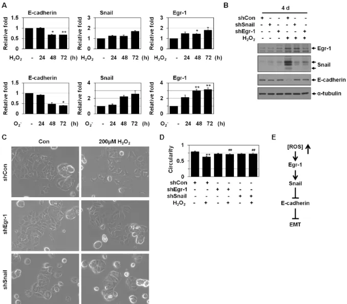
<0.01 versus mock. (D) MCF-7 cells were cotransfected with Egr-1 and Snail shRNA for the 4 days. The cells were then analyzed by Western blotting for Egr-1, Snail, E-cadherin, and α-tubulin.Egr-1-Snail cascade is also involved in ROS- induced EMT
Egr-1 and Snail expression has been shown to be redox- sensitive [10, 11]. As shown in Fig. 3A and 3B, Egr-1, and Snail levels were increased by ROS treatment. Egr-1 shRNA prevented ROS-induced Snail expression (Fig. 3B), indicating that Egr-1 acts as an upstream mediator for ROS-induced Snail expression. ROS is known to induce EMT [14]. Thus, we examined the relevance between ROS-induced EMT and these genes. Upon ROS treatment, MCF-7 cells exhibited the phenotypic changes including loss of cell polarity and inter- cellular adhesion and increased formation of pseudopodia (Fig. 3C). Spindle quantification also showed ROS induction of EMT (Fig. 3D). Both Egr-1 shRNA and Snail shRNA blocked ROS-induced EMT phenotype (Fig. 3C and 3D) and E-cadherin downregulation (Fig. 3B), indicating that the
Egr-1-Snail cascade is implicated in ROS-induced EMT.
Thus, Egr-1 appeared to regulate ROS-induced EMT via in- duction of the E-cadherin repressor Snail (Fig. 3E).
Egr-1 has been implicated in tumor cell death, growth, invasion, and angiogenesis. In this study, show a novel func- tion of Egr-1 that may contribute to tumor progression; to induce EMT (Fig. 1A and 3B). Egr-1 induces EMT with downregulation of E-cadherin, a hallmark of EMT (Fig. 1C and 3D). Snail appeared to act as a mediator of Egr-1-in- duced EMT (Fig. 1 and 2). Egr-1 has been implicated in HGF- induced cell scattering, migration, and invasion via Snail ac- tivation [8]. ROS have been demonstrated to induce EMT and Snail is known to be implicated in ROS-induced EMT.
We showed that the Egr-1-Snail axis is activated by ROS
and plays a critical role(s) in ROS-induced EMT. Recently,
we showed that Snail suppresses mitochondrial respiration
Fig. 2. Egr-1-induced Snail represses the expression of E-cadherin. (A) A schematic model of the human Snail proximal promoter region. The black boxes represent Egr-1 binding sites. MCF-7 cells were transfected with Egr-1 for the 2 days. ChIP assays were performed using IgG or Egr-1 antibodies and ChIP-enriched DNA was analyzed by PCR using the appropriate primers. (B) A schematic model of the human E-cadherin proximal promoter region.
The black boxes represent E-boxes. MCF-7 cells were transfected with Egr-1 for the 2 days. ChIP assays were performed using IgG or Snail anti- bodies and ChIP-enriched DNA was analyzed by PCR using the E-cadherin primer.
and cytochrome C oxidase (COX) activity by inhibiting the expression of 3 COX subunits, namely, COXVIc, COXVIIa, and COXVIIc. In addition, Snail induces a glycolytic switch via increased glucose consumption and lactate production [11]. Thus, Egr-1 may induce glycolytic switch by Snail acti- vation, which remains to be investigated.
Acknowledgements
This work was supported by a 2-Year Research Grant of Pusan National University. We thank Dr. Thomas E. Eling (Laboratory of Molecular Carcinogenesis, National Institute of Environmental Health Sciences, USA) for providing pcDNA3.1-Egr-1.
References
1. Adamson, E. D. and Mercola, D. 2002. Egr1 transcription factor: multiple roles in prostate tumor cell growth and survival.
Tumour Biol
23, 93-102.2. Ahmed, M. M. 2004. Regulation of radiation-induced apop- tosis by early growth response-1 gene in solid tumors.
Curr Cancer Drug Targets
4, 43-52.3. Baron, V., Adamson, E. D., Calogero, A., Ragona, G. and Mercola, D. 2006. The transcription factor Egr1 is a direct regulator of multiple tumor suppressors including TGFbeta1, PTEN, p53, and fibronectin.
Cancer Gene Ther
13, 115-124.4. Batlle, E., Sancho, E., Franci, C., Dominguez, D., Monfar, M., Baulida, J. and Garcia De Herreros, A. 2000. The tran- scription factor snail is a repressor of E-cadherin gene ex- pression in epithelial tumour cells.
Nat Cell Biol
2, 84-89.5. Blanco, M. J., Moreno-Bueno, G., Sarrio, D., Locascio, A., Cano, A., Palacios, J. and Nieto, M. A. 2002. Correlation of Snail expression with histological grade and lymph node status in breast carcinomas.
Oncogene
21, 3241-3246.6. Cannito, S., Novo, E., di Bonzo, L. V., Busletta, C., Colom- batto, S. and Parola, M. 2010. Epithelial-mesenchymal tran- sition: from molecular mechanisms, redox regulation to im- plications in human health and disease.
Antioxid Redox Signal
12, 1383-1430.7. Cat, B., Stuhlmann, D., Steinbrenner, H., Alili, L., Holtkotter, O., Sies, H. and Brenneisen, P. 2006. Enhancement of tumor invasion depends on transdifferentiation of skin fibroblasts mediated by reactive oxygen species.
J Cell Sci
119, 2727- 2738.8. Grotegut, S., von Schweinitz, D., Christofori, G. and Lehembre, F. 2006. Hepatocyte growth factor induces cell scattering through MAPK/Egr-1-mediated upregulation of Snail.
EMBO J
25, 3534-3545.Fig. 3. Egr-1-Snail cascade is also involved in ROS-induced EMT. (A) MCF-7 cells were treated with H2O2(200 μM) or menadione (O2-, 10 μM) for the indicated times and then analyzed by real-time PCR using primers for Egr-1, Snail, E-cadherin and β-actin. *
p
<0.05, **p
<0.01 versus untreated. (B) MCF-7 cells were transfected with control, Egr-1, or Snail shRNA and then treated with H2O2(200 μM) for 4 days. The cells were then analyzed by Western blotting for Egr-1, Snail, E-cadherin, and α-tubulin. (C) MCF-7 cells were transfected with control, Egr-1, or Snail shRNA and then treated with H2O2(200 μM) for 3 days. The cell morphology was then observed under a phase-contrast microscope (X200). (D) Images were then analyzed by circularity calculation. Circularity value of 1.0 indicates a perfect circle. As the value approaches 0.0, it indicates an increas- ingly elongated polygon. The results (58-118 cells in each group) are mean±SE. **p
<0.01 versus untreated;##p
<0.01 versus control shRNA. (E) Under oxidatively stressed conditions, Snail is activated by Egr-1. Egr-1 is induced by ROS and regulates ROS-induced EMT via induction of the E-cadherin repressor Snail.9. Hussain, S. P., Hofseth, L. J. and Harris, C. C. 2003. Radical causes of cancer.
Nat Rev Cancer
3, 276-285.10. Jeon, H. M., Lee, S. Y., Ju, M. K., Kim, C. H., Park, H. G.
and Kang, H. S. 2013. Early growth response 1 regulates glucose deprivation-induced necrosis.
Oncol Rep
29, 669-675.11. Lee, S. Y., Jeon, H. M., Ju, M. K., Kim, C. H., Yoon, G., Han, S. I., Park, H. G. and Kang, H. S. 2012. Wnt/Snail signaling regulates cytochrome c oxidase and glucose metabolism.
Cancer Res
72, 3607-3617.12. Lee, S. Y., Jeon, H. M., Kim, C. H., Ju, M. K., Bae, H. S.,
Park, H. G., Lim, S. C., Han, S. I. and Kang, H. S. 2011.
Homeobox gene Dlx-2 is implicated in metabolic stress-in- duced necrosis.
Mol Cancer
10, 113.13. Liao, H., Hyman, M. C., Lawrence, D. A. and Pinsky, D.
J. 2007. Molecular regulation of the PAI-1 gene by hypoxia:
contributions of Egr-1, HIF-1alpha, and C/EBPalpha.
FASEB J
21, 935-949.14. Lim, S. O., Gu, J. M., Kim, M. S., Kim, H. S., Park, Y. N., Park, C. K., Cho, J. W., Park, Y. M. and Jung, G. 2008.
Epigenetic changes induced by reactive oxygen species in
hepatocellular carcinoma: methylation of the E-cadherin promoter.
Gastroenterology
135, 2128-2140, e2121-2128.15. Liu, Y. N., Lee, W. W., Wang, C. Y., Chao, T. H., Chen, Y. and Chen, J. H. 2005. Regulatory mechanisms controlling human E-cadherin gene expression.
Oncogene
24, 8277-8290.16. Lucerna, M., Pomyje, J., Mechtcheriakova, D., Kadl, A., Gruber, F., Bilban, M., Sobanov, Y., Schabbauer, G., Breuss, J., Wagner, O., Bischoff, M., Clauss, M., Binder, B. R. and Hofer, E. 2006. Sustained expression of early growth re- sponse protein-1 blocks angiogenesis and tumor growth.
Cancer Res
66, 6708-6713.17. Mahalingam, D., Natoni, A., Keane, M., Samali, A. and Szegezdi, E. 2010. Early growth response-1 is a regulator of DR5-induced apoptosis in colon cancer cells.
Br J Cancer
102, 754-764.18. Moody, S. E., Perez, D., Pan, T. C., Sarkisian, C. J., Portocar- rero, C. P., Sterner, C. J., Notorfrancesco, K. L., Cardiff, R.
D. and Chodosh, L. A. 2005. The transcriptional repressor Snail promotes mammary tumor recurrence.
Cancer Cell
8, 197-209.19. Mori, K., Shibanuma, M. and Nose, K. 2004. Invasive poten- tial induced under long-term oxidative stress in mammary epithelial cells.
Cancer Res
64, 7464-7472.20. Nieto, M. A. 2002. The snail superfamily of zinc-finger tran- scription factors.
Nat Rev Mol Cell Biol
3, 155-166.21. Nishi, H., Nishi, K. H. and Johnson, A. C. 2002. Early Growth Response-1 gene mediates up-regulation of epi- dermal growth factor receptor expression during hypoxia.
Cancer Res
62, 827-834.22. Olmeda, D., Jorda, M., Peinado, H., Fabra, A. and Cano, A. 2007. Snail silencing effectively suppresses tumour growth and invasiveness.
Oncogene
26, 1862-1874.23. Pani, G., Galeotti, T. and Chiarugi, P. 2010. Metastasis: can- cer cell's escape from oxidative stress.
Cancer Metastasis Rev
29, 351-378.24. Pani, G., Giannoni, E., Galeotti, T. and Chiarugi, P. 2009.
Redox-based escape mechanism from death: the cancer lesson.
Antioxid Redox Signal
11, 2791-2806.25. Peinado, H., Olmeda, D. and Cano, A. 2007. Snail, Zeb and bHLH factors in tumour progression: an alliance against the epithelial phenotype?
Nat Rev Cancer
7, 415-428.26. Radisky, D. C., Levy, D. D., Littlepage, L. E., Liu, H., Nelson, C. M., Fata, J. E., Leake, D., Godden, E. L., Albertson, D.
G., Nieto, M. A., Werb, Z. and Bissell, M. J. 2005. Rac1b and reactive oxygen species mediate MMP-3-induced EMT and genomic instability.
Nature
436, 123-127.27. Rong, Y., Hu, F., Huang, R., Mackman, N., Horowitz, J. M., Jensen, R. L., Durden, D. L., Van Meir, E. G. and Brat, D.
J. 2006. Early growth response gene-1 regulates hypoxia-in- duced expression of tissue factor in glioblastoma multiforme through hypoxia-inducible factor-1-independent mecha- nisms.
Cancer Res
66, 7067-7074.28. Shi, D. Y., Xie, F. Z., Zhai, C., Stern, J. S., Liu, Y. and Liu, S. L. 2009. The role of cellular oxidative stress in regulating glycolysis energy metabolism in hepatoma cells.
Mol Cancer
8, 32.29. Sugimachi, K., Tanaka, S., Kameyama, T., Taguchi, K., Aishima, S., Shimada, M., Sugimachi, K. and Tsuneyoshi, M. 2003. Transcriptional repressor snail and progression of human hepatocellular carcinoma.
Clin Cancer Res
9, 2657- 2664.30. Thiel, G. and Cibelli, G. 2002. Regulation of life and death by the zinc finger transcription factor Egr-1.
J Cell Physiol
193, 287-292.31. Thiery, J. P. and Sleeman, J. P. 2006. Complex networks or- chestrate epithelial-mesenchymal transitions.
Nat Rev Mol Cell Biol
7, 131-142.32. Wagner, M., Schmelz, K., Dorken, B. and Tamm, I. 2008.
Transcriptional regulation of human survivin by early growth response (Egr)-1 transcription factor.
Int J Cancer
122, 1278-1287.33. Xie, B., Wang, C., Zheng, Z., Song, B., Ma, C., Thiel, G. and Li, M. 2011. Egr-1 transactivates Bim gene expression to pro- mote neuronal apoptosis.
J Neurosci
31, 5032-5044.34. Yamaguchi, H., Chen, C. T., Chou, C. K., Pal, A., Bornmann, W., Hortobagyi, G. N. and Hung, M. C. 2010. Adenovirus 5 E1A enhances histone deacetylase inhibitors-induced apoptosis through Egr-1-mediated Bim upregulation.
Onco- gene
29, 5619-5629.35. Yook, J. I., Li, X. Y., Ota, I., Hu, C., Kim, H. S., Kim, N.
H., Cha, S. Y., Ryu, J. K., Choi, Y. J., Kim, J., Fearon, E.
R. and Weiss, S. J. 2006. A Wnt-Axin2-GSK3beta cascade regulates Snail1 activity in breast cancer cells.
Nat Cell Biol
8, 1398-1406.36. Zagurovskaya, M., Shareef, M. M., Das, A., Reeves, A., Gupta, S., Sudol, M., Bedford, M. T., Prichard, J., Mohiud- din, M. and Ahmed, M. M. 2009. EGR-1 forms a complex with YAP-1 and upregulates Bax expression in irradiated prostate carcinoma cells.
Oncogene
28, 1121-1131.37. Zavadil, J. and Bottinger, E. P. 2005. TGF-beta and epi- thelial-to-mesenchymal transitions.
Oncogene
24, 5764-5774.38. Zhang, P., Tchou-Wong, K. M. and Costa, M. 2007. Egr-1 mediates hypoxia-inducible transcription of the NDRG1 gene through an overlapping Egr-1/Sp1 binding site in the promoter.