◈ Original Article ◈
The Relationship of Radiation Dose and Image Quality According to the Condition of Chest PA
4)
Jin Hyun Son
1·Jung Whan Min
1·Byung Sam Kang
1·Kyung Rae Dong
2,31
Department of Radiological Technology, Shingu University·
2
Department of Radiological Technology, Gwangju Health College University·
3
Department of Nuclear Engineering, Chosun University
Abstract
The purpose of this study is to compare the measurement result of radiation dose by using standard thoracic phantom and ionization chamber to advice proposal in the shooting condition of chest PA projection at hospitals recently. And to understand the change between radiation dose and resolution in different conditions. The period this study was from August 2010 to September 2010 and the subjects of the study was 3 general hospitals, 4 personal hospitals and 1 laboratory at the college. Finally we study with 6 DR, 1 CR, and 4 F/S equipments. Most hospitals met advice proposal, but some of the hospitals exceed advice dose from the result of our study. We can lower radiation dose about 25% when kVp is lowered about 20% in DR equipment. And we can lower radiation dose about 50% when mAs is lowered about 35%. The image quality was similar to the original in the study. Most hospitals which exceed advice dose were personal hospitals. The reason why it happened is that radiation dose for chest PA projection at personal hospitals is higher than general hospitals and the personal hospitals´ equipments are older than general hospitals´ equipments. We guess that patients´ radiation dose of chest PA projection can be lowered from the result.
Key word : Standard thoracic phantom, Ionization chamber, Radiation dose, chest PA
Ⅰ. Introduction
Usage of radiation has been contributed in medical field for 100 years since W.C. Röntgen discovered the X-ray in 1985. Recently, interests of health and well-being life are getting higher
Received September 27, 2011/ 1st Revised October 10, 2011/ 2nd Revised October 29, 2011/ Accepted for Publication November 12, 2011
Corresponding Author: Kyung Rae Dong
Department of Radiological Technology, Gwangju Health College University
(506-701) 683, Shinchang-dong, Gwangsan-gu, Gwangju, Republic of Korea
Tel: 062) 958-7668 Fax: 062) 958-7669 E-mail: [email protected]
due to development of the medical appliances and the facilities as well as national income level.
One research shows in case of being treated at the same radiation treatment in EC(European Comission) and OECD(Organization for Economic Cooperation and Development), however, there is 10 to 20 times difference of exposure dose.
Moreover, the radiation exposure dose in medical field takes around 90% of all artificial radiation.
Improvement of exposure for patients, an measu-
rement and an evaluation of exposure dose are
needed for certain patients who are receiving the
X-ray treatment in clinics. And also, establishing
and introducing the low cost program which is
suitable for our community so that the patients are able to receive the X-ray without any concerns.
1~4The purpose of this examination is to confirm if the diagnosis organizationin metro- politan area follow the international advice dose for the chest PA(Posterior-Anterior) radiography which is so called a fundamental of the radio therapeutics examination. The international orga- nization recommends the advicedose of the chest PA radiography for IAEA(International Atomic Energy Agency) is 0.4 mGy, EC 0.3 mGy, IPEM (Institute of Physics and Engineering in Medicine) 0.3 mGy, JRTA(Jejudo Radiological Technologists Association) 0.3 mGy which these numbers tell us 0.3 mGy is an optimal recommendation for the advice dose. Another purpose of this examination is to understand the condition of the chest PA which is able to satisfy the spatial resolution of diagnosis region and a reduction measurement of exposure.
Ⅱ. Material & Method
1. Object and examination equipment
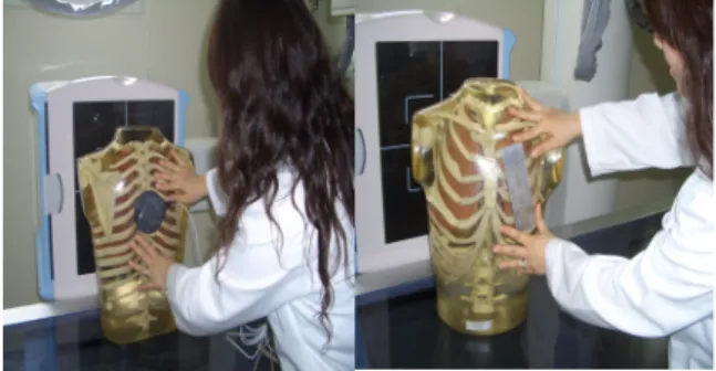
The examination was tested with 6 DR, 1 CR and 4 F/S from 3 general hospitals, 4 private hospitals and 1 university lavatory. The examin- ation wastaken from August, 2010 to September, 2010 with using the standard chest phantom, ion-chamber(2026C, USA) and the spatial resolu- tion pattern(Fig. 1).
2. Examination method
1) Attaching the chart of spatial resolution in area of 1/2 standard chest phantom heart shadow and attaching the ion-chamber in area of 6th thoracic spine(Fig. 2).
2) Taking the X-ray after setting up the standard chest phantom on a table and putting the chest PA radiography in position.
A
B
C