44 Copyright © 2013 The Korean Society of Cardiology Korean Circulation Journal
Introduction
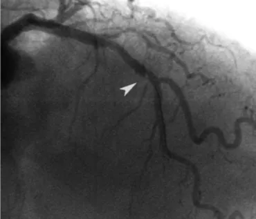
During percutaneuos coronary intervention (PCI) procedures, se- veral complications can occur, such as dissections, thrombi, ulcers or plaque, may appear as angiographic hazy or uncertain images. A pro- per diagnosis is important to guide the correct management in the catheterization laboratory and to improve patient outcomes. Angi- ography has important limitations to characterize those entities that, with contrast, can be diagnosed accurately thanks to intravascular imaging.
1)Among the intracoronary imaging techniques available, intravascular ultrasound (IVUS) and optical coherence tomography (OCT) are the most commonly used for research. Both techniques provide great value for characterization of coronary lesions and for guidance of PCI. IVUS is very useful to measure the vessel wall and to evaluate plaque burden, whereas the OCT (with almost microsco-
Case Report
http://dx.doi.org/10.4070/kcj.2013.43.1.44 Print ISSN 1738-5520 • On-line ISSN 1738-5555
Utility of Optical Coherence Tomography to Assess a Hazy Intracoronary Image after Percutaneous Coronary Intervention
Sebastian Carrizo, MD, Pablo Salinas, MD, Santiago Jimenez-Valero, MD, and Raul Moreno, MD
University Hospital La Paz, Interventional Cardiology Department, Madrid, Spain