Hemilaminectomy for Removal of Extramedullary or Extradural Spinal Cord Tumors: Medium
to Long-Term Clinical Outcomes
Toshitaka Naganawa,
1Kei Miyamoto,
2Hideo Hosoe,
1Naoki Suzuki,
1and Katsuji Shimizu
1Departments of 1Orthopaedic Surgery and 2Reconstructive Surgery for Spine, Bone, and Joint, Gifu University Graduate School of Medicine, Gifu, Japan.
Received: December 10, 2009 Revised: March 15, 2010 Accepted: March 19, 2010
Corresponding author: Dr. Kei Miyamoto, Department of Reconstructive Surgery for Spine, Bone, and Joint, Gifu University Graduate School of Medicine, 1-1 Yanagido, Gifu City, Gifu 501-1194, Japan.
Tel: 81-58-230-6333, Fax: 81-58-230-6334 E-mail: [email protected]
∙ The authors have no financial conflicts of interest.
© Copyright:
Yonsei University College of Medicine 2011 This is an Open Access article distributed under the terms of the Creative Commons Attribution Non- Commercial License (http://creativecommons.org/
licenses/by-nc/3.0) which permits unrestricted non- commercial use, distribution, and reproduction in any medium, provided the original work is properly cited.
Purpose: Laminectomy is generally the treatment of choice for removal of spinal tumors. However, it has been shown that laminectomy may cause instability due to damage of posterior elements of the spinal column, which may induce subsequent kyphosis in the future. Therefore, to reduce the risk of deformity and spinal instabil- ity after laminectomy, hemilaminectomy has been used. However, the medium to long-term effects of hemilaminectomy on spinal sagittal alignment is not well un- derstood. The present study was performed to evaluate the clinical outcomes, in- cluding spinal sagittal alignment of patients, associated with spinal cord tumors treated by surgical excision using hemilaminectomy. Materials and Methods:
Twenty hemilaminectomy operations at our institute for extramedullary or extradu- ral spinal cord tumors in 19 patients were evaluated retrospectively with an average follow-up of 85 months (range, 40-131 months). Neurological condition was evalu- ated using the improvement ratio of the Japanese Orthopaedic Association Score (JOA score) for cervical, thoracic myelopathy, or back pain, and sagittal alignment by sagittal Cobb angle of the hemilaminectomied area. Results: The mean im- provement ratio of neurological results was 56.7% in the cervical spine (p < 0.01, n
= 10), 26.3% in the thoracic spine (not significant, n = 5), and 48.6% in the lumbar spine (NS, n = 5). The sagittal Cobb angle was 4.3 ± 18.0° in the preoperative peri- od and 5.4 ± 17.6° at the latest follow-up, indicating no significant deterioration.
Conclusion: Hemilaminectomy is useful for extramedullary or extradural spinal cord tumors in providing fair neurological status and restoration of spinal sagittal alignment in medium to long-term follow-up.
Key Words: Hemilaminectomy, surgical treatment, spinal cord tumors, middle to long term clinical outcome, sagittal alignment
INTRODUCTION
A spinal tumor is defined as a growth of cells (mass) within or surrounding the spi- nal cord. In cases in which compression of the spinal cord is severe and the risk of neurological deterioration increases, surgery is needed to relieve the compression.
Bilateral laminectomy is generally the treatment of choice for removal of spinal
ing of surgery compared with other reports.
Surgical methods
All operations were performed by two surgeons belonging to our institution. A midline incision was made with the pa- tient in the prone position. Resection of bone and ligaments was restricted to the side of the tumor. The vertebral arch was drilled under a microscope using a high-speed drill.
The flavum was removed until the contralateral root or du- ral curve was exposed. The spinous process and its base, the contralateral lamina including the flavum and muscle were preserved (Fig. 1).12
Numbers of hemilaminectomied laminae
The numbers of hemilaminectomied laminae were 2 in 12 cases, 3 in 4 cases, 4 in 2 cases, 6 in 1 case, and 7 in 1 case, with an average of 2.9 ± 1.4 (Table 1).
Evaluation for operative outcomes Pathological diagnosis of tumors
Pathological diagnoses using specimens from resected tu- mors were identified (Table 1).
Invasiveness of the procedures
To evaluate the invasiveness of the operations, the amount tumors.1-3 However, it has been shown that laminectomy
may cause instability due to damage of posterior elements of the spinal column, which may induce subsequent kyphosis in the future.3-7 Therefore, to reduce the risk of deformity and spinal instability after laminectomy, hemilaminectomy has been used.1,8-11 However, the medium to long-term effects of hemilaminectomy on spinal sagittal alignment are not well understood. Therefore, the present study was performed to evaluate the clinical outcomes, including spinal sagittal alignment of patients, associated with spinal cord tumors treated by surgical excision using hemilaminectomy.
MATERIALS AND METHODS
Patients
Nineteen patients (9 female and 10 male) with spinal cord tumors treated surgically by hemilaminectomy (20 opera- tions) at our institute between 1997 and 2004 were fol- lowed-up and reviewed in a prospective study (Table 1).
The mean ± SD age at the time of surgery was 42.3 ± 16.4 years (range, 14-74 years), and the mean ± SD follow-up period was 85 ± 30 months (range, 40-131 months). One patient underwent hemilaminectomy twice for removal of tumors at the cauda equina and cervical region. All pa- tients reported local or radiating pain or sensory or motor disturbance of the extremities and were diagnosed as hav- ing spinal cord tumors by enhanced and plain magnetic resonance imaging (MRI). Hemilaminectomy was select- ed for resection of tumors with clear borders and extra- medullary or extradural dorsal and unilateral lesions. Tu- mors located anteriorly to the spinal cord and tumors appearing malignant radiologically were removed by total laminectomy, which provided a better view and safer re- moval of the tumors.
Levels and locations of tumors
The tumors were observed in the following regions: cervi- cal in 9 cases (45.0%), cervicothoracic in 1 (5.0%), thoracic in 5 (25.0%), and lumbar in 5 (25.0%). The locations of the tumors were extradural in 60.0% (n = 12) and intradural/
extramedullary in 40.0% (n = 8) (Table 1).
The timing of the surgery
Surgery was performed when imaging modalities showed that the tumors had grown, or when patients had a sensory or motor disorder. There were no obvious differences in tim-
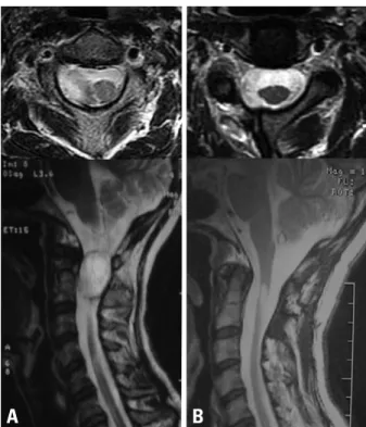
Fig. 1. (A) Preoperative MRI of Case 9 showing extradural neurinoma of the upper cervical spine. (B) Postoperative MRI showing tumor resection by hemilaminectomy. Upper panel, Axial image; Lower panel, Sagittal image.
A B
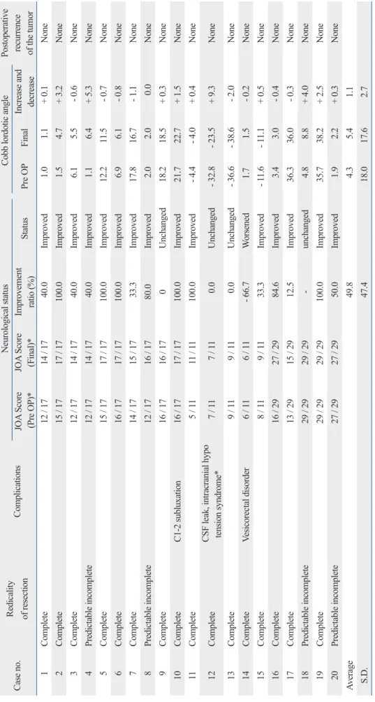
Table 1. Clinical Information of 20 Cases Case no.AgeSexLevels of tu- morsLocations of tumorsLevel for hemilami- nectomy Additional anterior operations Pathological diagnosis
Operation time (min)Blood loss (g) Follow- up period (months)TotalPer hemilaminec- tomied levelsTotalPer hemilaminec- tomied levels 155MC5-6Intradural / extramedullaryC5-6NoNeurinoma140 70 140 70131 235FC5Intradural / extramedullaryC5-6NoNeurinoma140 70 25 13130 367MC5-6Intradural / extramedullaryC4-6NoNeurinoma130 43 220 73120 439MC7-Th1Extradural (dumbbell)C7-T2YesNeurinoma390130 600200117 524FC1-4Extradural (dumbbell)C1-4NoNeurofibroma195 49 780195115 631MC2-3Extradural C2-3NoNeurinoma4002001,270635 94 763FC1-2ExtraduralC1-2NoNeurinoma150 75 250125 79 840MC2-3Extradural (dumbbell)C2-3YesNeurinoma2451231,420710 77 944FC1-2ExtraduralC1-2NoNeurinoma104 52 200100 41 1074MC2ExtraduralC1-3NoNeurinoma435145 890297 40 1125FT3-4ExtraduralT3-4NoNeurinoma300150 210105123 1224FT3-8Intradural / extramedullaryT3-8NoArachnoid cyst250 42 280 47 90 1352MT3-8Intradural / extramedullaryT2-8NoArachnoid cyst310 44 190 27 88 1460FT11-12ExtraduralT11-12NoNeurinoma275138 325163 87 1557MT3-6Intradural / extramedullaryT3-6NoArachnoid cyst200 50 250 63 87 1631ML2ExtraduralL2-3NoEpendymoma120 60 50 25 78 1714FL3-4Intradural / extramedullaryL3-4NoMeningioma290145 575288 62 1844FL3-4Intradural / extramedullary (dumbbell)L3-4NoNeurinoma125 63 180 90 60 1937ML4-5ExtraduralL4-SNoNeurinoma89 30 150 50 46 2030FL1-2Extradural (dumbbell)L1-2YesNeurofibroma205103 320160 43 Average42.3225 89 416172 85 S.D.16.4105 48 392190 30
predictably incomplete resection.9 Complications
Intraoperative and postoperative complications were ana- lyzed.
Changes in neurological status
Neurological status was evaluated using the Japanese Or- of blood loss during surgery and the duration of surgery
were noted. These values were standardized by the number of hemilaminectomied laminae (Table 1).
Radicality of resection
Radicality of resection was assessed by the surgeons as com- plete resection or incomplete resection. Cases of incomplete resection were classified into unexpectedly incomplete and
Table 2. The Japanese Orthopaedic Association Score for Cervical Myelopathy
Motor function of fingers Sensory function
1) Upper extremity
0
Unable to feed oneself w/any tableware including chopsticks, spoon, or fork, &/or unable to fasten buttons of any size
0 Complete loss of touch & pain sensation
1 Can manage to feed oneself w/spoon &/or fork
but not w/chopsticks 0.5 ≤ 50% normal sensation &/or severe pain or numbness
2
Either chopsticksfeeding of writhing is
possible but not practical, &/or large buttons can be fastened
1 > 60% normal sensation &/or moderate pain or numbness
3 Either chopstick feeding or writing is clumsy but
practical, &/or cuff buttons can be fastened 1.5 Subjective numbness of slight degree w/out any objective sensory deficit
4 Normal 2 Normal
Shoulder & elbow: evaluated by MMT score of the
deltoid or biceps muscles, whichever is weaker 2) Trunk
- 2 MMT 2 or below 0 Complete loss of touch & pain sensation
- 1 MMT 3 0.5 ≤ 50% normal sensation &/or severe pain or
numbness
- 0.5 MMT 4 1 > 60% normal sensation &/or moderate pain
or numbness
0 MMT 5 1.5 Subjective numbness of slight degree w/out
any objective sensory deficit
2 Normal
Lower extremity 3) Lower extremity
0 Unable to stand & walk by any means 0 Complete loss of touch & pain sensation 0.5 Able to stand but unable to walk 0.5 ≤ 50% normal sensation &/or severe pain or
numbness 1 Unable to walk w/out a cane or other support on a
level 1 > 60% normal sensation &/or moderate pain
or numbness
1.5 Able to walk w/out support but w/a clumsy gait 1.5 Subjective numbness of slight degree w/out any objective sensory deficit
2 Walks independently on a level surface but needs
support on stairs 2 Normal
2.5 Walks independently when going upstairs,
but needs support when going downstairs Bladder function
3 Capable of fast but clumsy walking 0 Urinary retention &/or incontinence
4 Normal 1 Sense of retention &/or dribbling &/or thin stream
&/or incomplete continence 2 Urinary retardation &/or pollakiuria
3 Normal
maximum score: 17 points MMT, manual muscle test.
Improvement ratio of JOA-B: (postoperative score-preop- erative score) × 100 / [29 (full score) -preoperative score] (%)
Changes in the neurological status were classified into three grades: improved, unchanged, and worsened.
Effects of hemilaminectomy of postoperative spinal align- ment
Cobb sagittal angle between the vertebral bodies at the up- per and lower ends of the area of hemilaminectomy was measured on plain X-ray films preoperatively and at the fi- nal follow-up.
Postoperative recurrence of the tumor
At the final follow-up, MRI was used to assess the presence or absence of the recurrences of tumors.
Statistical analyses
The neurological improvement ratio was compared among patients with lesions of the cervical, thoracic, and lumbar spine by one-way analysis of variance.
thopaedic Association Scores (JOA score) for cervical my- elopathy (JOA-C),13,14 thoracic myelopathy (JOA-T),15-17 and back pain (JOA-B).18,19 The JOA-C, JOA-T, and JOA-B were recorded within one month before surgery and at the final follow-up. The increases in these scores, i.e., the dif- ference between the final and preoperative scores, were also evaluated. A full score of JOA-C was defined as 17 points: 8 for upper and lower motor functions, 6 for sensory functions, and 3 for bladder-rectal function (Table 2). JOA- T, consisting of 11 points, was obtained after subtracting the parameters on the upper extremities from the JOA-C (Table 2). A full score of JOA-B was defined as 29 points: 9 for 3 subjective symptoms, 6 for 3 clinical signs, and 14 for 7 activities of daily living (Table 3). The improvement ratio of these scores,17 which indicates the degree of normaliza- tion after surgery, was calculated as follows:
Improvement ratio of JOA-C: (postoperative score - preop- erative score) × 100 / [17 (full score) - preoperative score] (%) Improvement ratio of JOA-T: (postoperative score - preop- erative score) × 100 / [11 (full score) - preoperative score] (%) Table 3. The Japanese Orthopaedic Association Score for Back Pain
Symptoms and signs Evaluation and scores Symptoms and signs Evaluation and scores
I Subjective symptoms III Activity of daily living Severe Moderate None
Lower back pain None 3 Turn over while lying down
Occasional mild pain 2 0 1 2
Occaional severe pain 1 Standing 0 1 2
Continuous severe pain 0 Washing 0 1 2
Leg pain and/or tingling None 3 Leaning forwards 0 1 2
Occaional slight symptoms 2 Standing (about 1 hour) 0 11 2
Occaional severe symptoms 1 Lifting or holding heavy object 0 1 2
Continuous severe symptoms 0 Walking 0 1 2
Gait Nomal 3 IV Urinary Bladder Function
Able to walk farther than 500 m
although it results in symptoms 2 Normal 0
Unable to walk farther than 500 m 1 Mild dysuria - 3
Unable to walk farther than 100 m 0 Severe dysuria - 6
II Clinical signs
Straight-leg-raising test Normal 2
30 - 70° 1
Less than 30° 0
Sensory disturbance None 2
Silght disturbance (not subjective) 1
Marked disturbance 0
Motor disturbance Normal 2
Slight weakness (MMT 4) 1
Marked weakness (MMT 3 to 0) 0 MMT, manual muscle test.
Table 4. Clinical Outcomes of 20 Cases Case no.Redicality of resectionComplicationsNeurological statusCobb lordotic anglePostoperative recurrence of the tumorJOA Score (Pre OP)*JOA Score (Final)*Improvement ratio (%)StatusPre OPFinalIncrease and decrease 1Complete12 / 1714 / 17 40.0Improved 1.0 1.1+ 0.1None 2Complete15 / 1717 / 17100.0Improved 1.5 4.7+ 3.2None 3Complete12 / 1714 / 17 40.0Improved 6.1 5.5 - 0.6None 4Predictable incomplete12 / 1714 / 17 40.0Improved 1.1 6.4+ 5.3None 5Complete15 / 1717 / 17100.0Improved 12.2 11.5 - 0.7None 6Complete16 / 1717 / 17100.0Improved 6.9 6.1 - 0.8None 7Complete14 / 1715 / 17 33.3Improved 17.8 16.7 - 1.1None 8Predictable incomplete12 / 1716 / 1780.0Improved 2.0 2.0 0.0None 9Complete16 / 1716 / 170Unchanged 18.2 18.5+ 0.3None 10CompleteC1-2 subluxation16 / 1717 / 17100.0Improved 21.7 22.7+ 1.5None 11Complete 5 / 1111 / 11100.0Improved - 4.4 - 4.0+ 0.4None 12CompleteCSF leak, intracranial hypo tension syndrome* 7 / 11 7 / 11 0.0Unchanged- 32.8- 23.5+ 9.3None 13Complete 9 / 11 9 / 11 0.0Unchanged- 36.6- 38.6 - 2.0None 14CompleteVesicorectal disorder 6 / 11 6 / 11- 66.7Worsened 1.7 1.5 - 0.2None 15Complete 8 / 11 9 / 11 33.3Improved- 11.6- 11.1+ 0.5None 16Complete16 / 2927 / 29 84.6Improved 3.4 3.0 - 0.4None 17Complete13 / 2915 / 29 12.5Improved 36.3 36.0 - 0.3None 18Predictable incomplete29 / 2929 / 29-unchanged 4.8 8.8+ 4.0None 19Complete29 / 2929 / 29100.0Improved 35.7 38.2+ 2.5None 20Predictable incomplete27 / 2927 / 29 50.0Improved 1.9 2.2+ 0.3None Average 49.8 4.3 5.4 1.1 S.D. 47.4 18.0 17.6 2.7 CSF, cerebrospinal fluid; OP, operation; JOA score, Japanese Orthopaedic Association Score. *Case1-10, JOA score for cervical myelopathy. Case 11-15, JOA score for Thoracic myelopathy. Case 16-20, JOA score for Back pain.
ma, C1-3 levels), slight subluxation at C1-2 occurred after the operation. However, the subluxation was asymptomatic.
Case 12 suffered from intracranial hypotension syndrome due to cerebrospinal fluid leakage, which was successfully managed conservatively. Case 14 developed vesicorectal disorder after the resection of thoracic neurinoma. At the fi- nal follow-up, the symptoms had recovered almost com- pletely (Table 4).
Changes in the neurological status
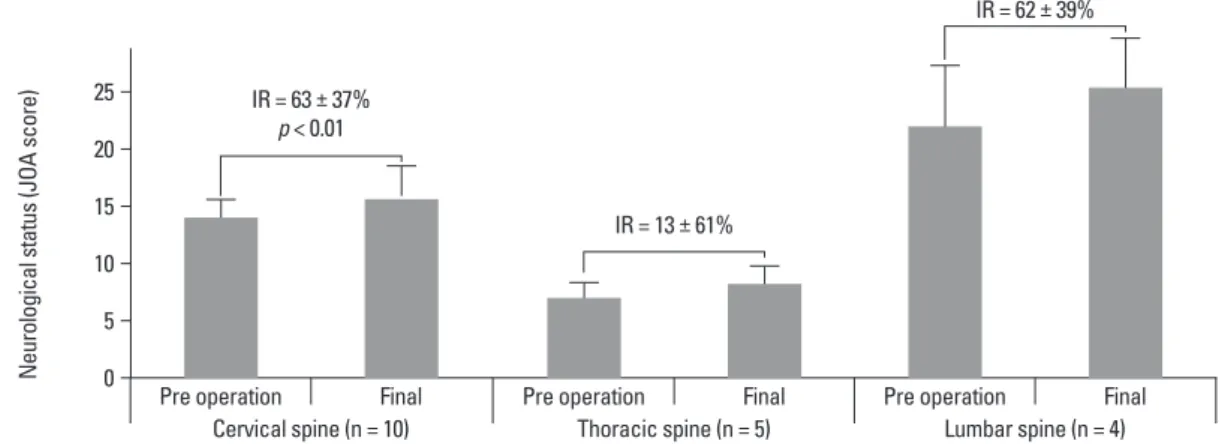
Postoperative neurological status improved in 16 cases (80%), unchanged in 3 (15%), and worsened in 1 (5%). Case 14 suffering from vesicorectal dysfunction showed worsen- ing of the neurological status. The mean improvement ratio in neurological status scores was 49.8% (Table 4). When the scores of the three spinal regions were analyzed separately, we found that the improvement ratios were 56.7% in the cer- vical spine (p < 0.01), 26.3% in the thoracic spine not signifi- cant (NS), and 48.6% in the lumbar spine (NS) (Fig. 2).
There were no significant differences in improvement ratios among the three groups (one-way analysis of variance).
Postoperative spinal alignment
The Cobb sagittal angle was 4.3 ± 18.0° lordosis (range, - 36.6°
to 35.7°) preoperatively and 5.4 ± 17.6° lordosis (range, - 38.6° to 38.2°) at the final follow-up. The change in the lor- dotic angle ranged from 2.0° decrease to 9.3° increase, show- ing no significant changes (Table 2).
Postoperative recurrence of the tumor
There were no cases of tumor recurrence in the postopera- tive period in this series, and none received adjuvant thera- py (Table 2).
RESULTS
Pathological diagnosis of tumors
Pathological examination revealed neurinoma in 12 cases (54.5%), arachnoid cyst in 3 (13.6%), neurofibroma in 2 (10.0%, n = 2), meningioma in 1 (5.0%), chondroma in 1 (5%), and ependymoma in 1 (5%) (Table 1).
Invasiveness of the procedures
The duration of surgery was 225 ± 105 min (average ± SD). When divided by the number of hemilaminectomied levels, the duration was 89 ± 48 min. The amount of blood loss during surgery was 416 ± 392 g. When divided by the number of hemilaminectomied levels, the amount of blood loss was 172 ± 190 g (Table 1).
Radicality of resection
Radicality of resection was “complete” in 16 patients (80.0%) and “predictably incomplete” in 4 patients (20.0%). There were no “unexpected incomplete” resections. The four pa- tients with predictably incomplete resections were those with dumbbell-shaped tumors; three of these patients un- derwent additional resections using the anterior approach.
These patients, however, did not require any form of instru- mented fusion (Table 4). Patient No.13 had a huge arach- noid cyst from T3 to T8, compressing the spinal cord. Total removal of this cyst required multilevel hemilaminectomy from T2 to T8. Conversion to conventional laminectomy was not required in any of the cases in the present study.
Complications
Three complications were recorded. In Case 10 (chondro-
Fig. 2. The average improvement ratio of postoperative neurological status was 63.3% in tumors of the cervical region (p < 0.01), 13.3% in tumors of the thoracic region, and 61.8% in tumors of the lumbar region. IR, improvement ratio; JOA score, Japanese Orthopaedic Association Score.
25 20 15 10 5
0 Pre operation Pre operation Pre operation
Cervical spine (n = 10) Thoracic spine (n = 5) Lumbar spine (n = 4) IR = 63 ± 37%
p < 0.01
IR = 13 ± 61%
IR = 62 ± 39%
Final Final Final
Neurological status (JOA score)
with this concept and have altered our treatment strategy in accordance with it. Hemilaminectomy would be optimal for tumors with clear borders, extramedullary and extradu- ral tumors, and dorsal and unilateral lesions throughout the spine. In contrast, we chose conventional laminectomy for removal of tumors with unclear borders, and for intramed- ullary, ventral, and bilateral spreading lesions. We observed a high radicality ratio (80.0%), no incidence of intraopera- tive conversion from hemilaminectomy to conventional lam- inectomy, and no postoperative tumor recurrence. Consis- tent with previous findings,23 the radicality of resection was predictably incomplete in 4 of the 5 patients with dumbbell- shaped tumors. We found cerebrospinal fluid (CSF) leakage resulting from one of the 20 operations (5%), higher than reported in patients who underwent either hemilaminecto- my (0.7%) or total laminectomy (3%).14 The CSF leakage we observed in one of our patients was deemed minor and was managed conservatively. Definitions of CSF leakage should be standardized, in order to assess differences in rates of CSF leakage. During preoperative screening, we excluded patients suspected of having malignant tumors or tumors located anteriorly to the spinal cord. Those tumors were surgically removed via total laminectomy. Conse- quently, we did not convert these patients from hemilami- nectomy to total laminectomy during surgery. Pathological analysis showed that all of these tumors were benign. These findings suggest that spinal tumor removal by hemilami- nectomy through strict preoperative assessment using im- aging modalities9 can guarantee a successful clinical out- come.
There were several limitations in the design of this study.
First, in this study, a single cohort that underwent a single surgical strategy was followed-up prospectively. Therefore, a comparative study with similar patients treated using other strategies in a randomized manner must be performed. Sec- ond, the mean overall final follow-up period was 85 months, ranging from 40 to 131 months. Evaluations at consistent time periods are required in future studies to obtain more clinically relevant data. Third, the patients in this study were relatively young (median age, 42.3 years), indicating that they are not representative of a generalized patient popula- tion. Inclusion of elderly and/or osteopenic patients may have altered our results. Fourth, our patient population was skewed, in having more cervical patients then other regions.
A laminectomy lower down in the spine would probably have had more destabilizing effects, thus altering the results of postoperative spinal alignment. Finally, our results may
DISCUSSION
In the present study, clinical outcomes of the removal of spinal tumors by hemilaminectomy in 20 cases were re- viewed with an average follow-up of 85 months. While several authors have reported the usefulness of this surgical method,1,8-11 medium to long-term follow-up results have rarely been reported. The present results with a medium to long-term follow-up showed a relatively low level of oper- ative invasiveness, good improvement ratio of neurological status, no significant deterioration in spinal sagittal align- ment, and no recurrence of tumors. Importantly, as hemi- laminectomy was originally adopted for spinal tumor re- moval due to its possible advantage in preserving the sagittal alignment,20 the present results actually confirmed the ad- vantage of this approach.
To reduce the risk of deformity and spinal instability after laminectomy, Raimondi, et al.2 and Parkinson21 recom- mended osteoplastic laminectomy, originally described by Bickham,22 to reconstruct the structures of the posterior col- umn. However, this technique is somewhat difficult, and is therefore time consuming,2,21 To avoid subsequent compli- cations in spinal sagittal alignment, the hemilaminectomy approach that can preserve interspinous ligaments, interver- tebral joints, and paravertebral muscles of the contralateral side was then indicated for resection of spinal cord tu- mors.19 Although the usefulness of tumor removal by hemi- laminectomy in maintenance of sagittal alignment in cervi- cal regions has been reported previously by Asazuma, et al.20 the present medium to long-term results from cases with hemilaminectomy of a number of different levels and spinal regions with no deterioration in the spinal sagittal alignment represent a significant addition to the literature.
While hemilaminectomy is advantageous in preserving posterior spinal structures, the hemilaminectomy approach provides a relatively narrow view of the spinal intracanalar regions.12 Ozawa, et al.23 noted several limitations and dis- advantages of hemilaminectomy in removal of spinal tu- mors. They suggested that additional foraminotomies and reconstructions using interspinous wiring are necessary for radical resection of dumbbell tumors of Eden type 2 and 3.23 They also suggested that huge tumors with scalloping of vertebrae, midline tumors that require resection and re- construction of the dural sac, easy bleeding tumors spread- ing to both sides, malignant lymphomas, and hemangiomas are difficult to manage by hemilaminectomy.23 We agree
11. Sario-glu AC, Hanci M, Bozkuş H, Kaynar MY, Kafadar A. Uni- lateral hemilaminectomy for the removal of the spinal space-occu- pying lesions. Minim Invasive Neurosurg 1997;40:74-7.
12. Oktem IS, Akdemir H, Kurtsoy A, KoçRK, Menkü A, Tucer B.
Hemilaminectomy for the removal of the spinal lesions. Spinal Cord 2000;38:92-6.
13. Hosono N, Yonenobu K, Ono K. [Japanese Orthopedic Associa- tion: Scoring system for cervical myelopathy]. J Jpn Orthop Assoc 1994;68:490-503.
14. Yonenobu K, Abumi K, Nagata K, Taketomi E, Ueyama K. In- terobserver and intraobserver reliability of the japanese orthopae- dic association scoring system for evaluation of cervical compres- sion myelopathy. Spine (Phila Pa 1976) 2001;26:1890-4.
15. Yonenobu K, Ebara S, Fujiwara K, Yamashita K, Ono K, Yamamo- to T, et al. Thoracic myelopathy secondary to ossification of the spi- nal ligament. J Neurosurg 1987;66:511-8.
16. Fujimura Y, Nishi Y, Nakamura M, Toyama Y, Suzuki N. Long- term follow-up study of anterior decompression and fusion for thoracic myelopathy resulting from ossification of the posterior longitudinal ligament. Spine (Phila Pa 1976) 1997;22:305-11.
17. Ohnishi K, Miyamoto K, Kanamori Y, Kodama H, Hosoe H, Shi- mizu K. Anterior decompression and fusion for multiple thoracic disc herniation. J Bone Joint Surg Br 2005;87:356-60.
18. Miyakoshi N, Abe E, Shimada Y, Okuyama K, Suzuki T, Sato K.
Outcome of one-level posterior lumbar interbody fusion for spon- dylolisthesis and postoperative intervertebral disc degeneration adjacent to the fusion. Spine (Phila Pa 1976) 2000;25:1837-42.
19. Yorimitsu E, Chiba K, Toyama Y, Hirabayashi K. Long-term out- comes of standard discectomy for lumbar disc herniation: a fol- low-up study of more than 10 years. Spine (Phila Pa 1976) 2001;
26:652-7.
20. Asazuma T, Nakamura M, Matsumoto M, Chibo K, Toyama Y.
Postoperative changes of spinal curvature and range of motion in adult patients with cervical spinal cord tumors: analysis of 51 cases and review of the literature. J Spinal Disord Tech 2004;17:178-82.
21. Parkinson D. Replacement laminotomy. Surg Neurol 1977;8:277-9.
22. Bickham WS. III. Technique of Exposure of the Spinal Cord and Canal; Osteoplastic Resection and Laminectomy. Ann Surg 1905;
41:372-98.
23. Ozawa H, Kokubun S, Aizawa T, Hoshika T, Kawahara C. Spinal dumbbell tumors: an analysis of a series of 118 cases. J Neurosurg Spine 2007;7:587-93.
have been more convincing had the patient cohort been more limited relative to the types of tumor.
In conclusion, twenty cases of spinal tumor excision by hemilaminectomy were reviewed. This surgical method provided satisfactory outcomes with a good neurological status, maintenance of sagittal alignment, and little compli- cation over medium to long-term follow-up.
REFERENCES
1. Eggert HR, Scheremet R, Seeger W, Gaitzsch J. Unilateral micro- surgical approaches to extramedullary spinal tumours. Operative technique and results. Acta Neurochir (Wien) 1983;67:245-53.
2. Raimondi AJ, Gutierrez FA, Di Rocco C. Laminotomy and total reconstruction of the posterior spinal arch for spinal canal surgery in childhood. J Neurosurg 1976;45:555-60.
3. Yasuoka S, Peterson HA, Laws ER Jr, MacCarty CS. Pathogene- sis and prophylaxis of postlaminectomy deformity of the spine af- ter multiple level laminectomy: difference between children and adults. Neurosurgery 1981;9:145-52.
4. Bradford DS. Spinal instability: orthopedic perspective and pre- vention. Clin Neurosurg 1980;27:591-610.
5. Cattell HS, Clark GL Jr. Cervical kyphosis and instability follow- ing multiple laminectomies in children. J Bone Joint Surg Am 1967;49:713-20.
6. Panjabi MM, White AA 3rd. Basic biomechanics of the spine.
Neurosurgery 1980;7:76-93.
7. Reimer R, Onofrio BM. Astrocytomas of the spinal cord in chil- dren and adolescents. J Neurosurg 1985;63:669-75.
8. Bertalanffy H, Mitani S, Otani M, Ichikizaki K, Taya S. Useful- ness of hemilaminectomy for microsurgical management of intra- spinal lesions. Keio J Med 1992;41:76-9.
9. Chiou SM, Eggert HR, Laborde G, Seeger W. Microsurgical uni- lateral approaches for spinal tumour surgery: eight years’ experi- ence in 256 primary operated patients. Acta Neurochir (Wien) 1989;100:127-33.
10. Oleshkevich FV, Rozhanets NI, Volkovets NN. [Hemilaminecto- my in the removal of spinal cord tumors]. Zh Vopr Neirokhir Im N N Burdenko 1988:30-2.