Effects of the long-term use of maxillary protraction facemasks with skeletal anchorage on pharyngeal airway dimensions in growing patients with
cleft lip and palate
Objective: To investigate the effects of the long-term use of a maxillary protraction facemask with miniplate (FM-MP) on pharyngeal airway dimensions in growing patients with cleft lip and palate (CLP). Methods: The study included 24 boys with CLP (mean age, 12.2 years; mean duration of FM-MP therapy, 4.9 years), divided into two groups according to the amount of A point advancement to the vertical reference plane (VRP): Group 1, > 4 mm; Group 2, < 2 mm; n = 12/group.
After evaluating the skeletodental and airway variables using lateral cephalograms acquired before and after FM-MP therapy, statistical analyses were performed.
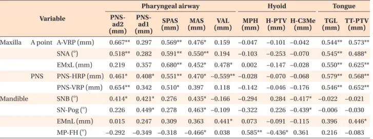
Results: Group 1 showed greater forward and downward displacements of the posterior maxilla (posterior nasal spine [PNS]-horizontal reference plane [HRP]; PNS- VRP), greater increase in ANB, more forward tongue position (tongue tip-Pt vertical line to Frankfort horizontal plane), and greater increase in the oropharynx (superior posterior airway space [SPAS]; middle airway space [MAS]) and upper nasopharynx (PNS-adenoid2) than did Group 2. While maxillary advancement (A-VRP and PNS-VRP) correlated with increases in SPAS, MAS, and PNS-adenoid2, downward displacement of the PNS (PNS-HRP) correlated with increases in SPAS, MAS, PNS- adenoid1, and PNS-adenoid2, and with a decrease in vertical airway length (VAL).
Mandibular forward displacement and decrease in mandibular plane correlated with increases in MAS. Conclusions: FM-MP therapy had positive effects on the oropharyngeal and nasopharyngeal airway spaces without increases in VAL in Group 1 rather than in Group 2. However, further validation using an untreated control group is necessary.
[Korean J Orthod 2020;50(4):238-248]
Key words: Growing patients with cleft lip and palate, Maxillary protraction, Facemask with miniplate, Pharyngeal airway dimension
Jung-Eun Kim
aSunjin Yim
bJin-Young Choi
cSukwha Kim
dSu-Jung Kim
eSeung-Hak Baek
fa
Department of Dentistry, Graduate School, Kyung Hee University, Seoul, Korea
b
Department of Dentistry, Graduate School, Seoul National University, Seoul, Korea
c
Department of Oral and Maxillofacial Surgery, School of Dentistry, Seoul National University, Seoul, Korea
d
Department of Plastic and Reconstructive Surgery, College of Medicine, Seoul National University, Seoul, Korea
e
Department of Orthodontics, School of Dentistry, Kyung Hee University, Seoul, Korea
f
Department of Orthodontics, School of Dentistry, Seoul National University, Seoul, Korea
Received October 21, 2019; Revised February 27, 2020; Accepted March 3, 2020.
Corresponding author: Su-Jung Kim.
Professor, Department of Orthodontics, School of Dentistry, Kyung Hee University, 26, Kyungheedae-ro, Dongdaemun-gu, Seoul 02447, Korea.
Tel +82-2-958-9458 e-mail [email protected] Corresponding author: Seung-Hak Baek.
Professor, Department of Orthodontics, School of Dentistry, Seoul National University, 101, Daehak-ro, Jongno-gu, Seoul 03080, Korea.
Tel +82-2-2072-3952 e-mail [email protected]
How to cite this article: Kim JE, Yim S, Choi JY, Kim S, Kim SJ, Baek SH. Effects of the long-term use of maxillary protraction facemasks with skeletal anchorage on pharyngeal airway dimensions in growing patients with cleft lip and palate. Korean J Orthod 2020;50:238-248.
© 2020 The Korean Association of Orthodontists.
This is an Open Access article distributed under the terms of the Creative Commons Attribution Non-Commercial License (http://creativecommons.org/licenses/by-nc/4.0) which permits unrestricted non-commercial use, distribution, and reproduction in any medium, provided the original work is properly cited.
pISSN 2234-7518 • eISSN 2005-372X
https://doi.org/10.4041/kjod.2020.50.4.238
INTRODUCTION
Patients with cleft lip and palate (CLP) have been reported to present impaired craniofacial and upper air- way development.
1,2They have craniofacial deformities characterized by nasomaxillary deficiency mostly due to the inhibitory effects of scar tissues from primary sur- geries.
3-5The maxillomandibular skeletal discrepancies relative to the cranium influence the morphology and dimension of the upper airway, which encompasses the nasal cavity and pharyngeal airway spaces.
6,7A previous study demonstrated that nasal airway patency was sig- nificantly lower on the cleft side than on the non-cleft side.
8A recent cone-beam computed tomography study also revealed that oropharyngeal airway dimension and volume were significantly reduced in patients with CLP than in age-matched controls without CLP.
1Accordingly, patients with CLP are liable to experience respiratory functional disorders like mouth breathing, snoring, and obstructive sleep apnea.
9,10As a treatment modality for skeletal modification in preadolescent or adolescent patients with CLP, the maxillary protraction facemask has been commonly supplemented with or without maxillary expansion.
The main skeletal and dental effects of the protraction facemask include forward and downward displacements of the maxilla, counterclockwise rotation of the palatal plane, clockwise rotation of the mandible that increases lower facial height, proclination of the upper incisors, retroclination of the lower incisors, and decrease of overbite.
11,12A few studies elucidated that short-term application of the facemask increased nasopharyngeal airway dimensions, including increasing airway length, without causing significant changes in oropharyngeal or hypopharyngeal airway spaces in children with Class III malocclusion.
13-15However, only few studies have re- ported the effects of the facemask on skeletal and upper airway morphology in patients with CLP by comparing them to controls without CLP. Moreover, the scar tissues affect not only abnormal growth and development but also treatment response and stability, and the treatment effect of maxillary protraction in patients with CLP may differ from that of patients with Class III malocclusion without CLP.
Recently, the introduction of skeletal anchorage to maxillary protraction has increased the range of skel- etal effects up to post-adolescent age groups with less dentoalveolar effects.
16Baek et al.
17suggested facemask with miniplate (FM-MP) therapy as an effective option for maxillary deficiency in patients with CLP. Based on the findings of previous studies reporting more favor- able skeletodental effects of FM-MP therapy over those of conventional facemask therapy, representing greater maxillary advancement with lesser clockwise rotation
of the mandible in adolescent patients with CLP,
18,19we anticipated a different influence on the upper airway dimension and morphology. Accordingly, the aim of this retrospective study was to investigate the effects of the long-term use of the FM-MP on pharyngeal airway di- mensions in growing patients with CLP who are liable to experience upper airway impairment.
MATERIALS AND METHODS
Patients
Seventy-five patients with CLP who underwent FM- MP therapy at the Department of Orthodontics, Seoul National University Dental Hospital, were initially recruited. The inclusion criteria were as follows: (1) patients diagnosed with non-syndromic unilateral or bilateral CLP (UCLP or BCLP, respectively); (2) patients treated using an identical surgical technique performed by a single surgeon (Millard rotation and advancement flap for cheiloplasty at 3 to 5 months after birth or Fur- low double-opposing Z-plasty for one-stage palatorrha- phy at 12 to 18 months after birth); (3) patients whose miniplates were installed by a single surgeon and who underwent FM-MP therapy for at least or more than 2 years under a single orthodontist; (4) boys in the growth stage with a skeletal maturation index of less than 5 before the start of FM-MP therapy;
20and (5) patients who had skeletal Class III relationship with maxillary hypoplasia (SNA < 78
oand ANB < 0
o). The exclusion cri- teria were as follows: (1) patients who had craniofacial anomalies; (2) patients who had a history of primary gingivoperiosteoplasty or velopharyngoplasty; (3) pa- tients who were girls, in order to avoid the influence of sex; and (4) patients with A point advancement ranging between 2 mm and 4 mm. Patients who had more than 4 mm of A point advancement were included in the ex- perimental group, because this amount of advancement shows a therapeutic effect of one cusp width correction.
Since less than 2 mm of A point advancement might be comparable to natural growth changes in patients with CLP,
21these patients were included in the positive con- trol group.
The final study sample comprised 24 boys with CLP
(13 with UCLP and 11 with BCLP; mean age, 12.2 ±
2.2 years; mean duration of FM-MP therapy, 4.9 ± 1.6
years). They were divided into two groups according to
the amount of A point advancement to the vertical ref-
erence plane (VRP): Group 1 included 12 boys with ad-
vancement > 4 mm, mean age of 11.7 years, and mean
duration of FM-MP therapy of 4.9 years; Group 2 in-
cluded 12 boys with advancement < 2 mm, mean age of
12.1 years, and mean duration of FM-MP therapy of 4.8
years (Table 1). This study was reviewed and approved
by the Institutional Review Board of Seoul National Uni-
versity School of Dentistry (ERI 18019).
Protocol of FM-MP therapy
The protocol of FM-MP therapy was as follows.
17,18After placement of curvilinear surgical miniplates (KLS Martin, Tuttlingen, Germany) on the infrazygomatic crest area (1 per side), the ends of the miniplates were exposed through the attached gingiva between the maxillary canine and first premolar. At 8 weeks after the placement of the miniplates, a Petit-type facemask (Kwang Myung DAICOM, Seoul, Korea) was delivered.
Patients were instructed to apply the elastics from the miniplates to the facemask hooks for at least 12 to 14 hours per day with 500 g of force on each side (Figure 1).
Cephalometric analysis
Lateral cephalograms were assessed before (T0) and at the time of completion of FM-MP therapy (T1). While acquiring the lateral cephalograms, the patients were asked to stand with a natural head position, to occlude slightly after a usual swallow, and to hold their breath after the end of expiration.
22The reference planes, land-
marks, and cephalometric variables used in this study are shown in Figures 1 and 2, and Table 2. Since the nasion area showed forward growth during FM-MP therapy, the actual sagittal and vertical changes of point A and the posterior nasal spine (PNS) had to be evaluated using the VRP and horizontal reference plane (HRP) passing the Sella point.
19A single investigator traced the lateral cephalograms and measured the cephalometric variables using the V- ceph software ver. 7.0 (Osstem Implant Co., Seoul, Ko- rea). All variables from randomly selected patients were remeasured by the same operator after a 2-week interval.
The intraoperator measurement error was assessed using the intraclass correlation coefficient. Since no significant differences were observed between the first and second measurements, the first set of measurements was used for analysis.
Statistical analysis
The power analysis was performed to determine the sample size with G*Power version 3.1.9.4 (Kiel University, Kiel, Germany; 0.05 two-sided significance level) using
500 gm/
side
A B
Vertical reference plane
Horizontal reference
plane (SN-7 ) S
N 7
A-VRP A point SN plane
Figure 1. A, An example of facemask with miniplate (FM- MP) therapy. B, Reference lines. The horizontal reference plane (HRP) passes the Sella (S) at an angle of 7 degree clockwise to the Sella-Nasion (SN) plane. The vertical refer- ence plane (VRP) is perpen- dicular to the HRP passing through the S. A-VRP is the horizontal distance from the VRP to A point.
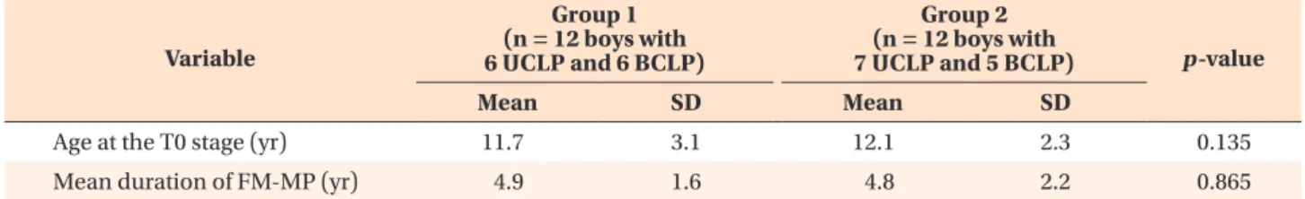
Table 1. Demographic data of patients Variable
Group 1 (n = 12 boys with 6 UCLP and 6 BCLP)
Group 2 (n = 12 boys with
7 UCLP and 5 BCLP) p-value
Mean SD Mean SD
Age at the T0 stage (yr) 11.7 3.1 12.1 2.3 0.135
Mean duration of FM-MP (yr) 4.9 1.6 4.8 2.2 0.865
Independent t-test was performed.
Group 1, A point advancement > 4 mm; Group 2, A point advancement < 2 mm; UCLP, unilateral cleft lip and palate; BCLP,
bilateral cleft lip and palate; SD, standard deviation; T0, before treatment; FM-MP, facemask with miniplate.
the mean and standard deviation values of SNA from a previous study.
18Since the sample size required for 80%
power for the significance levels of representative ceph- alometric parameters was more than 10 per group, we selected 12 patients per group to account for potential dropouts.
All data were analyzed using IBM SPSS Statistics for Windows, version 22.0 (IBM Corp., Armonk, NY, USA).
Following the Shapiro–Wilk test to assess the normal- ity of data distribution, the paired t-test was performed to assess the treatment changes in each group, and the independent t-test was performed to compare the treat- ment changes in each cephalometric variable between the two groups. To identify correlation factors contrib- uting to the treatment changes in pharyngeal airway dimensions, Pearson’s correlation analysis was used. The level of significance was established as p < 0.05.
RESULTS
Comparison of the demographic data and cephalometric variables at T0
No significant differences were observed in age, mean duration of FM-MP therapy (Table 1), and skeletodental and airway space variables at T0 between groups 1 and
2 (all p > 0.05; Table 3).
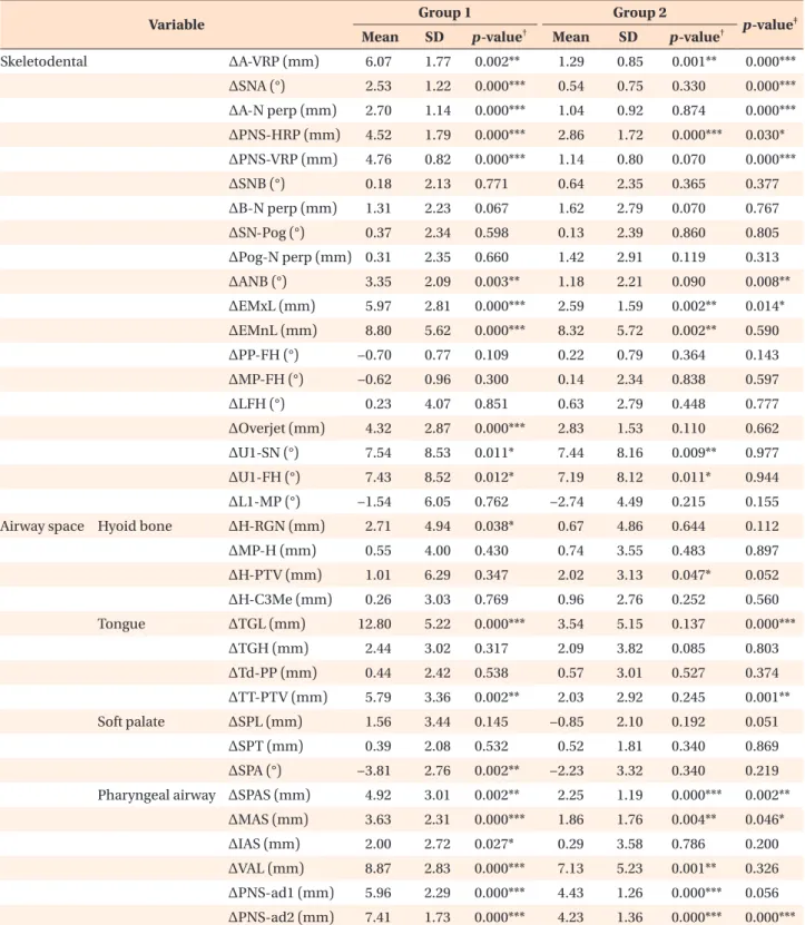
Evaluation of the change in cephalometric variables during T0–T1 in each group (Table 4)
Group 1 showed advancement of A point (A-VRP, 6.1 mm, p < 0.01; SNA, 2.5
oand A-N perp, 2.7 mm, all p < 0.001), downward and forward displacements of the PNS (PNS-HRP, 4.5 mm; PNS-VRP, 4.8 mm, all p <
0.001), increase in ANB (3.4
o, p < 0.01), increase in ef- fective maxillary and mandibular lengths (EMxL, 6.0 mm and EMnL, 8.8 mm, all p < 0.001), increase in overjet (4.3 mm, p < 0.001) and labioversion of the upper incisors (U1-FH, 7.4
oand U1-SN, 7.5
o, all p < 0.05), increase in tongue length and forward repositioning of the tongue tip (TGL, 12.8 mm, p < 0.001; TT-PTV, 5.8 mm, p <
0.01), decrease in soft palate angle (SPA, –3.8
o, p < 0.01), and increase in the pharyngeal airway spaces (superior posterior airway space [SPAS], 4.9 mm, p < 0.01; middle airway space [MAS], 3.6 mm, p < 0.001; inferior airway space [IAS], 2.0 mm, p < 0.05; and nasopharyngeal air- way space [PNS-ad1, 6.0 mm and PNS-ad2, 7.4 mm], all p < 0.001).
Group 2 showed advancement of A point (A-VRP, 1.3 mm, p < 0.01), downward displacement of the PNS (PNS-HRP, 2.9 mm, p < 0.001), increase in effective Figure 2. Cephalometric landmarks and measurements. A, Landmarks. S, Sella; N, nasion; Ba, basion; Or, orbitale; Po, porion; Pt, pterygoid point; R, midpoint between sella and basion; AD1, adenoid point 1 (adenoid tissue on the PNS-Ba line); AD2, adenoid point 2 (adenoid tissue on the R-PNS line); Co, condylion; Go, gonion; ANS, anterior nasal spine; PNS, posterior nasal spine; A, point A; B, point B; TT, tongue tip; Pog, pogonion; Me, menton; P, tip of the soft palate; Eb, base of the epiglottic fold; H, hyoidale (the most anterosuperior point on the body of the hyoid bone); RGN, retrognathion;
Pm, protuberance menti; Xi, geometric center of the ramus; FH, Frankfort horizontal; C3, the third vertebrae. B, Skeleto- dental variables. 1, SNA; 2, SNB; 3, SN-Pog; 4, ANB; 5, effective maxillary length (EMxL); 6, effective mandibular length (EMnL); 7, ANS-Me; 8, lower facial height (ANS-Xi-Pm); 9, overjet; 10, U1-SN; 11, U1-FH; 12, L1-MP. C, Airway-related variables. hyoid bone 1, H-RGN; 2, MP-H; 3, H-PTV; 4, H-C3Me; tongue 5, TGL; 6, TGH; 7, Td-PP; 8, TT-PTV; soft palate 9, SPL; 10, SPT; 11, SPA; pharyngeal airway 12, SPAS; 13, MAS; 14, IAS; 15, PNS-AD1; 16, PNS-AD2.
See Table 2 for definitions of each landmark or measurement.
A B C
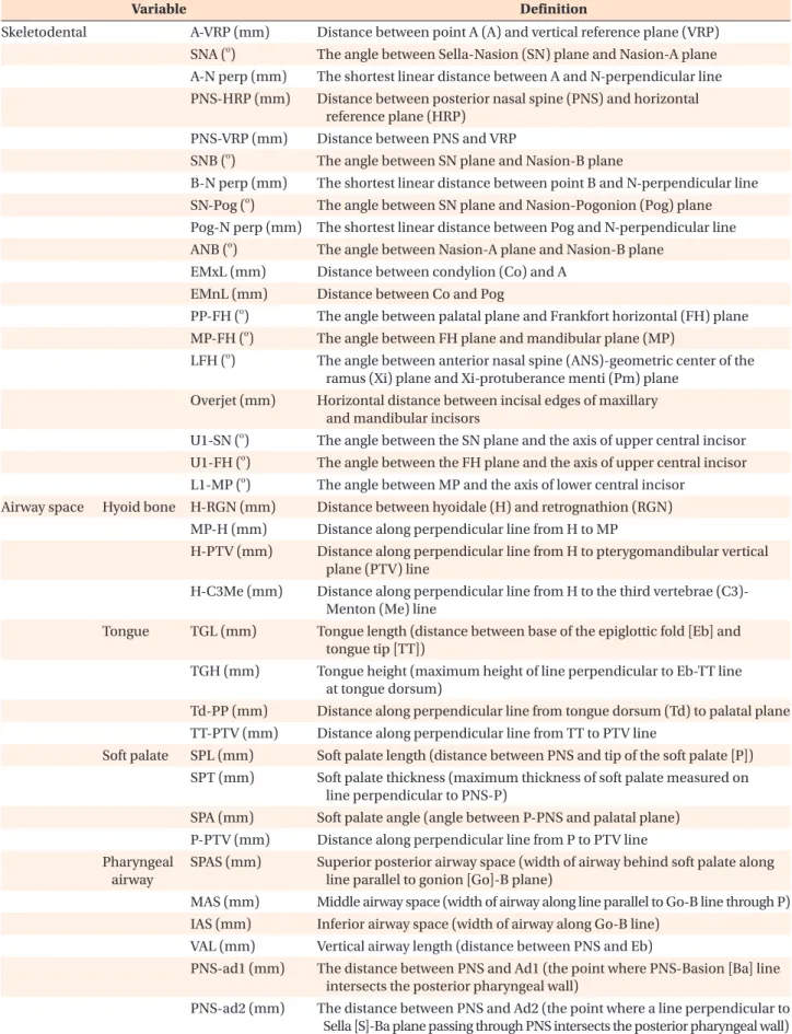
Table 2. Definition of the cephalometric skeletodental and airway variables
Variable Definition
Skeletodental A-VRP (mm) Distance between point A (A) and vertical reference plane (VRP) SNA (
o) The angle between Sella-Nasion (SN) plane and Nasion-A plane A-N perp (mm) The shortest linear distance between A and N-perpendicular line PNS-HRP (mm) Distance between posterior nasal spine (PNS) and horizontal
reference plane (HRP) PNS-VRP (mm) Distance between PNS and VRP
SNB (
o) The angle between SN plane and Nasion-B plane
B-N perp (mm) The shortest linear distance between point B and N-perpendicular line SN-Pog (
o) The angle between SN plane and Nasion-Pogonion (Pog) plane Pog-N perp (mm) The shortest linear distance between Pog and N-perpendicular line ANB (
o) The angle between Nasion-A plane and Nasion-B plane
EMxL (mm) Distance between condylion (Co) and A EMnL (mm) Distance between Co and Pog
PP-FH (
o) The angle between palatal plane and Frankfort horizontal (FH) plane MP-FH (
o) The angle between FH plane and mandibular plane (MP)
LFH (
o) The angle between anterior nasal spine (ANS)-geometric center of the ramus (Xi) plane and Xi-protuberance menti (Pm) plane
Overjet (mm) Horizontal distance between incisal edges of maxillary and mandibular incisors
U1-SN (
o) The angle between the SN plane and the axis of upper central incisor U1-FH (
o) The angle between the FH plane and the axis of upper central incisor L1-MP (
o) The angle between MP and the axis of lower central incisor
Airway space Hyoid bone H-RGN (mm) Distance between hyoidale (H) and retrognathion (RGN) MP-H (mm) Distance along perpendicular line from H to MP
H-PTV (mm) Distance along perpendicular line from H to pterygomandibular vertical plane (PTV) line
H-C3Me (mm) Distance along perpendicular line from H to the third vertebrae (C3)- Menton (Me) line
Tongue TGL (mm) Tongue length (distance between base of the epiglottic fold [Eb] and tongue tip [TT])
TGH (mm) Tongue height (maximum height of line perpendicular to Eb-TT line at tongue dorsum)
Td-PP (mm) Distance along perpendicular line from tongue dorsum (Td) to palatal plane TT-PTV (mm) Distance along perpendicular line from TT to PTV line
Soft palate SPL (mm) Soft palate length (distance between PNS and tip of the soft palate [P]) SPT (mm) Soft palate thickness (maximum thickness of soft palate measured on
line perpendicular to PNS-P)
SPA (mm) Soft palate angle (angle between P-PNS and palatal plane) P-PTV (mm) Distance along perpendicular line from P to PTV line Pharyngeal
airway
SPAS (mm) Superior posterior airway space (width of airway behind soft palate along line parallel to gonion [Go]-B plane)
MAS (mm) Middle airway space (width of airway along line parallel to Go-B line through P) IAS (mm) Inferior airway space (width of airway along Go-B line)
VAL (mm) Vertical airway length (distance between PNS and Eb)
PNS-ad1 (mm) The distance between PNS and Ad1 (the point where PNS-Basion [Ba] line intersects the posterior pharyngeal wall)
PNS-ad2 (mm) The distance between PNS and Ad2 (the point where a line perpendicular to
Sella [S]-Ba plane passing through PNS intersects the posterior pharyngeal wall)
maxillary and mandibular lengths (EMxL, 2.6 mm and EMnL, 8.3 mm, all p < 0.01), increase in labioversion of the upper incisors (U1-FH, 7.2
o, p < 0.05; U1-SN, 7.4
o,
p < 0.01), and increase in the pharyngeal airway spaces (SPAS, 2.3 mm, p < 0.001; MAS, 1.9 mm, p < 0.01;
PNS-ad1, 4.4 mm and PNS-ad2, 4.2 mm, all p < 0.001).
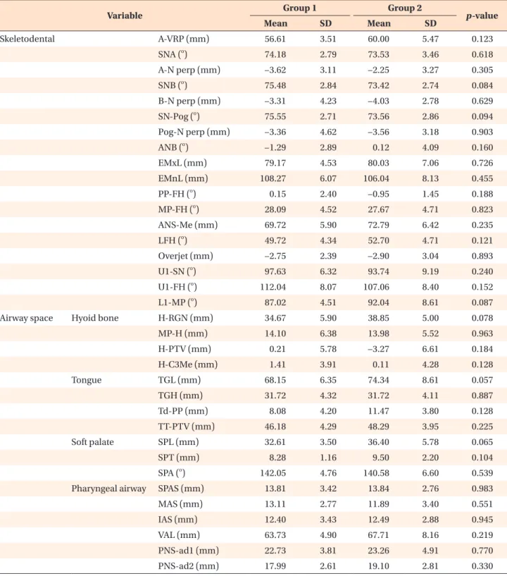
Table 3. Comparison of the cephalometric variables between groups 1 and 2 at T0
Variable Group 1 Group 2
p-value
Mean SD Mean SD
Skeletodental A-VRP (mm) 56.61 3.51 60.00 5.47 0.123
SNA (
o) 74.18 2.79 73.53 3.46 0.618
A-N perp (mm) −3.62 3.11 −2.25 3.27 0.305
SNB (
o) 75.48 2.84 73.42 2.74 0.084
B-N perp (mm) −3.31 4.23 −4.03 2.78 0.629
SN-Pog (
o) 75.55 2.71 73.56 2.86 0.094
Pog-N perp (mm) −3.36 4.62 −3.56 3.18 0.903
ANB (
o) −1.29 2.89 0.12 4.09 0.160
EMxL (mm) 79.17 4.53 80.03 7.06 0.726
EMnL (mm) 108.27 6.07 106.04 8.13 0.455
PP-FH (
o) 0.15 2.40 −0.95 1.45 0.188
MP-FH (
o) 28.09 4.52 27.67 4.71 0.823
ANS-Me (mm) 69.72 5.90 72.79 6.42 0.235
LFH (
o) 49.72 4.34 52.70 4.71 0.121
Overjet (mm) −2.75 2.39 −2.90 3.04 0.893
U1-SN (
o) 97.63 6.32 93.74 9.19 0.240
U1-FH (
o) 112.04 8.07 107.06 8.40 0.152
L1-MP (
o) 87.02 4.51 92.04 8.61 0.087
Airway space Hyoid bone H-RGN (mm) 34.67 5.90 38.85 5.00 0.078
MP-H (mm) 14.10 6.38 13.98 5.52 0.963
H-PTV (mm) 0.21 5.78 −3.27 6.61 0.184
H-C3Me (mm) 1.41 3.91 0.11 4.28 0.128
Tongue TGL (mm) 68.15 6.35 74.34 8.61 0.057
TGH (mm) 31.72 4.32 31.72 4.11 0.887
Td-PP (mm) 8.08 4.20 11.47 3.80 0.128
TT-PTV (mm) 46.18 4.29 48.29 3.95 0.225
Soft palate SPL (mm) 32.61 3.50 36.40 5.78 0.065
SPT (mm) 8.28 1.16 9.50 2.20 0.104
SPA (
o) 142.05 4.76 140.58 6.60 0.539
Pharyngeal airway SPAS (mm) 13.81 3.42 13.84 2.76 0.983
MAS (mm) 13.11 2.77 11.89 3.40 0.551
IAS (mm) 12.40 3.43 12.49 2.88 0.945
VAL (mm) 63.73 4.90 67.71 8.16 0.219
PNS-ad1 (mm) 22.73 3.81 23.26 4.91 0.770
PNS-ad2 (mm) 17.99 2.61 19.10 2.81 0.330
Independent t-test was performed.
Group 1, A point advancement > 4 mm; Group 2, A point advancement < 2 mm; SD, standard deviation.
See Table 2 for definitions of each landmark or measurement.
Table 4. Comparison of the treatment changes in skeletodental and airway variables between groups 1 and 2 (T1–T0)
Variable Group 1 Group 2
p-value
‡Mean SD p-value
†Mean SD p-value
†Skeletodental ΔA-VRP (mm) 6.07 1.77 0.002** 1.29 0.85 0.001** 0.000***
ΔSNA (°) 2.53 1.22 0.000*** 0.54 0.75 0.330 0.000***
ΔA-N perp (mm) 2.70 1.14 0.000*** 1.04 0.92 0.874 0.000***
ΔPNS-HRP (mm) 4.52 1.79 0.000*** 2.86 1.72 0.000*** 0.030*
ΔPNS-VRP (mm) 4.76 0.82 0.000*** 1.14 0.80 0.070 0.000***
ΔSNB (°) 0.18 2.13 0.771 0.64 2.35 0.365 0.377
ΔB-N perp (mm) 1.31 2.23 0.067 1.62 2.79 0.070 0.767
ΔSN-Pog (°) 0.37 2.34 0.598 0.13 2.39 0.860 0.805
ΔPog-N perp (mm) 0.31 2.35 0.660 1.42 2.91 0.119 0.313
ΔANB (°) 3.35 2.09 0.003** 1.18 2.21 0.090 0.008**
ΔEMxL (mm) 5.97 2.81 0.000*** 2.59 1.59 0.002** 0.014*
ΔEMnL (mm) 8.80 5.62 0.000*** 8.32 5.72 0.002** 0.590
ΔPP-FH (°) −0.70 0.77 0.109 0.22 0.79 0.364 0.143
ΔMP-FH (°) −0.62 0.96 0.300 0.14 2.34 0.838 0.597
ΔLFH (°) 0.23 4.07 0.851 0.63 2.79 0.448 0.777
ΔOverjet (mm) 4.32 2.87 0.000*** 2.83 1.53 0.110 0.662
ΔU1-SN (°) 7.54 8.53 0.011* 7.44 8.16 0.009** 0.977
ΔU1-FH (°) 7.43 8.52 0.012* 7.19 8.12 0.011* 0.944
ΔL1-MP (°) −1.54 6.05 0.762 −2.74 4.49 0.215 0.155
Airway space Hyoid bone ΔH-RGN (mm) 2.71 4.94 0.038* 0.67 4.86 0.644 0.112
ΔMP-H (mm) 0.55 4.00 0.430 0.74 3.55 0.483 0.897
ΔH-PTV (mm) 1.01 6.29 0.347 2.02 3.13 0.047* 0.052
ΔH-C3Me (mm) 0.26 3.03 0.769 0.96 2.76 0.252 0.560
Tongue ΔTGL (mm) 12.80 5.22 0.000*** 3.54 5.15 0.137 0.000***
ΔTGH (mm) 2.44 3.02 0.317 2.09 3.82 0.085 0.803
ΔTd-PP (mm) 0.44 2.42 0.538 0.57 3.01 0.527 0.374
ΔTT-PTV (mm) 5.79 3.36 0.002** 2.03 2.92 0.245 0.001**
Soft palate ΔSPL (mm) 1.56 3.44 0.145 −0.85 2.10 0.192 0.051
ΔSPT (mm) 0.39 2.08 0.532 0.52 1.81 0.340 0.869
ΔSPA (°) −3.81 2.76 0.002** −2.23 3.32 0.340 0.219
Pharyngeal airway ΔSPAS (mm) 4.92 3.01 0.002** 2.25 1.19 0.000*** 0.002**
ΔMAS (mm) 3.63 2.31 0.000*** 1.86 1.76 0.004** 0.046*
ΔIAS (mm) 2.00 2.72 0.027* 0.29 3.58 0.786 0.200
ΔVAL (mm) 8.87 2.83 0.000*** 7.13 5.23 0.001** 0.326
ΔPNS-ad1 (mm) 5.96 2.29 0.000*** 4.43 1.26 0.000*** 0.056 ΔPNS-ad2 (mm) 7.41 1.73 0.000*** 4.23 1.36 0.000*** 0.000***
Group 1, A point advancement > 4 mm; Group 2, A point advancement < 2 mm; SD, standard deviation.
See Table 2 for definitions of each landmark or measurement.
*p < 0.05, **p < 0.01, ***p < 0.001.
†
Paired t-test was performed.
‡