• 검색 결과가 없습니다.

Anti-Inflammatory Effects of Tongbi-san(通痺散) Extract on RAW264.7 Macrophages

N/A
N/A
Protected

Academic year: 2021

Share "Anti-Inflammatory Effects of Tongbi-san(通痺散) Extract on RAW264.7 Macrophages"

Copied!
10
0
0

로드 중.... (전체 텍스트 보기)

전체 글

(1)

pISSN 1738-6640 eISSN 2234-4020 http://dx.doi.org/10.6114/jkood.2016.29.4.024

Original Article / 원저

Anti-Inflammatory Effects of Tongbi-san (通痺散) Extract on RAW264.7 Macrophages

Yong-Min Kim

1)

·Hee-Taek Kim

2)

·Ee-Hwa Kim

3)

1

School of Oriental Medicine and Bio Convergence Sciences, Semyung University

2

Dept. of Korean Medical Ophthalmology & Otolaryngology & Dermatology, College of Korean Medicine, Semyung University

3

Dept. of Meridian & Acupoint, College of Korean Medicine, Semyung University

통비산(通痺散) 열수추출물의 항염증반응 및 항산화활성에 대한 연구

김용민

1)

·김희택

2)

·김이화

3)

1 세명대학교 한방바이오융합과학부

2 세명대학교 한의과대학 한방안이비인후피부과학과

3 세명대학교 한의과대학 경락경혈학교실

Abstract

Objectives : This study is to investigate the anti-inflammatory and anti-oxidant effects of Tongbi-san extract (TS) on RAW264.7 macrophages using by cell cytotoxicity, Nitric Oxide (NO) and Prostaglandin E

2

(PGE

2

) production and 1,1-diphenyl-2-picryl ghdrazyl (DPPH) free radical scavenging capability.

Methods : Cell cytotoxicity was measured by 3-(4,5-dimethylthiazol-2-yl)-2,5-diphenyltetrazolium bromide (MTT) assay. The production of NO was measured by Griess assay. The production of PGE
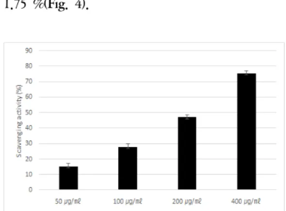
2

was measured by immunoassay. And the anti-oxidant activity was measured by the DPPH method.

Results : TS did not increased significantly compared to the TS untreated group in the cell cytotoxicity. TS inhibited NO and PGE

2

production in lipopolysaccharide-stimulated RAW 264.7 cells. TS had the DPPH free radical scavenging capability.

Conclusion : The anti-inflammtory and anti-oxidant effects of TS may be use for a treatment of anti-inflammatory diseases.

Key words : Anti-inflammation; Anti-oxidant activity; Nitric Oxide; Prostaglandin E

2

; reactive oxygen species;

Tongbi-san

ⓒ 2016 the Society of Korean Medicine Ophthalmology & Otolaryngology & Dermatology

This is an Open Access journal distributed under the terms of the Creative Commons Attribution Non-Commercial License (http://creativecommons.org/license/by-nc/3.0/) which permits unrestricted non-commercial use, distribution, and reproduction in any medium, provided the original work is properly cited.

(2)

Ⅰ. Introduction

Traditional medicine is an important natural source of phytochemical compounds with substantial therapeutic effects and represents the primary health resource to many people

1)

. World Health Organization (WHO) estimates that 80 % of people indeveloping countries use traditional medicine as primary healthcare

2)

. Therefore, it is important to assess and validate the traditional effects of plants to assure people that consume them

3)

.

Inflammation is a local, protective response of the immune system. Excessive inflammatory responses can be harmful, as in diseases such as rheumatoid arthritis, Alzheimer’s disease and septic shock syndrome

4)

. Lipopolysaccharide (LPS), a component of the cell wall of Gram-negative bacteria, stimulates macrophages to produce pro-inflammatory mediators such as tumor necrosis factor alpha, interleukin-6, and inducible nitric oxide synthase, which trigger a cascade responsible for the inflammatory response

5)

. Antioxidants can protect against the damage caused by free radicals that have been implicated in the etiology of large number of major diseases

6)

.

Nitric oxide (NO) is synthesized from amino acid, arginine, by nitric oxide synthase (NOS). NO plays an important role as a vasodilator, neurotransmitter and in the immunological system as a defense against tumor cells, parasites and

bacteria

7)

. However, NO production is increased by the inducible isoform of NOS (iNOS), subsequently, brings about cytotoxicity and tissue damage

8)

. Therefore, much attention has focused on how to decease the NO production generated by iNOS.

In addition, cyclooxygenase 2 (COX-2) is the rate limiting enzyme and responsible for the catalysis of prostaglandin E

2

(PGE

2

) from arachidonic acid

9)

. Chang et al. noted that the induction of COX-2 activity and subsequent generation of PGE

2

are closely related to the NO production

10)

. Thus, reduce the levels of PGE

2

and the levels of COX-2 may be an effective strategy for inhibiting the inflammation and carcinogenesis.

Tongbi-san (TS) has been used in pain control, improve blood circulation, and mediate injury healing. It is consistently used in the clinical treatment of pain and bone injury

11)

. In the present study, protective effect of Tongbi-san on the effects of anti-inflammation and and-oxidation by modulation of nitric oxide and PGE

2

production in LPS-stimulated RAW264.7 macrophages were investigated.

Ⅱ. Materials & Methods

1. Cell culture

Cells of the murine macrophage RAW 264.7 were purchased from the Korean Cell Line Bank (KCLB, Seoul, Korea). Cells were cultrued in Dulbecco’s Modified Eagle Medium (DMEM) (Gibco BRL, Grand Island, NY, USA) supplemented with 10% heat-inactivated fetal

Corresponding Author : Ee-Hwa Kim, Dept. of Meridian & Acupoint, College of Korean Medicine, Semyung University, 65 Semyung-ro, Jecheon, Chungbuk, Korea.

(Tel : 043-649-1348, E-mail : [email protected])

Recieved 2016/9/26

Revised 2016/11/3

Accepted 2016/11/10

(3)

bovine serum (FBS) (Gibco BRL) at 37℃ in 5%

CO

2

, 95% O

2

in a humidified cell incubator. Cells were plated in culture dish (Corning Incorporated, Corning, NY, USA) at a density of 1 × 10

6

cells per dish, and the media was changed once every 2 days.

2. Preparation of extract

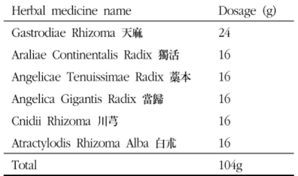
Tongbi-san (TS) were obtained from Semyung Korean medical hospital (Chungbuk, Korea). The procedure in brief is as follow: each medicinal plants were performed reflux extraction with distilled water (D.W) for 3 h at 100℃. Filtration and evaporation were performed with rotary vacuum evaporator (N-N series, EYELA, Japan) at 60℃. The solution was freeze dried for 24 h at 80℃ and lyophilized to yield. The composition and dosage of Tongbi-san (TS) are epitomized in Table 1.

Herbal medicine name Dosage (g) Gastrodiae Rhizoma 天麻 24 Araliae Continentalis Radix 獨活 16 Angelicae Tenuissimae Radix 藁本 16 Angelica Gigantis Radix 當歸 16

Cnidii Rhizoma 川芎 16

Atractylodis Rhizoma Alba 白朮 16

Total 104g

Table 1. The Composition of Tongbi-san (TS)

3. MTT cytotoxicity assay

Cell viability was determined by the MTT assay kit using as per the manufactures protocol. Cells were cultured in 96 well plates. Experimental groups are exposed to TS at final concentrations of 50, 100, 200 and 400 ㎍/㎖ for 24 hrs, and

saline of an equal volume was added to untreated group. Ten ㎖ of the MTT labeling reagent was then added to each well, and the plates were incubated for 4 h. After the cells were incubated in 100 ㎖ of the solubilization solution for 12 h, the absorbance was measured with a microtiter plate reader (Bio-Tek, Winooski, VT, USA) at a test wavelength of 595 ㎚ with a reference wavelength of 690 ㎚. The optical density (O.D.) was calculated as the result of the subtraction of the absorbance at the reference wavelength from that of the test wavelength. Percent viability was calculated as (O.D. of drug-treated sample/control O.D.) × 100.

4. NO assay

The concentration of NO in the culture supernatants was determined by measuring nitrite, a major stable product of NO, using the Griess reagent. Briefly, Cells were plated onto 24-well plates and pretreated with the various concentrations of TS 1 h prior to stimulation with 1 ㎍/㎖ of LPS for 24 h. Supernatant samples were mixed with equal volume of Griess reagent (1% sulfanilamide and 0.1% naphthyl ethylene diamine dihydro chloride in 5% phosphoric acid) and the incubated at room temperature for 10 min. The absorbance was measured at 540 ㎚ on a microplate reader (Thermo electron corporation, Marietta, OH).

5. Measurement of PGE2

Enzyme-linked immunosorbent assay (ELISA)

kit (R&D Systems, Minneapolis, MN) was used to

measure PGE

2

production according to the

(4)

manufacturer’s instructions. Cells were plated in 24-well plates and pretreated with the indicated concentrations of TS 1 h prior to stimulation with 1 ㎍/㎖ LPS for 24 h. One hundred microliters of culture media were collected for the determination of PGE

2

concentrations by ELISA according to the manufacturer’s instructions.

6. Assessment of DPPH radical scavenging activity

The DPPH radical scavenging activity was mesured according to previous studies with a few modifications. Briefly, 2 ㎖ of 0.2 mM methanolic solution of DPPH radicals were added to 2 ㎖ of water-solution of TS at various concentrations.

The absorbance of the mixture was measured at 517 nm after 30 min of incubation at 37℃ in the dark. Ascobic acid was used as the control and distilled water as the blank. The scavenging effect was calculated according to the following equation: Scavenging rate %=(1-As/A0)×100%, where as is the absorbance obtained for a sample and A0, the absorbance of the blank.

7. Statistical analysis

Statistical analysis was performed using Student's t-test (SPSS ver 12.0) and the results were expressed as mean ± S.E.M. Differences were considered significant for p < 0.05.

Ⅲ. Results

1. Cell viability assay in RAW 264.7 macrophages

In order to find out the concentration at which

the cytotoxic effect of TS on the RAW 264.7 cell line become evident, cells were cultured with TS at final concentrations of 50 ㎍/㎖, 100 ㎍/㎖, 200

㎍/㎖, and 400 ㎍/㎖ for 24 hrs, and MTT assays were carried out, with cells cultured in TS-free media as the control. The viabilities of cells incubated with TS at concentrations of 50 ㎍/㎖, 100 ㎍/㎖, 200 ㎍/㎖ and 400 ㎍/㎖ were 99.41 ± 1.60 %, 97.47 ± 1.87 %, 94.39 ± 2.88 % and 88.50 ± 5.71 % of the control (100 ± 1.63 %) value respectively. As shown in Fig. 1, TS, at concentrations 50-400 ㎍/㎖, showed no obious cytotoxicity on RAW 264.7 cells.

Fig. 1. Cytotoxic effects of Tongbi-san aqueous extract (TS). RAW 264.7 cells were incubated with TS at various concentrations (0-400 ㎍/㎖) prior to the determination of cellular viability through MTT assay. Results were represented as mean

± standard error.

2. Effect of Tongbi-san (TS) on NO release

in LPS-stimulated RAW 264.7 macrophages

To determine the effects of TS on NO

production in RAW 264.7 cells, the cells were

pre-incubated with various concentrations of TS

for 1 h and then stimulated with 1 ㎍/㎖ of LPS

for 24 h. The control group was not treated with

(5)

LPS or TS. Supernatant from cell culture media was collected, and NO levels were determined with the Griess assay. TS was found to inhibit LPS-induced NO productions in a dose-dependent manner. The NO production of TS at concentrations of 50 ㎍/㎖, 100 ㎍/㎖, 200 ㎍/㎖

and 400 ㎍/㎖ were 98.46 ± 3.35 %, 93.85 ± 7.02

%, 90.49 ± 2.14 % and 78.01 ± 3.95 % of the control (100 ± 4.87 %) value respectively. TS at 200 ㎍/㎖ and 400 ㎍/㎖ significantly inhibited NO production(Fig. 2).

Fig. 2. Effects of Tongbi-san aqueos extract (TS) on the nitric oxide (NO) production in RAW 264.7 cells. Control : treated with LPS (1 ㎍/㎖) ; 50-400 : treated with LPS and TS (50-400 ㎍/

㎖). *represents p<0.05 compared to the Control group.

3. Effect of Tongbi-san (TS) on PGE2 release in LPS-stimulated RAW 264.7 macrophages To determine the effects of TS on PGE

2

production in RAW 264.7 cells, the cells were pre-incubated with various concentrations of TS for 1 h and then stimulated with 1 ㎍/㎖ of LPS for 24 h. The control group was not treated with LPS or TS. Supernatant from cell culture media was collected, and PGE

2

levels were determined

with the EIA kit. TS was found to inhibit LPS-induced PGE

2

productions in a dose-dependent manner. The PGE

2

production of TS at concentrations of 50 ㎍/㎖, 100 ㎍/㎖, 200

㎍/㎖ and 400 ㎍/㎖ were 739.81 ± 11.45 pg/well, 732.92 ± 21.11 pg/well, 706.58 ± 14.92 pg/well, 675.25 ± 13.90 pg/well of the control (742.24 ± 11.63 pg/well) value respectively. TS at 200 ㎍/㎖

and 400 ㎍/㎖ significantly inhibited PGE

2

production(Fig. 3).

Fig. 3. Effects of Tongbi-san aqueos extract (TS) on the prostaglandin E2 (PGE2) production in RAW 264.7 cells. Control : treated with LPS (1 ㎍/㎖)

; 50-400 : treated with LPS and TS (50-400

㎍/㎖). *represents p<0.05 compared to the Control group.

4. Effect of Tongbi-san (TS) on DPPH radical scavenging activity

Scavenging of DPPH radicals is the basis of a common antioxidant assay. Antioxidants can protect against the damage caused by free radicals that have been implicated in the etiology of large number of major diseases. TS displayed concentration dependent radical scavenging effects. TS at concentrations of 50 ㎍/㎖, 100 ㎍/

㎖, 200 ㎍/㎖ and 400 ㎍/㎖ were 15.34 ± 1.98 %,

(6)

27.86 ± 2.03 %, 47.03 ± 1.54 % and 75.27 ± 1.75 %(Fig. 4).

Fig. 4. DPPH free radical scavenging capability of Tongbi-san aqueos extract (TS). The absorbance of the TS (50-400 ㎍/㎖) was measured at 517 ㎚. Results were represented as mean ± standard error.

Ⅳ. Discussion

The purpose of this study is to investigate the effects of TS on the production of NO and PGE

2

induced by LPS-stimulated RAW 264.7 cells and DPPH free radical scavenging capability.

It is well known that Korean herbal medicine, TS, which is comprised six herbs of Gastrodiae Rhizoma, Araliae Continentalis Radix, Angelicae Tenuissimae Radix, Angelica Gigantis Radix, Cnidii Rhizoma and Atractylodis Rhizoma Alba

11)

. These herbs are effective for the treatment of inflammation, hyperlipidemia, arteriosclerosis, and gynecological disease. However, there was no research about the anti-inflammatory and anti-oxidant effects of TS

12-15)

.

In this study, we used TS as the potent NO and PGE

2

production inhibitor to investigate the involvement of anti-inflammation and anti-oxidant

effect. The TS reduced NO production and PGE

2

production induced by the induction of LPS, and increased the radical scavenging activity. The radical scavenging activity of TS is and indication that it is a potential natural antioxidant.

It is well known that chronic low grade inflammation is involved in a range of pathophysiologic metabolic disorders and diseases such as obesity, insulin resistance, cardiovascular disease and cancer. In obesity, low grade inflammation underlies with an aberrant infiltration of activated macrophages in dysfunctional adipose tissue. This systemic process involves activation of intracellular signaling pathways able to release arrested NF-κB by the inhibitor of κB (IκB) proteins, allowing its nuclear translocation, and development of the expression of genes (iNOS/NO, COX-2/PGE

2

) involved in inflammation as well as cytokines, chemokines and grow factors. This process plays an important function in the control of apoptosis, oxidative/nitrosative stress and proliferation

3)

. Furthermore, activation of this signaling pathway is associated with persistent release of pro-inflammatory mediators, which drive to insulin resistance and chronic diseases

16-18)

. Hence, alternative phytochemical compounds targeting intracellular NF-κB signaling pathway could contribute to the anti-inflammatory process and inhibition of its associated pathologies

3)

.

Inflammation is a complex process regulated by a variety of immune cells and effector molecules.

NO, PGE

2

, and pro-inflammatory cytokines are

important mediators of macrophage-mediated

inflammation

19)

. Therefore, the inhibition of these

mediators with pharmacological modulators may

(7)

be an effective therapeutic strategy for preventing inflammatory reactions and diseases

20)

.

Macrophages play critical roles in immune reactions, allergy, and inflammation

21)

. These cells induce inflammatory reactions, and initiate and maintain specific immune responses by releasing different types of cytokines. LPS, a comonent of the gram-negative bacterial cell wall, has often been used in inflammatory response because it can activate macrophages

22)

.

In general, NO plays an important role in the antitumour, antivirus replication and other diseases

23)

, the overproduction of NO is harmful to the host, leading to rheumatoid arthritis

24)

and allograft rejection

25)

. NO production from macrophages can be induced by inflammatory cytokines or bacterial products, including LPS, IFN-γ, or TNF-α

26)

.

PGE

2

is considered the one of the strongest inflammatory mediators in inflammatory response.

It was transformed from arachidonic acid via the cyclooxygenase-2 (COX-2) catalytic reaction.

Nonsteroidal anti-inflammatory drugs (NSAIDs), which were used widely in current clinical, play their antipyretic, anti-inflammatory and analgesic effects through the inhibition of COX activity and the reduction of inflammatory mediator production such as PGE

227)

.

In no stimulated cells, ROS are generated by normal metabolism and include hydrogen peroxide, hydroxylradical and superoxide anions radicals. In basal ROS production, NADP+/NADPH oxidase complex maintains redox homeostasis, but in overproduction, numerous detoxifying/antioxidative enzymatic systems are developed such as superoxide

dismutase, catalase and glutathione peroxidase and hemeoxigenase-1. However, an imbalance between ROS production and inadequate anti-oxidant mechanism results in an oxidative stress state. Several researchers have reported that anti-oxidant properties of polyphenols can produce their chemo-preventive effects through NF-κB and nuclear factor E

2

related factor (Nrf2)/anti-oxidant response element (ARE) pathways activation

28)

.

Oxidative stress is an important factor in the genesis of most pathologies, ranging from cancer to cardiovascular and degenerative diseases

29)

. In order to protect the body against the consequences of oxidative stress, an efficacious approach consists in improving the anti-oxidant nutrition. Anti-oxidants from natural sources have a higher bioavailability and therefore higher protective efficacy than synthetic anti-oxidants

30)

. Free radicals are chemical species with one or two unpaired electrons in their outermost layer, which can be created in a multiple ways. They can be exogenic or endogenic. A lack of anti-oxidant or an overproduction in free radicals cab lead to an imbalance between the oxidant and anti-oxidant system. One of the most significant factors in the production of free radicals is oxidative stress

31)

. Oxidative stress is involved in a several illnesses, including diabetes

32)

, atherosclereosis, Alzheimer’s disease, Parkinson’s disease, glaucoma and age-related macular degeneration

33)

. The provision of anti-oxidants through diet or herb-medicine is a simple means to reduce the development of illnesses brought on by oxidative stress

34)

.

TS has effects on reinforcing kidney, improving

(8)

circulation, reduce bleeding, air wound healing, etc

35)

. In the clinical setting, TS is consistently used for the treatment of inflammation as well as pain control. Under these influences, modern pharmacological studies mainly focus on the anti-inflammatory effect and anti-oxidant effect.

Ⅴ. Conclusion

1. This study is to investigate the anti- inflammatory and anti-oxidant effects of TS extract on RAW264.7 macrophages using by cell cytotoxicity, NO and PGE

2

production and DPPH free radical scavenging capability.

2. TS did not increased significantly compared to the untreated group in the cell cytotoxicity.

3. TS inhibited NO and PGE

2

production in lipopolysaccharide-stimulated RAW264.7 cells.

4. TS had the DPPH free radical scavenging capability.

5. The anti-inflammtory and anti-oxidant effects of TS may be use for a treatment of inflammatory diseases.

Acknowledgement

This paper was supported by the Semyung University Research Grant of 2014.

References

1. Cordell GA. Phytochemistry and traditional medicine-the revolution continues. Phytochem Lett. 2014;10:28-40.

2. World Health Organization. WHO Traditional medicine strategy. 2013. Availble from:URL:

http://www.who.int/medicines/

publications/traditional/trm-strategy14_23.

3. Torres-Rodrigues ML, Garcia-Chavez E, Berhow M, de Mejia EG. Anti-inflammatory and anti-oxidant effect of Calea urticifolia lyophilized aqueous extract on lipopolysaccharide-stimulated RAW 264.7 macrophages. J Ethnopharmacol. 2016;188:

266-74.

4. Tracey KJ. The inflammatory reflex. Nature.

2002;420:853-9.

5. Chen X, Miao J, Wang H, Zhao F, Hu J, Gao P, et al. The anti-inflammatory activities of Ainsliaea fragrans Champ. extract and its components in lipopolysaccharide-stimulated RAW264.7 macrophages through inhibition of NF-kB pathway. J Ethnopharmacol. 2015;

170:72-80.

6. Devasagayam TPA, Tilak JC, Boloor KK, Sane KS, Ghaskadbi SS, Lele RD. Free radicals and antioxidants in human health:

current status and future prospects. Journal of the Association of Physicians of India.

2004;52:794-804.

7. Nakagawa T, Yokozawa T. Direct scavenging of nitric oxide and superoxide by green tea.

Food Chem. Toxicol. 2002;40:1745–50.

8. Kim HK, Cheon BS, Kim YH, Kim SY, Kim HP. Effects of naturally occurring flavonoids on nitric oxide production in the macrophage cell line RAW 264.7 and their structure–

activity relationships. Biochem. Pharmacol.

1999;58:759–65.

9. Surh YJ, Chun KS, Cha HH, Han SS, Keum

(9)

Y, Park K, et al. Molecular mechanisms underlying chemopreventive activities of anti-inflammatory phytochemicals: down- regulation of COX-2 and iNOs through suppression of NFκB activation. Mutat Res.

2001;480–481:243–68.

10. Chang YC, Li PC, Chen BC, Chang MS, Wang JL, Chiu WT, et al. Lipoteichoic acid-induced nitric oxide synthase expression in RAW 264.7 macrophages is mediated by cyclooxygenase-2, prostaglandin E

2

, protein kinase A, p38 MAPK, and nuclear factor-kappa B pathways. Cell Signal.

2006;18:1235–43.

11. Zhang L. Zhang‘s Treatise on general medicine. 2nd ed. Beijing:Traditional Chinese Medical Publishing Company in China.

1995;809-10.

12. Lim HH, Kim YO, Seo MJ, Choi SW.

Anti-Inflammatory Effects of Volatile Flavor Extract from Herbal Medicinal Prescriptions Including Cnidium officinale Makino and Angelica gigas Nakai. Journal of the society of cosmetics scientists of korea. 2011;37:199-210.

13. Han KS, Kim KC, Wang JH, Kim HJ. Effect of Unfermented and Fermented Atractylodes macrocephalae on Gut Permeability and Lipopolysaccharide-Induced Inflammation.

Journal of Society of Korean Medicine for Obesity Research. 2013;13:24-32.

14. Cho JH, Kwon JE, Cho Y, Kim I, Kang SC.

Anti-inflammatory effect of Angelica gigas via Heme Oxygenase-1 expression. Nutrients.

2015;15:4862-74.

15. Chen LG, Jan YS, Tsai PW, Norimoto H, Michihara S, Murayama C, et al. Anti-

inflammatory and Antinociceptive constituents of Atractylodes joponica koidzumi. J Agric Food Chem. 2016;64:2254-62.

16. Xu L, Kitade H, Ni Y, Ota T. Roles of chemokines and chemokine receptors in obesity-associated insulin resistance and nonalcoholic fatty liver disease. Biomolecules.

2015;5:1563-79.

17. Tilg H, Moschen AR. Inflammatory mechanisms in the regulation of insulin resistance. Mol Med. 2008;14:222-31.

18. Hotamisligil GS. Inflammation and metabolic disorders. Nature. 2006;444:860-7.

19. Reed GA, Arneson DW, Putnam WC, Smith HJ, Gray JC, Sullivan DK, et al. Single-dose and multiple-dose administration of indole-3- carbinol to women: pharmacokinetics based on 3,3’-diindolymethane. Cancer Epidermiol.

Biomarkers Prev. 2006;15:2477-81.

20. Xia Z, Triffitt JT. A review on macrophage response to biomaterials. Biomed. Mater.

2006;1:1-9.

21. Ross JA, Auger MJ, Burke B, Lewis CE. The biology of the macrophage. In:Burker B, Lewis CE(Eds.). The Macrophage. 2nd ed.

Oxford:Oxford Medical Publications. 2002:

1-72.

22. Poltorak A, He X, Smirnova I, Liu MY, Van Huffel C, Du X, et al. Defective LPS signaling in C3H/Hej and C57BL/10ScCr mice.

Mutations in Tlr4 gene. Science. 1998;

282;2085-8.

23. Schmidt HH, Walter U. NO at work. Cell.

1994;78:919-25.

24. St Clair EW, Wilkinson WE, Lang T, Sanders

L, Misukonis MA, Gilkeson GS, et al.

(10)

Increased expression of blood mononuclear cell nitric oxide synthase type 2 in rheumatoid arthritis patients. Journal of Experimental Medicine. 1996;184:1173-8.

25. Worrall NK, Lazenby WD, Misko TP, Lin TS, Rodi CP, Manning PT, et al. Modulation of in vivo alloreactivity by inhibition of inducible nitric oxide synthase. Journal of Experimental Medicine. 1995;181:63-70.

26. Chiou WF, Chou CJ, Chen CF. Camptothecin suppresses nitric oxide biosynthesis in RAW 264.7 macrophages. Life Sciences. 2001;69:

625-35.

27. Hu XD, Yang Y, Zhong XG, Zhang XH, Zhang YN, Zheng ZP, et al. Anti- inflammatory effects of Z23 on LPS-induced inflammatory responses in RAW264.7 macrophages. Journal of Ethnopharmacology.

2008;120:447-51.

28. Bak MJ, Truong BL, Kang HS, Jun M, Jeong WS. Anti-inflammatory effect of procyanidins from wild grape (Vitis amurensis) seeds in LPS-induced RAW264.7 cells. Oxid Med Cell Longev. 2013:36;340-8.

29. Parthasarathy S, Khan-Marchant N, Penumeteha M, Santanam N. Oxidative stress in cardiovascular disease. J Nuclear Cardiol.

2001;8(3):379-89.

30. Benedetti S, Benvenah F, Pagharami S, Francegh S, Stephano S, Canestrari F.

Antioxidant properties of a novel phycocyania extract from the blue-green alga Aphani- zomenonflos-aquae. Life sci. 2004;75:2353-62.

31. Guerci B, Bohme P, Kearney-Schwartz A, Zannad F, Drouin P. Endothelial dysfunction and type 2 diabetes. Diabetes Metab. 2001;

27:436-47.

32. Pincemail J, Meurisse M, Limet R, Defraigne JO. L’evaluation du stress oxydatif d’un individu : une realite pour le medecin.

Vaisseaux Coeur Poumons. 1999;4(5):12-23.

33. Drobek-Slowik M, Karezewicz DK. The potential role of oxidative stress in the pathogenesis of the age-related macular degeneration (AMD). Postepy Hig Med Dosw.

2007;61:28-37.

34. Zafra-Stones S, Yasmin T, Bagchi M, Chattejee A, Vinson JA, Bagchi D. Berry anthocyanins as novel antioxidants in human health and disease prvention. Mol Nutr Food Res. 2007;51:675-83.

35. Tang Q, Chen L, Jie Y. Effects of traditional

Chinese medicine Drynaria fortuneismith on

promoting the proliferation, differentiation

and calcification of mouse osteoblastic

MC3T3-E1 cells. China Journal of Chinese

Mareria Medica. 2004;29(2):64-168.

수치

Table 1. The Composition of  Tongbi-san  (TS)
Fig. 1. Cytotoxic  effects  of  Tongbi-san   aqueous  extract  (TS).  RAW  264.7  cells  were  incubated  with  TS  at  various  concentrations  (0-400  ㎍/㎖)  prior  to  the  determination  of  cellular  viability  through  MTT  assay
Fig. 2. Effects  of  Tongbi-san   aqueos  extract  (TS)  on  the  nitric  oxide  (NO)  production  in  RAW  264.7 cells
Fig. 4. DPPH  free  radical  scavenging  capability  of  Tongbi-san   aqueos  extract  (TS)

참조

관련 문서

Since it was reported that COX2 expression is regulated by NF-κB, whose binding ability is enhanced by APE in response to oxidative stresses, we examined

The present study was designed to investigate the role in intracellular calcium mobilization of two representative endoplasmic reticulum (ER) proteins, STIM1

Conclusions: Thus, 25-HC induced odontoclast differentiation through the odontoclastogenesis factor-mediated activation of NF-κB and upregulated inflammatory

Conclusions : No significant corelation of the patency rate was shown in the study on the effects of local irrigation and systemic application of heparin

addition of each monomer molecules to the chain end addition of each monomer molecules to the chain end involves an attack by the radical site on the unsaturated

This study researched the possibility of using natural products for hair growth agents and hair loss prevention materials based on the test for the antioxidant and

In the present study we investigated the proliferation effect of human oral cancer KB cell treated with pulsatilla koreana extract.. We analyzed the effects of this

This study was conducted to investigate the effects of a 12-week swimming exercise program on body composition, blood lipids and inflammatory indicators in