https://doi.org/10.4174/astr.2017.93.5.252 Annals of Surgical Treatment and Research
Prognostic factors after curative resection hepatocellular carcinoma and the surgeon’s role
Dong Do You, Dong Goo Kim1, Chang Ho Seo1, Ho Joong Choi1, Young Kyung Yoo1, Yong Gyu Park2
Department of Surgery, St. Vincent’s Hospital, College of Medicine, The Catholic University of Korea, Suwon, 1Department of Surgery, Seoul St. Mary’s Hospital, College of Medicine, The Catholic University of Korea, Seoul, 2Department of Medical Life Science, The Catholic University of Korea, Seoul, Korea
INTRODUCTION
Hepatocellular carcinoma (HCC) is the most common primary cancer of the liver and one of the most frequent neoplasms worldwide [1]. Most cases of HCC are accompanied by liver disease induced by viral hepatitis or alcohol. It is necessary to consider both the tumor characteristics and hepatic function to determine the most appropriate treatment method, such as liver resection or liver transplantation. Hepatic resection is the treatment of choice if the patient can tolerate surgery.
The majority of patients with HCC have liver cirrhosis and this makes liver resection technically demanding, and at
times risky, depending on the extent of the remnant liver and functional hepatic reserve [2,3]. Nevertheless, the results of hepatic resection for HCC have improved markedly due to increased surgical skill and perioperative management [4,5].
Various prognostic factors affect the outcomes of HCC; pa- tient factors (age, sex, laboratory findings, cirrhosis, and hepa- titis virus), tumor factors (tumor diameter, number of tumors, histological grade, microvascular invasion, capsule formation, serosa invasion, and serum α-FP and proteins induced by vita min K antagonist or absence-II [PIVKA-II]), and surgical factors (extent of resection, estimated blood loss [EBL], blood transfusion, and surgical resection margin) [6]. Of these, Purpose: Patient, surgical, and tumor factors affect the outcome after surgical resection for hepatocellular carcinoma (HCC). The surgical factors are only modifiable by the surgeon. We reviewed our experience with curative resection for HCC in terms of surgical factors.
Methods: After analyses of the prospectively collected clinical data of 256 consecutive patients undergoing surgical resec- tion for HCC, prognostic factors for disease-free survival (DFS) and overall survival (OS) were identified; all patients were stratified by tumor diameters > or <5 cm and their outcomes were compared.
Results: Multivariate analyses showed that microvascular invasion, estimated blood loss, blood transfusion, and the number of tumors were independent adverse prognostic factors for DFS, whereas microvascular invasion, serum alpha feto protein, and tumor diameter were independent adverse prognostic factors for OS. Blood transfusion had borderline signi ficance (P = 0.076). After stratification by tumor diameter, blood transfusion was only associated with poor DFS and OS in patients with tumor diameters > 5 cm.
Conclusion: Tumor recurrence after liver resection for HCC depends on tumor status, bleeding, and transfusions, which subsequently lead to poor patient survival. Surgeons can help improve the prognosis of patients by minimizing blood loss and transfusion, particularly in patients with larger tumors.
[Ann Surg Treat Res 2017;93(5):252-259]
Key Words: Hepatocellular carcinomas, Surgeons, Liver cirrhosis, Prognosis, Hepatectomy
Reviewed January February March April May June July August September October November December
Received February 13, 2017, Revised April 18, 2017, Accepted April 25, 2017 Corresponding Author: Dong Goo Kim
Department of Surgery, Seoul St. Mary’s Hospital, College of Medicine, The Catholic University of Korea, 222 Banpo-daero, Seocho-gu, Seoul 06591, Korea
Tel: +82-2-2258-6096, Fax: +82-2-595-2822 E-mail: [email protected]
Copyright ⓒ 2017, the Korean Surgical Society
cc Annals of Surgical Treatment and Research is an Open Access Journal. All articles are distributed under the terms of the Creative Commons Attribution Non- Commercial License (http://creativecommons.org/licenses/by-nc/4.0/) which permits unrestricted non-commercial use, distribution, and reproduction in any medium, provided the original work is properly cited.
surgical factors, such as surgical method, extent of resection, surgical margin, intraoperative bleeding, and blood transfusion are modifiable only by the surgeon; patients and tumor factors cannot be altered. Therefore, the purpose of this study was to review our experience with curative resection for HCC in terms of surgical factors.
METHODS
Patients
We prospectively collected the clinical data of 271 conse cu- tive patients who underwent surgical resection for HCC from January 2010 to December 2014 by 2 surgeons (DGK, YKY) at Seoul St. Mary Hospital. In total, 256 consecutive patients were enrolled after applying the following exclusion criteria:
palliative resec tion such as tumor-involved surgical margin (n
= 10), incom plete removal of tumor/thrombus from the portal vein or bile duct (n = 1), HCC-cholangiocarcinoma mixed tumor (n = 3), and perioperative mortality within 30 days of surgery (n
= 1). The clinical data were reviewed after approval by the Insti- tu tional Review Board of Seoul St. Mary Hospital (KC16RISI1021).
Patients were followed until March 2016.
Perioperative evaluation and surgical procedure
Preoperative liver biochemistry tests were performed. Child- Pugh score and model for end-stage liver disease (MELD) score were also calculated. The indocyanine green (ICG) test was performed to evaluate residual hepatic function. Serum α-FP and PIVKA-II were assessed as tumor markers. All patients were staged before surgery using abdominal and chest CT, MRI, and 2-18F-fluoro-2-deoxy-d-glucose positron emission tomography (FDG-PET). If extrahepatic metastases or tumor thrombi were identified in the main portal vein, the patients were excluded from curative resection. Patients with a large volume of ascites or hyperbilirubinemia, as well as those who corresponded to Child class C, were also excluded; however, partial hepatectomy was performed in Child class B patients. The safe limit for the ICG retention value on the ICG test was <15% at 15 min for major hepatectomy. We performed a partial hepatectomy for patients with an ICG retention value >15%. Liver resection was performed in accordance with the Couinaud segmentation to implement hepatic segmentectomy or combined resection for adjacent liver segments (anatomical resection), or partial hepatectomy containing tumor (nonanatomical resection).
Major hepatectomy was defined as resection of 2 hepatic sec- tions/3 segments or more, and minor hepatectomy was resec- tion of 1 section or less. Laparoscopic hepatectomy was per- formed in selected patients. During the operation, we do not use the Pringle maneuver routinely. The largest tumor diameter was chosen in cases of multiple HCC. EBL was collected from the anesthetic record. Blood transfusion was defined as a
transfusion of red blood cells, whereas transfusions of other blood products, such as fresh-frozen plasma, platelets or albu- min, were not considered. Curative resection was defined as complete removal of the tumor with a clear microscopic margin.
Tumor stages were based on the 7th edition of the American Joint Committee on Cancer TNM staging system.
Each patient was managed with a standardized treatment protocol. A follow-up abdominal CT scan was performed on day 7 after surgery to evaluate intra-abdominal status. After discharge, we assessed tumor markers, such as α-FP and PIVKA- II, in the outpatient clinic at intervals of 4 months for the first year after surgery. During the second year after surgery, tumor markers were evaluated at intervals of 3 months, and CT was performed every 6 months for the next year and then annually thereafter. If recurrence was suspected or other abnormal find- ings were noted, liver MRI and PET-CT were performed.
Statistical analyses
Continuous data are provided as medians with ranges. The Mann-Whitney U-test or Student t-test was used to analyze the continuous data, and the chi square test or Fisher exact test was employed to assess categorical data. The primary and second endpoints were overall survival (OS) and disease-free sur vival (DFS), respectively. Survival curves were generated using the Kaplan-Meier method, and the log-rank test was used to compare survival. Only variables with P-values < 0.1 in the univariate analysis were included in the multivariate analy sis, which was performed using Cox proportional hazards regres- sion model. A P-value < 0.05 was considered significant. All statistical analyses were performed using IBM SPSS Statistics ver. 22.0 (IBM Co., Armonk, NY, USA).
RESULTS
Patients’ characteristics
The preoperative features of the 256 patients according to tumor recurrence are described in Table 1. Of the 256 patients, 224 (88%) were diagnosed with HCC during regular health screening, routine follow-up for liver disease, or work-up for another disease. Sixty-seven patients underwent preoperative treat ment such as transarterial chemoembolization, percuta- neous ethanol injection, radiotherapy or a combination of them. Most patients were in Child class A (n = 242, 95%), and the others were in Child class B.
The extent of hepatic resection included extended right hemi- hepa tectomy (n = 3, 1%), hemihepatectomy (n = 105, 41%), sectionectomy (n = 40, 16%), and partial hepatectomy (n = 108, 42%). Anatomical resection including caudate lobectomy was performed in 162 patients (63%) and laparoscopic liver resection was performed in 47 patients (18%). A laparoscopic approach was used for the hemihepatectomy (n = 2), sectionectomy (n = 10),
and partial hepatectomy (n = 35). Of the 256 patients, 67 (26%) had tumors > 5 cm and 37 (15%) had multiple HCCs. Micro- vascular and serosal invasion was identified in 62 (24%) and 69 (27%) patients, respectively. Continuous variables, such as MELD score, serum AFP and PIVKA-II levels, and EBL differed between patients with and without recurrence, with the exception of
age (P < 0.05). Number and diameter of the tumor, presence of microvascular invasion, EBL, and trans fusion were correlated with tumor recurrence (P < 0.05) (Table 1).
Outcomes
During a median follow-up duration of 31.2 months (range, Table 1. Clinical features of patients according to recurrence
Variable No recurrence (n = 137) Recurrence (n = 119) P-value
Age (yr) 57 (29–81) 57 (34–80) 0.701
Sex
Male 105 (77) 95 (80) 0.542
Female 32 (23) 24 (20)
HBs Ag positive 95 (69) 90 (76) 0.273
MELD score 7 (2–14) 8 (6–15) 0.042
ICG-R15 (%) 11.0 (0.8–44.5) 10.5 (0.3–74.1) 0.671
α-FP 10.45 (0.4–124,167) 24.7 (1.7–198,080) 0.012
PIVKA-II 43.5 (1.1–80,603) 102.5 (2.5–204,121) 0.033
No. of tumors
Single 124 (91) 95 (80) 0.021
Multiple 13 (9) 25 (20)
Tumor diameter (cm)
>5 28 (20) 39 (33) 0.032
≤5 109 (80) 80 (67)
Differentiation
Edmondson 1/2 72 (56) 63 (57) 1.000
Edmondson 3/4 56 (44) 48 (43)
Microvascular invasion
Yes 21 (15) 41 (35) 0.005
No 116 (85) 78 (65)
Serosal invasion
Yes 30 (22) 39 (33) 0.071
No 104 (78) 78 (67)
Operations method
Major resection 54 (39) 54 (45) 0.372
Minor resection 83 (61) 65 (55)
Anatomical 78 (57) 70 (59) 0.802
Nonanatomical 59 (43) 49 (41)
Open 117 (85) 92 (77) 0.113
Laparoscopic 20 (15) 27 (23)
EBL 600 (30–3,500) 700 (30–20,000) 0.041
Transfusion
Yes 44 (32) 63 (53) 0.004
No 93 (68) 56 (47)
Margin status (mm)
≤5 58 (42) 47 (39) 0.502
>5, ≤10 23 (17) 27 (23)
>10 56 (41) 45 (38)
TNM stage
I 106 (77) 63 (53) 0.006
II 28 (20) 44 (37)
III 3 (2) 12 (10)
Values are presented as median (range) or number (%).
MELD, model for end-stage liver disease; ICG-R15, indocyanine green retention rate at 15 min; PIVKA-II, proteins induced by vitamin K antagonist or absence-II; EBL, estimated blood loss.
Disease-freesurvival
A
0 80
Months after surgery 0.0
1.0
0.8
0.6
0.4
0.2
B
60 40
20
Overallsurvival
0 80
Months after surgery 0.0
1.0
0.8
0.6
0.4
0.2
60 40
20 69%
52%
38%
90%
78%
74%
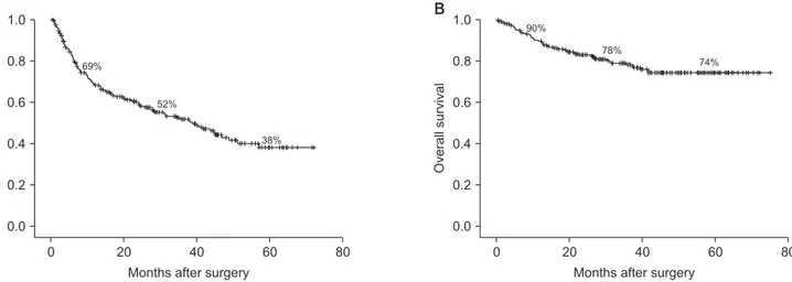
Fig. 1. Disease-free survival (A) and overall survival (B) of all patients at 1, 3, and 5 years were 69%, 52%, 38% and 90%, 78%, 74%, respectively.
Table 2. Multivariate analyses of factors independently associated with disease-free survival and overall survival
Variable Disease-free survival Overall survival
Hazard ratio 95% CI P-value Hazard ratio 95% CI P-value
Sex
Female 0.53 0.23–1.19 0.126
Male
MELD score 0.229
α-FP 0.278 0.004
No. of tumors
Multiple 3.10 1.39–6.93 0.006
Single
Tumor diameter (cm)
>5 1.15 0.67–1.97 0.618 1.83 0.90–3.70 0.094
≤5
Microvascular invasion
Yes 5.92 2.33–15.01 0.000 4.55 1.33–15.57 0.016
No
Serosal invasion
Yes 1.41 0.89–2.25 0.147 1.25 0.66–2.37 0.498
No
Operation method Major resection
Minor resection 0.88 0.58–1.35 0.560 0.92 0.48–1.75 0.800
EBL 0.003
Transfusion
Yes 1.81 1.78–2.78 0.007 1.73 0.92–3.23 0.088
No
TNM stage 0.260 0.500
II 0.43 0.15–1.18 0.50 0.14–1.77
III 0.45 0.12–1.72 0.79 0.25–2.54
CI, confidence interval; MELD, Model for End-Stage Liver Disease; EBL, estimated blood loss.
1–75 months), tumor recurrence occurred in 119 patients (46%), and 53 patients (21%) died. The most common site of tumor recurrence was the remnant liver (n = 110, 92%), lung (n = 16, 13%), bone (n = 7, 6%), peritoneal seeding (n = 4, 3%), and lymph node (n = 3, 2%). DFS and OS of all patients at 1, 3, and 5 years were 69%, 52%, and 38% and 90%, 78%, and 74%, respectively (Fig. 1).
In the univariate analysis for DFS, MELD score, α-FP, number and diameter of tumors, microvascular and serosal invasion, major/minor resection, EBL, and transfusion were significant, whereas in the univariate analysis for OS, α-FP, tumor diameter, microvascular and serosal invasion, major/minor resection trans fusion were significant (P < 0.05). Multivariate analyses showed that microvascular invasion, blood transfusion, EBL, and the number of tumors were independent adverse prog- nostic factors for DFS, whereas microvascular invasion and α-FP were independent adverse prognostic factors for OS (Table 2).
Blood transfusion and tumor diameter had borderline signifi- cance in the multivariate analysis for OS (P = 0.088, P = 0.094, respectively).
Surgeon-correctable factors
We subclassified patients according to tumor diameter of 5 cm and compared the outcomes between groups for sur- geon-correctable factors, such as detailed operation method (major/minor resection, anatomical/nonanatomical, and open/
laparoscopic approach), tumor margin status, EBL and trans fu- sion. Operation method had no effect on DFS or OS in the uni- va riate analysis, regardless of tumor diameter (data not shown).
The extent of the tumor margin did not contribute to survival in either subgroup. In univariate analyses for DFS and OS, EBL and blood transfusion were associated with poor outcomes in pa tients with tumor diameters > 5 cm (Fig. 2), and then we per- formed multivariate analysis for DFS and OS in that group (Table
Disease-freesurvival
A
0 80
Months after surgery 0.0
1.0
0.8
0.6
0.4
0.2
B
60 40
20
Overallsurvival
0 80
Months after surgery 0.0
1.0
0.8
0.6
0.4
0.2
60 40
20 No transfusion (n = 22)
Transfusion (n = 45) P = 0.003
No transfusion (n = 22)
Transfusion (n = 45)
P = 0.02
Fig. 2. Disease-free survival (A) and overall survival (B), according to transfusion in patients with tumor > 5 cm.
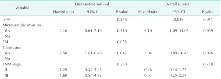
Table 3. Multivariate analyses of factors associated with recurrence and overall survival in patients with tumor >5 cm
Variable Disease-free survival Overall survival
Hazard ratio 95% CI P-value Hazard ratio 95% CI P-value
α-FP 0.278 0.036 0.011
Microvascular invasion
Yes 2.16 0.64–7.19 0.210 6.10 1.09–34.05 0.039
No
EBL 0.058
Transfusion
Yes 2.58 1.03–6.46 0.042 3.04 0.89–10.43 0.076
No
TNM stage 0.550 0.710
II 1.29 0.31–5.45 0.46 0.14–1.77
III 1.68 0.57–4.92 0.63 0.25–2.54
CI, confidence interval; AFP, alpha-fetoprotein; EBL, estimated blood loss.
3). Blood transfusion was only the independent risk factor for DFS and had borderline significance in multivariate analyses for OS.
DISCUSSION
Surgical resection is a curative treatment modality for HCC;
however, the major obstacle to improved survival and prognosis in patients with HCC is the high recurrence rate after surgery.
The life expectancy of patients with HCC is hard to predict, making it difficult to determine the patient’s prognosis. Many factors, such as the patient’s general condition (age, sex, coexist- ing hepatitis, liver function, and α-FP level), tumor status (tumor dia meter, number, capsule formation, vessel invasion, and differ entiation) are proven significant prognostic factors [6].
Opera tion-related factors, such as anatomical/nonanatomical resec tion, open/laparoscopic resection, extent of resection, surgi cal margin, intraoperative bleeding, and blood transfusion, are only modifiable by the surgeon. We attempted to clarify the risk factors for HCC recurrence and patient survival after hepatic resection in terms of surgical factors.
Tumor factors and survival
Microvascular invasion and number of tumors were inde- pendent adverse prognostic factors for DFS, while microvascular invasion and α-FP were independent adverse prognostic factors for OS. Tumor factors have been mostly proven to be independent prognostic factors for the DFS and OS of patients.
It is reasonable to consider that multiple tumors, microvascular invasion, tumor size, and serum α-FP index the aggressiveness of the tumor, consequently affecting surgical results. However, a few studies have reported conflicting results [7,8].
Surgical factors and survival
Notably, anatomic resection did not significantly affect tumor recurrence or the survival of patients in the present study.
Although some authors have reported that anatomic resec tion achieves better DFS and OS than nonanatomic resec tion [9-11], other reports are consistent with our results [12-14]. Authors insisting superiority of anatomic resection hypo thesized that systematic removal of a hepatic segment confined by tumor- bearing portal tributaries effectively eradicates intra hepatic HCC metastases because of the high likelihood of cancer cells from HCC spreading through the portal venous system.
However, spreading through the portal venous system cannot be completely blocked by anatomic resection, as mobilizing the liver for a good surgical view through the laparotomy site may squeeze the tumor and dislodge tumor cells into the portal venous or hepatic venous tributaries.
A resection margin of at least 1 cm is commonly used by many surgeons; however, the role of the resection margin in
con tri buting to the long-term survival of patients remains con- tro versial. Although some authors advocate a definite resection margin > 1 cm and reported that this could definitely prolong OS of patients [15-17], others found no significant effect of the surgical margin on tumor recurrence and survival [11,14,18], consistent with our study. As many patients with HCC have coexisting hepatitis or cirrhosis, hepatic function reserve is frequently suboptimal, which is an obstacle for major and/or anatomic resection. In addition, a cirrhotic liver due to chronic hepatitis has a likelihood of multicentric carcinogenesis. In our study, 46% of all patients experienced recurrence after curative resection and the majority of these recurrences were at multicentric locations away from the resection margin, as previous studies [14,19,20]. A detailed, balanced and deliberate decision is necessary because increasing the tumor-free margin will lead to resecting more nontumorous liver.
Considerable interest has arisen on the effect of blood trans- fusions on HCC recurrence after hepatectomy, with respect to improvement of the postoperative prognosis. A blood trans- fusion may have a deleterious effect on recurrence and the sur vival of patients due to immunosuppression, which is not fully understood, several studies have suggested that blood trans fusions suppress host immunity via a toxic T-cell function, increased numbers of suppressor T cells and decreased function of macrophages and monocytes [21-23]. Several studies have reported that HCC frequently recurs after a perioperative blood transfusion [24-26]. In our study, EBL and blood transfusion were independent factors determining the recurrence of HCC.
In addition, blood transfusion had borderline significance as a prognostic factor for OS. Furthermore, in our subgroup analyses according to a tumor diameter of 5 cm, blood transfusion had an adverse effect on DFS and OS, mainly in patients with tumor diameters > 5 cm. Thus, the primary aim of the surgeon during hepatectomy should be to achieve the least bleeding and blood transfusion, particularly for patients with large tumors through hepatic inflow control (Pringle maneuver). However, if using the Pringle maneuver, there might be ischemia-reperfusion injury. There has been long debated for the potential of liver remnant ischemia-reperfusion injury and its resultant impact on tumor progression [27-30]. Most of studies arguing that ischemia-reperfusion injury may promote progression of HCC were experimental for microenvironmental condition such as disrupting hepatic microvasculature, antiapoptosis induced by proinflammatory cytokine. As a clinical surgeon, it is more reasonable to practice as clinical studies rather than to follow experimental studies on condition that the hypothesis would not be proven.
In conclusion, the surgical technique is as important as preoperative liver function and tumor status in terms of tumor recurrence, which leads to poor patient survival. The ability of the surgeon to minimize bleeding and blood transfusion
improves the outcomes of patients, particularly in cases with a large tumor diameter. After carefully selecting patients through liver function screening, a meticulous surgical technique is highly important for an improved hepatectomy outcome for HCC.
CONFLICTS OF INTEREST
No potential conflict of interest relevant to this article was reported.
REFERENCES
1. El-Serag HB. Hepatocellular carcinoma. N Engl J Med 2011;365:1118-27.
2. European Association For The Study Of The Liver; European Organisation For Research And Treatment Of Cancer.
EASL-EORTC clinical practice guidelines:
management of hepatocellular carcinoma.
J Hepatol 2012;56:908-43.
3. Korean Liver Cancer Study Group (KLCSG);
National Cancer Center, Korea (NCC). 2014 Korean Liver Cancer Study Group-National Cancer Center Korea Practice Guideline for the Management of Hepatocellular Carcinoma. Korean J Radiol 2015;16:465- 522.
4. Fan ST, Mau Lo C, Poon RT, Yeung C, Leung Liu C, Yuen WK, et al. Continuous improvement of survival outcomes of resection of hepatocellular carcinoma: a 20-year experience. Ann Surg 2011;253:
745-58.
5. Han DH, Choi GH, Park JY, Ahn SH, Kim KS, Choi JS, et al. Lesson from 610 liver resections of hepatocellular carcinoma in a single center over 10 years. World J Surg Oncol 2014;12:192.
6. Qin LX, Tang ZY. The prognostic signifi- cance of clinical and pathological features in hepatocellular carcinoma. World J Gastroenterol 2002;8:193-9.
7. Fan ST, Ng IO, Poon RT, Lo CM, Liu CL, Wong J. Hepatectomy for hepatocellular carcinoma: the surgeon's role in long-term survival. Arch Surg 1999;134:1124-30.
8. Sim HG, Ooi LL. Results of resections for hepatocellular carcinoma in a new hepatobiliary unit. ANZ J Surg 2003;73:8- 13.
9. Hasegawa K, Kokudo N, Imamura H, Matsuyama Y, Aoki T, Minagawa M, et al.
Prognostic impact of anatomic resection for hepatocellular carcinoma. Ann Surg 2005;242:252-9.
10. Hwang S, Lee YJ, Kim KH, Ahn CS, Moon DB, Ha TY, et al. The impact of tumor size on long-term survival outcomes after resection of solitary hepatocellular car ci- noma: single-institution experience with 2558 patients. J Gastrointest Surg 2015;
19:1281-90.
11. Imamura H, Matsuyama Y, Tanaka E, Ohkubo T, Hasegawa K, Miyagawa S, et al. Risk factors contributing to early and late phase intrahepatic recurrence of hepatocellular carcinoma after hepa tec- tomy. J Hepatol 2003;38:200-7.
12. Park JH, Koh KC, Choi MS, Lee JH, Yoo BC, Paik SW, et al. Analysis of risk factors asso ciated with early multinodular recur- rences after hepatic resection for hepa to- cellular carcinoma. Am J Surg 2006;192:
29-33.
13. Tanaka K, Shimada H, Matsumoto C, Matsuo K, Nagano Y, Endo I, et al. Ana to- mic versus limited nonanatomic resection for solitary hepatocellular carcinoma.
Surgery 2008;143:607-15.
14. Zhang XF, Meng B, Qi X, Yu L, Liu C, Liu XM, et al. Prognostic factors after liver re- sec tion for hepatocellular carcinoma with hepatitis B virus-related cirrhosis: sur- geon’s role in survival. Eur J Surg Oncol 2009;35:622-8.
15. Predictive factors for long term prognosis after partial hepatectomy for patients with hepatocellular carcinoma in Japan.
The Liver Cancer Study Group of Japan.
Cancer 1994;74:2772-80.
16. Ikai I, Arii S, Kojiro M, Ichida T, Makuuchi M, Matsuyama Y, et al. Reevaluation of
prognostic factors for survival after liver resection in patients with hepatocellular car cinoma in a Japanese nationwide sur- vey. Cancer 2004;101:796-802.
17. Lise M, Bacchetti S, Da Pian P, Nitti D, Pilati PL, Pigato P. Prognostic factors affecting long term outcome after liver resec tion for hepatocellular carcinoma:
results in a series of 100 Italian patients.
Cancer 1998;82:1028-36.
18. Poon RT, Fan ST, Ng IO, Wong J. Signifi- cance of resection margin in hepatectomy for hepatocellular carcinoma: a critical reappraisal. Ann Surg 2000;231:544-51.
19. Adachi E, Maeda T, Matsumata T, Shirabe K, Kinukawa N, Sugimachi K, et al. Risk fac tors for intrahepatic recurrence in hu man small hepatocellular carcinoma.
Gastroenterology 1995;108:768-75.
20. Belghiti J, Panis Y, Farges O, Benhamou JP, Fekete F. Intrahepatic recurrence after resec tion of hepatocellular carcinoma com plicating cirrhosis. Ann Surg 1991;
214:114-7.
21. Blumberg N, Heal JM. Effects of trans fu- sion on immune function. Cancer re cur- rence and infection. Arch Pathol Lab Med 1994;118:371-9.
22. Gascon P, Zoumbos NC, Young NS. Immu- no logic abnormalities in patients recei- v ing multiple blood transfusions. Ann Intern Med 1984;100:173-7.
23. Kaplan J, Sarnaik S, Gitlin J, Lusher J.
Diminished helper/suppressor lympho- cyte ratios and natural killer activity in recipients of repeated blood transfusions.
Blood 1984;64:308-10.
24. Asahara T, Katayama K, Itamoto T, Yano M, Hino H, Okamoto Y, et al. Perioperative blood transfusion as a prognostic indi ca-
tor in patients with hepatocellular car ci- noma. World J Surg 1999;23:676-80.
25. Hanazaki K, Kajikawa S, Shimozawa N, Matsushita A, Machida T, Shimada K, et al. Perioperative blood transfusion and survival following curative hepatic resec tion for hepatocellular carcinoma.
Hepatogastroenterology 2005;52:524-9.
26. Tung-Ping Poon R, Fan ST, Wong J. Risk fac tors, prevention, and management of post operative recurrence after resection of hepatocellular carcinoma. Ann Surg
2000;232:10-24.
27. Arii S, Teramoto K, Kawamura T. Current progress in the understanding of and thera peutic strategies for ischemia and reperfusion injury of the liver. J Hepa- tobiliary Pancreat Surg 2003;10:189-94.
28. Man K, Ng KT, Lo CM, Ho JW, Sun BS, Sun CK, et al. Ischemia-reperfusion of small liver remnant promotes liver tumor growth and metastases: activation of cell invasion and migration pathways. Liver Transpl 2007;13:1669-77.
29. Ozaki M, Todo S. Surgical stress and tumor behavior: impact of ischemia-reper- fusion and hepatic resection on tumor progression. Liver Transpl 2007;13:1623-6.
30. Shimoda M, Iwasaki Y, Sawada T, Kubota K. Protective effect of ischemic pre con- di tion ing against liver injury after major hepa tec tomy using the intermittent prin gle maneuver in swine. Pathobiology 2007;74:42-9.