CASE REPORT
J Korean Surg Soc 2012;82:185-189
http://dx.doi.org/10.4174/jkss.2012.82.3.185
JKSS
Journal of the Korean Surgical Society pISSN 2233-7903ㆍeISSN 2093-0488
Received May 24, 2011, Revised June 11, 2011, Accepted July 6, 2011 Correspondence to: Myung-Chul Chang
Department of Surgery, Dankook University Hospital, Dankook University College of Medicine, 201 Manghyang-ro, Dongnam-gu, Cheonan 330-715, Korea
Tel: +82-41-550-3930, Fax: +82-41-556-3878, E-mail: [email protected]
cc Journal of the Korean Surgical Society is an Open Access Journal. All articles are distributed under the terms of the Creative Commons Attribution Non-Commercial License (http://creativecommons.org/licenses/by-nc/3.0/) which permits unrestricted non-commercial use, distribution, and reproduction in any medium, provided the original work is properly cited.
Bilateral adrenal pheochromocytoma with a germline L790F mutation in the RET oncogene
Jun Won Min, Youn Joon Park, Hee Jin Kim
1, Myung-Chul Chang
Departments of Surgery and 1Internal Medicine, Dankook University College of Medicine, Cheonan, Korea
About ten percent of pheochromocytomas are associated with familial syndrome. Hereditary pheochromocytoma has char- acteristics of early onset, multifocality and bilaterality. We experienced a case of 44-year-old man with bilateral pheochromo- cytoma without evidence of medullary thyroid cancer. Genetic test detected a L790F germline mutation of RET oncogene.
The author found a necessity for genetic tests in cases of young-age, bilateral pheochromocytoma.
Key Words: Pheochromocytoma, RET, Germ-line mutation
INTRODUCTION
Pheochromocytoma was well known as “10 percent tu- mor”. It is widely believed that approximately 10% of pheochromocytomas are associated with familial syn- dromes. However, it is now recognized that the frequency of germline mutations in apparently sporadic pheochro- mocytoma is as high as 24% [1]. Hereditary pheochromo- cytoma could be a phenotype of multiple endocrine neo- plasia (MEN) type 2, Von Hipple-Lindau disease, familial pheochromocytoma-paraganglioma and neurofibroma- tosis type 1. The causal genes were discovered as RET, VHL, SDHB, SDHD and NF1 respectively. Each genetic syndromes share common characteristics such as early on- set, multifocality and bilaterality. So in case of young- aged, bilateral pheochromocytoma, searching for genetic mutation is recommended [2]. We report a case of a
44-year-old man with bilateral pheochromocytomas whose genetic study shows a rare germline L790F muta- tion of RET oncogene.
CASE REPORT
A 44-year-old man was admitted for incidentally found bilateral bulky adrenal mass lesions. He daily drank one or two bottles of Soju (Korean distilled spirits). A computed tomography (CT) scan was taken for evaluation of alco- holic liver disease. He felt paroxysmal attack of palpitation and sweating since last year, but had no medication. His systolic blood pressure was 110 mmHg and diastolic blood pressure was 65 mmHg. His heart rate was 70 per minute. On the physical examination, no mass was pal- pable in abdomen and neck lesion.
Fig. 1. Abdominal computed tomography scan shows huge mass in both adrenal glands.
Fig. 2. Single photon emission computed tomography image of I-123 metaiodobenzylguanidine scan shows bilateral adrenal uptake.
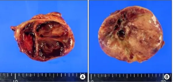
Fig. 3. (A) Cut surface of right adrenal gland shows well‐demarcated, multilocular cystic mass.
It contains bloody, dark red brown coloredfluid.
(B) Cut surface of left adrenal gland shows soft, variegated, red- dish pink mass with a few small cysts.
Fig. 4. Genetic testing detected mutation in codon 790 (L790F) of RET oncogene.
In the hormonal studies, 24 hours urinary meta- nephrine and vanillylmandelic acid was elevated as 6.12 mg/day (normal value, less than 1.3 mg/day) and 23.38 mg/day (normal value, less than 8 mg/day). Twenty-four hours urinary cortisol was normal as 45.9 ug/day (normal value, 20 to 90 ug/day). Plasma rennin activity and aldos- terone level was also normal as 0.44 ng/mL/hr (normal val- ue, 0.15 to 2.33 ng/mL/hr) and 65.0 pg/mL (normal value, 35.7 to 240.0 pg/mL).
On the CT scan, right adrenal mass was 6 cm sized, well defined and mostly necrotic. The left adrenal mass was 8 cm, hypervascular and centrally necrotic (Fig. 1). I-123 metaiodobenzylguanidine scan showed bilateral adrenal uptake with no evidence of metastasis (Fig. 2).
The patient was diagnosed as bilateral pheochromo- cytomas. After prescribing alpha-blocker terazosin for twelve days, bilateral adrenalectomy was done under gen- eral anesthesia. On the operation, both adrenal masses
Fig. 5. Family pedigree shows no evidence of medullary thyroid cancer or endocrine disease. All of his family members are alive.
Arrow indicates index patient.
were well marginated without invasion and lymph node metastasis (Fig. 3). The pathological diagnosis was also pheochromocytoma. He was recovered uneventfully, dis- charged with gluco-corticoid and mineralo-corticoid replacement.
We tested germline mutation of RET oncogene with pa- tient’s informed consent. Germline mutation was ana- lyzed by direct sequencing of exons 10, 11, 13, 14, 15, 16 of RET oncogene. At the codon 790 of exon 13, missense mu- tation of L790F was found. It was a point mutation of DNA change from TTG to TTT, resulted amino acid change from Leucine to Phenylalanine (Fig. 4).
After the result of germline mutation of RET oncogene, we diagnosed the patient as multiple endocrine neoplasia type 2A, and searched an evidence of medullary thyroid cancer in the patient and his family members. In his family members, there was no history of thyroid cancer or endo- crine diseases (Fig. 5). We checked an ultrasonography of patient’s thyroid, but there was no evidence of thyroid nodule. The patient’s calcitonin level was 3.7 pg/mL (normal value, less than 20 pg/mL). We checked the stimu- lated calcitonin level, but the result was also normal range.
We explained the results and recommended prophylactic total thyroidectomy and genetic test about his parents, but he rejected. So we made a plan to check the calcitonin and thyroid ultrasonography regularly.
DISCUSSION
About 10 percent of pheochromocytomas were as- sumed to be hereditary. However, recent advances of ge- netic studies show much higher germline mutation rates than that of previous reports. According to the European Network for the Study of Adrenal Tumors Pheochromocy- toma Working Group [2], germline mutation rate was 25.9% from 642 pheochromocytoma and paraganglioma patients. In the other study [1], 24.3% of the 271 sporadic pheochromocytoma patients had germline mutations. It is now estimated that about 20 to 30% of pheochromocyto- mas have hereditary tendency.
Hereditary pheochromocytoma is a phenotype of five genes; VHL, RET, SDHB, SDHD and NF1. From the reports of the European Network [2], each mutation rates were 8.7% in VHL gene, 5.3% in SDHB, 4.8% in SDHD, 4.8% in RET and 3.7% in NF1 respectively. Other European study [1] shows similar results; 11.1% in VHL, 4.4% in SDHB, 4.1% in SDHD, 4.8% in RET.
Each genetic syndrome has characteristic phenotypes. It is possible to anticipate genotype by characteristic pheno- type, so identification of clinical characteristics is very important. Genetic testing for NF1 gene is not routinely carried out. Diagnosis of neurofibromatosis type 1 is pos- sible based on the typical clinical features; café-au-lait spots, neurofibromas and axillary and inguinal freckling.
On the contrary, genetic test of NF1 gene is difficult and costly due to large sized NF1 gene, composed of 57 exons without hot spots. In this case we can exclude NF1 muta- tion based on the clinical features.
Von Hippel-Lindau disease is characterized by he- mangioblastomas, renal tumors, pancreatic and endolym- phatic sac tumors. Pheochromocytomas occur in about 26% of von Hippel-Lindau patients [3]. Associated pheo- chromocytoma is typically lack of phenylethanolamine- N-methyltransferase which converts noradrenaline to adrenaline, resulted in high normetanephrine and normal metanephrine [4]. In this case, we can rule out VHL muta- tion due to high level of metanephrine.
SDHB and SDHD gene are recently identified as para- ganglioma syndrome type 4 and type 1. These genes are subunits of the mitochondrial complex II, which is in-
volved in the Krebs cycle as succinate dehydrogenase.
Pheochromocytoma with SDHB and SDHD germline mu- tation is frequently malignant, extra-adrenal or bilateral.
So in that phenotype, genetic test about SDHB and SDHD gene are necessary [2].
Germline mutation of RET oncogene is associated with MEN type 2 which has typical character of genotype-phe- notype correlation and mutation hot spots. Medullary thyroid cancer is the most frequent and malignant tumor in MEN type 2. MEN type 2 was classified according to the aggressiveness and onset of medullary thyroid cancer. The penetrance of medullary thyroid cancer is different ac- cording to the mutation site. Patients with level 1 muta- tions (codons 609, 768, 790, 791, 804 and 891) have the low- est risk for medullary thyroid cancer, patients with level 2 mutations (codons 611, 618, 620 and 634) are intermediate risk, and patient with level 3 mutations (codons 883 and 918) have the highest risk for medullary thyroid cancer [5].
Medullary thyroid cancer was developed in 100% of level 3, 73% of level 2 and only 45% of level 1 RET gene mutation [6].
In this case, L790F mutation was found in RET onco- gene, which was not reported before in Korea. 1998, Berndt et al. [7] first described a new hot spot for muta- tions affecting codon 790 of RET oncogene. They reported that nine (69%) of 13 carriers with L790F mutation had de- veloped medullary thyroid cancer. Initially, L790F muta- tion was reported to be associated with pheochromocyto- ma, but in the following study [8], L790F mutation rarely associated with pheochromocytoma.
Interestingly, this case shows bilateral pheochromocy- tomas which was rare phenotype of L790F and does not show medullary thyroid cancer which was common phe- notype of L790F. Machens et al. [6] reported that mean di- agnostic age of pheochromocytoma was 46.5 years and medullary thyroid cancer was 51.6 years in level 1 RET mutation. We can expect patient’s occurrence of medullary thyroid cancer in the near future.
Like as risk classification of medullary thyroid cancer, risk of pheochromocytoma also can be classified. The highest-risk category of pheochromocytoma includes SDHB, SDHD and the level 3 risk of RET; the high-risk cat- egory includes VHL missense mutation and the level 2 risk
of RET; the least-high risk category includes VHL truncat- ing mutations and level 1 risk of RET [9].
The incidence of germline mutation of multifocal or bi- lateral pheochromocytomas was much higher than that of unilateral pheochromocytoma. In the 26 patients of multi- focal or bilateral pheochromocytomas, 80% of them had a mutation (46% in VHL, 19% in RET, 15% in SDHD, none in SDHB). In the other study [10], 12 RET, 1 VHL, 1 SDHD gene mutations were found in the 23 bilateral pheochro- mocytomas. They recommended sequential mutational analysis of RET, followed by VHL and SDHD in bilateral pheochromocytoma.
This case is the first report of L790F RET germline muta- tion in Korea. In case of bilateral pheochromocytoma, germline mutation test for hereditary pheochromocytoma is necessary.
CONFLICTS OF INTEREST
No potential conflict of interest relevant to this article was reported.
ACKNOWLEDGEMENTS
The present research was conducted by the research fund of Dankook University in 2010.
REFERENCES
1. Neumann HP, Bausch B, McWhinney SR, Bender BU, Gimm O, Franke G, et al. Germ-line mutations in non- syndromic pheochromocytoma. N Engl J Med 2002;346:
1459-66.
2. Gimenez-Roqueplo AP, Lehnert H, Mannelli M, Neumann H, Opocher G, Maher ER, et al. Phaeochromocytoma, new genes and screening strategies. Clin Endocrinol (Oxf) 2006;65:699-705.
3. Walther MM, Reiter R, Keiser HR, Choyke PL, Venzon D, Hurley K, et al. Clinical and genetic characterization of pheochromocytoma in von Hippel-Lindau families: com- parison with sporadic pheochromocytoma gives insight into natural history of pheochromocytoma. J Urol 1999;
162(3 Pt 1):659-64.
4. Eisenhofer G, Lenders JW, Linehan WM, Walther MM,
Goldstein DS, Keiser HR. Plasma normetanephrine and metanephrine for detecting pheochromocytoma in von Hippel-Lindau disease and multiple endocrine neoplasia type 2. N Engl J Med 1999;340:1872-9.
5. Brandi ML, Gagel RF, Angeli A, Bilezikian JP, Beck-Peccoz P, Bordi C, et al. Guidelines for diagnosis and therapy of MEN type 1 and type 2. J Clin Endocrinol Metab 2001;86:
5658-71.
6. Machens A, Brauckhoff M, Holzhausen HJ, Thanh PN, Lehnert H, Dralle H. Codon-specific development of pheo- chromocytoma in multiple endocrine neoplasia type 2. J Clin Endocrinol Metab 2005;90:3999-4003.
7. Berndt I, Reuter M, Saller B, Frank-Raue K, Groth P, Grussendorf M, et al. A new hot spot for mutations in the ret protooncogene causing familial medullary thyroid car-
cinoma and multiple endocrine neoplasia type 2A. J Clin Endocrinol Metab 1998;83:770-4.
8. Gimm O, Niederle BE, Weber T, Bockhorn M, Ukkat J, Brauckhoff M, et al. RET proto-oncogene mutations affect- ing codon 790/791: A mild form of multiple endocrine neo- plasia type 2A syndrome? Surgery 2002;132:952-9.
9. Machens A, Brauckhoff M, Gimm O, Dralle H. Risk-ori- ented approach to hereditary adrenal pheochromocytoma.
Ann N Y Acad Sci 2006;1073:417-28.
10. Korpershoek E, Petri BJ, van Nederveen FH, Dinjens WN, Verhofstad AA, de Herder WW, et al. Candidate gene mu- tation analysis in bilateral adrenal pheochromocytoma and sympathetic paraganglioma. Endocr Relat Cancer 2007;14:
453-62.