Background and PurposezzThe benefit of carotid endarterectomy (CEA) is directly influ- enced by the risk of perioperative adverse outcomes. However, patient-level risks and predictors including coronary stenosis are rarely evaluated, especially in Asian patients. The aim of this study was to determine the relationship between the vascular risk factors underlying CEA, in- cluding coronary stenosis, and postoperative outcome.
MethodszzOne hundred and fifty-three consecutive CEAs from our hospital records were in- cluded in this analysis. All patients underwent coronary computed tomography angiography before CEA. Data were analyzed to determine the vascular outcomes in patients with mild-to- moderate vs. severe coronary stenosis and high vs. standard operative risk, based on the criteria for high operative risk defined in the Stenting and Angioplasty with Protection in Patients at High Risk for Endarterectomy (SAPPHIRE) trial. The vascular outcome was defined as the oc- currence of postoperative (≤30 days) stroke, myocardial infarction (MI), or death.
ResultszzAn adverse vascular outcome occurred in 8 of the 153 CEAs, with 6 strokes, 2 MIs, and 3 deaths. The vascular outcome differed significantly between the groups with mild-to- moderate and severe coronary stenosis (p=0.024), but not between the high- and standard-op- erative-risk groups (stratified according to operative risk as defined in the SAPPHIRE trial).
Multivariable analysis adjusting for potent predictors revealed that severe coronary stenosis (odds ratio, 6.87; 95% confidence interval, 1.20–39.22) was a significant predictor of the early vascular outcome.
ConclusionszzSevere coronary stenosis was identified herein as an independent predictor of an adverse early vascular outcome.
Key Wordszz carotid endarterectomy, coronary artery disease, coronary angiography, risk assessment.
Preoperative Coronary Stenosis Is a Determinant of Early Vascular Outcome after Carotid Endarterectomy
INTRODUCTION
Atherosclerosis is common in the intracranial artery than the extracranial artery in Asian patients; as a result, carotid endarterectomy (CEA) has not frequently been performed in Korea. However, according to a recent nationwide, hospital-based stroke registry study in Korea, it appears that the prevalence of intracranial cerebral artery disease has been declin- ing, while that of extracranial disease has been increasing.1,2
Prospective randomized studies have demonstrated that CEA reduces the incidence of stroke in symptomatic patients with ipsilateral carotid artery stenosis of ≥50%, and in as- ymptomatic patients with internal carotid stenosis of ≥70%.3,4 However, there is a trend to- ward minimizing intervention for asymptomatic carotid stenosis in favor of medical treat- ment because of the significant advances in such therapies for vascular disease.5,6 The ultimate goal of the treatment of carotid artery disease is prolongation of healthy life; there- Jung Hwa Kima,b
Sung Hyuk Heoa Hyo Jung Nama Hyo Chul Younc Eui-Jong Kimd Ji Sung Leee Young Seo Kimf Hyun Young Kimf Seong-Ho Kohf Dae-Il Changa
aDepartments of Neurology,
cThoracic and Cardiovascular Surgery, and dRadiology, College of Medicine, Kyung Hee University, Seoul, Korea
b Department of Neurology, Seoul Bukbu Hospital, Seoul, Korea
e Clinical Research Center, Asan Medical Center, Seoul, Korea
f Department of Neurology,
College of Medicine, Hanyang University, Seoul, Korea
pISSN 1738-6586 / eISSN 2005-5013 / J Clin Neurol 2015;11(4):364-371 / http://dx.doi.org/10.3988/jcn.2015.11.4.364
Received February 9, 2015 Revised May 7, 2015 Accepted May 8, 2015 Correspondence Sung Hyuk Heo, MD, PhD Department of Neurology, College of Medicine, Kyung Hee University,
23 Kyungheedae-ro, Dongdaemun-gu, Seoul 02447, Korea
Tel +82-2-958-8499 Fax +82-2-958-8490 E-mail [email protected]
cc This is an Open Access article distributed under the terms of the Creative Commons Attribution Non-Com- mercial License (http://creativecommons.org/licenses/by-nc/3.0) which permits unrestricted non-commercial use, distribution, and reproduction in any medium, provided the original work is properly cited.
JCN
Open Access ORIGINAL ARTICLEKim JH et al.
JCN
fore, caution is necessary when selecting an invasive treatment approach, and perioperative complications should be prevent- ed wherever possible for interventions such as CEA. Decision- making regarding CEA is dependent upon whether the post- operative risk of stroke, myocardial infarction (MI), or death exceeds the risk of these outcomes under medical manage- ment. Defining the relationship between various risk factors and outcomes after CEA is important for appropriate patient selection for this procedure. Several studies have shown that certain risk factors are associated with adverse postoperative vascular outcomes such as stroke, MI, or death after CEA.7-9
A link between carotid and coronary arterial diseases has been proposed. Atherosclerosis of the carotid arteries often occurs concomitantly with coronary stenosis, and both dis- eases are believed to be associated with similar risk factors and pathological mechanisms. Atherosclerotic stroke occurs as a result rupture of atherosclerotic plaques at the site of arterial occlusion or in the proximal relevant arteries, and may be di- rectly relevant to atherosclerotic diseases in other organs.10 Furthermore, coronary artery disease (CAD) is a very im- portant cause of death during the perioperative and follow- up periods after CEA.11-14 However, the relationship between preoperative coronary stenosis and postoperative vascular outcome in patients treated by CEA is not well understood.
The aim of this study was to determine whether preopera- tive coronary stenosis can influence early outcomes after CEA, and to identify the factors that may affect major adverse events (MAEs) following that procedure.
METHODS
Subjects
Routine preoperative coronary evaluation by coronary com- puted tomography angiography (CCTA) was adopted at our hospital in July 2007. Patients who underwent CEA after that time were included in this study. One hundred and seventy- three consecutive CEAs were performed by a single experi- enced surgeon at Kyung Hee University Hospital between July 2007 and December 2013. Twenty procedures were ex- cluded because of a lack of CCTA data; further evaluation was not needed in these patients because their electrocardiogra- phy and echocardiography findings were normal and they had no cardiac symptoms. This study ultimately included 141 patients who underwent a total of 153 CEA procedures (12 patients underwent bilateral CEA for separate ipsilateral symp- tomatic events or severe stenosis). Patients with neurological symptoms referable to the ipsilateral carotid territory within 6 months before surgery were classified as symptomatic. Sur- gery was indicated for symptomatic patients with a carotid ste- nosis of ≥50% or severe ulceration of the carotid artery, and
for asymptomatic patients with a carotid stenosis of ≥70%.
All surgeries were performed under general anesthesia, and simultaneous electroencephalography was performed and monitored during the procedure to determine the status of intraluminal shunting.
This study was approved by an independent ethics com- mittee at Kyung Hee University Medical Center (KMC IRB 1414-02).
Risk factors and outcome measurement
Data on patient demographics, surgical indications, operative details, and postoperative outcomes were obtained from hos- pital records. Postoperative MAEs included any episodes of stroke, MI, or death within 30 days after the operation. Upon appearance of new neurological symptoms or signs, strokes were identified and confirmed by formal neurological exami- nation by neurologists, and by brain imaging. The association between these outcomes and various demographic and pre- operative factors that could influence their likelihood were evaluated. These included age, sex, hypertension, dyslipid- emia, diabetes mellitus, other cerebral arterial lesions (occlu- sion or ≥50% stenosis), smoking, and atrial fibrillation.
Definitions of high and standard coronary and operative risks
CCTA was performed prior to CEA and the results used to determine coronary risk; this was in turn used to predict post- operative outcome. The degree of coronary stenosis, as as- sessed by CCTA, was determined using the scoring system described by Cury et al.,15 which defines three categories of stenosis: mild (0–40%), moderate (41–70%), and severe (71–
100%). Patients were examined using a 64-channel multide- tector computed tomography (MDCT) system (Brilliance 64, Philips Medical Systems, Best, The Netherlands). All quanti- tative measurements were made in a semiautomatic maneu- ver using the FD10 software provided with Allura 9 (Philips, Eindhoven, The Netherlands) in orthogonal projections at end-diastole, when this was possible. In addition, the severity of coronary stenosis was determined by coronary angiogra- phy (CAG) and graded by an experienced angiographer. The degree of stenosis of each of the three major blood vessels (left anterior descending artery, left circumflex artery, and right coronary artery) was graded by both CCTA and CAG. The presence of severe stenosis in at least one of the three vessels resulted in classification into the high-coronary-risk (HCR) group. If the results of the two tests were inconsistent, those obtained using CAG were prioritized; for example, a subject with severe stenosis based on CCTA and with mild-to-mod- erate stenosis based on CAG was categorized into the stan- dard-coronary-risk (SCR) group.
Coronary Stenosis and Early Outcome of CEA
JCN
The high-operative-risk (HOR) and standard-operative- risk (SOR) groups were also defined using the following crite- ria for high risk in the Stenting and Angioplasty with Protec- tion in Patients at High Risk for Endarterectomy (SAPPHIRE) trial: significant cardiac disease (congestive heart failure, ab- normal stress test, or need for open-heart surgery), severe pulmonary disease, contralateral carotid occlusion, contralat- eral laryngeal nerve palsy, previous radical neck surgery/radi- ation, tracheostomy, recurrent carotid stenosis, or age greater than 80 years.16 The SOR group included all procedures not included in the HOR group.
Statistical analysis
The data are presented as mean±SD or median (interquartile range) values (for continuous variables), or as the number (%) of subjects (for categorical variables). Categorical variables were compared using Fisher’s exact test, and continuous vari- ables were compared using Student’s t-test and Wilcoxon’s rank-sum test. Preoperative variables positively associated with postoperative outcome at p<0.2 were included in a mul- tivariable analysis using logistic regression with Firth’s bias correction estimation method. All statistical analyses were performed using SAS version 9.3 (SAS Institute, Cary, NC,
USA). The threshold for statistical significance was set at p<
0.05 (two-sided).
RESULTS
Patient characteristics
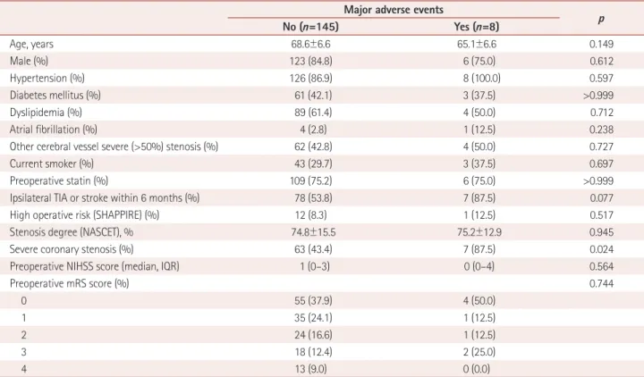
One hundred and fifty-three CEA procedures were per- formed in 141 patients. The baseline characteristics of those patients are given in Table 1. Only the preexistence of severe coronary stenosis significantly affected the postoperative out- come. Preoperative CCTA was performed before CEA in all patients, and CAG was performed in 68 CEAs (44.4%). CAG was performed in 39 of the 57 subjects with severe coronary stenosis based on CCTA. Of those, 35 subjects (89.7%) had severe coronary stenosis based on CAG, and were treated with a percutaneous coronary intervention (PCI). In 96 sub- jects without severe coronary stenosis based on CCTA, 29 subjects underwent CAG in response to a history of chest pain or a positive stress test. Of those, 17 subjects (58.6%) were observed to have coronary stenosis based on CAG. Of the 52 procedures in subjects with severe coronary stenosis based on CAG, preoperative PCI was performed in 44 pro- cedures. Eight subjects did not undergo PCI because of long-
Table 1. Baseline characteristics of all subjects undergoing carotid endarterectomy
Major adverse events
No (n=145) Yes (n=8) p
Age, years 68.6±6.6 65.1±6.6 0.149
Male (%) 123 (84.8) 6 (75.0) 0.612
Hypertension (%) 126 (86.9) 8 (100.0) 0.597
Diabetes mellitus (%) 61 (42.1) 3 (37.5) >0.999
Dyslipidemia (%) 89 (61.4) 4 (50.0) 0.712
Atrial fibrillation (%) 4 (2.8) 1 (12.5) 0.238
Other cerebral vessel severe (>50%) stenosis (%) 62 (42.8) 4 (50.0) 0.727
Current smoker (%) 43 (29.7) 3 (37.5) 0.697
Preoperative statin (%) 109 (75.2) 6 (75.0) >0.999
Ipsilateral TIA or stroke within 6 months (%) 78 (53.8) 7 (87.5) 0.077
High operative risk (SHAPPIRE) (%) 12 (8.3) 1 (12.5) 0.517
Stenosis degree (NASCET), % 74.8±15.5 75.2±12.9 0.945
Severe coronary stenosis (%) 63 (43.4) 7 (87.5) 0.024
Preoperative NIHSS score (median, IQR) 1 (0–3) 0 (0–4) 0.564
Preoperative mRS score (%) 0.744
0 55 (37.9) 4 (50.0)
1 35 (24.1) 1 (12.5)
2 24 (16.6) 1 (12.5)
3 18 (12.4) 2 (25.0)
4 13 (9.0) 0 (0.0)
Except where indicated otherwise, the data are mean±SD, numbers of patients (%), or median (IQR) values. p values were calculated using Student’s t-test, Fisher’s exact test, or Wilcoxon’s rank-sum test, as appropriate.
IQR: interquartile range, mRS: modified Rankin Scale, NASCET: North American Symptomatic Carotid Endarterectomy Trial, NIHSS: National Institutes of Health Stroke Scale, SHAPPIRE: Stenting and Angioplasty with Protection in Patients at High Risk for Endarterectomy, TIA: transient ischemic attack.
Kim JH et al.
JCN
segment stenosis, or other specific reasons, such as patient refusal. The patterns of the coronary lesions revealed by pre- operative coronary workups are summarized in Fig. 1.
Early outcomes
Postoperative (≤30 days) MAEs occurred in eight CEA pro- cedures (5.2%): six strokes (3.9%), two MIs (1.3%), and three
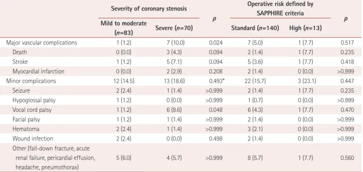
deaths (2.0%; caused by one major stroke and two MIs). The early outcome differed significantly between the HCR and SCR groups (p=0.024). There were no statistically significant differences between the HOR and SOR groups, as defined in the SAPPHIRE trial, with respect to stroke (p=0.418), death (p=0.235), MI (p>0.999), or all MAEs (p=0.517). Vocal cord palsy was the only minor complication that differed signifi-
Fig. 1. Preoperative coronary workups for all procedures (n=153). CAG: coronary angiography, CCTA: coronary computed tomography angiogra- phy, CEA: carotid endarterectomy, PCI: percutaneous coronary intervention.
All CEA procedures from Jul. 2007 to Dec. 2013 (n=173)
CEA procedures without CCTA data (n=20)
Mild to moderate coronary stenosis (n=83) 1. Mild to moderate in CCTA but no CAG data (n=67) 2. Mild to moderate in both CCTA and CAG (n=12) 3. Mild to moderate in CAG but severe in CCTA (n=4)
Treatment of coronary stenosis 1. Preoperative PCI (n=44) 2. No preoperative PCI (n=26) Severe coronary stenosis (n=70) 1. Severe in both CCTA and CAG (n=35) 2. Severe in CCTA but no CAG data (n=18) 3. Severe in CAG but mild to moderate in CCTA (n=17)
Total CEA procedures for analysis (n=153)
Table 2. Early outcome (≤30 days) stratified according to severity of coronary stenosis Severity of coronary stenosis
p
Operative risk defined by SAPPHIRE criteria Mild to moderate p
(n=83) Severe (n=70) Standard (n=140) High (n=13)
Major vascular complications 1 (1.2) 7 (10.0) 0.024 7 (5.0) 1 (7.7) 0.517
Death 0 (0.0) 3 (4.3) 0.094 2 (1.4) 1 (7.7) 0.235
Stroke 1 (1.2) 5 (7.1) 0.094 5 (3.6) 1 (7.7) 0.418
Myocardial infarction 0 (0.0) 2 (2.9) 0.208 2 (1.4) 0 (0.0) >0.999
Minor complications 12 (14.5) 13 (18.6) 0.493* 22 (15.7) 3 (23.1) 0.447
Seizure 2 (2.4) 1 (1.4) >0.999 2 (1.4) 1 (7.7) 0.235
Hypoglossal palsy 1 (1.2) 0 (0.0) >0.999 1 (0.7) 0 (0.0) >0.999
Vocal cord palsy 1 (1.2) 6 (8.6) 0.048 6 (4.3) 1 (7.7) 0.470
Facial palsy 1 (1.2) 1 (1.4) >0.999 2 (1.4) 0 (0.0) >0.999
Hematoma 2 (2.4) 1 (1.4) >0.999 3 (2.1) 0 (0.0) >0.999
Wound infection 2 (2.4) 0 (0.0) 0.498 2 (1.4) 0 (0.0) >0.999
Other (fall-down fracture, acute renal failure, pericardial effusion, headache, pneumothorax)
5 (6.0) 4 (5.7) >0.999 8 (5.7) 1 (7.7) 0.560
Data are n (%) values.
*p was calculated using Fisher’s exact test and chi-square test.
SHAPPIRE: Stenting and Angioplasty with Protection in Patients at High Risk for Endarterectomy.
Coronary Stenosis and Early Outcome of CEA
JCN
cantly between the HCR and SCR groups (Table 2). No sig- nificant differences were found in early outcome between vascular territories of coronary stenosis (Table 3). Four MAEs (15.4%) occurred in 26 procedures in patients with severe coronary stenosis who received PCI prior to CEA, and 3 MAEs (6.8%) occurred in 44 patients with severe coronary stenosis who did not receive PCI. However, the difference was not statistically significant because both the sample size and number of events were small (p=0.411).
Candidate variables for multivariable analysis using logistic regression were identified, including age, atrial fibrillation, neurological symptoms (transient ischemic attack or stroke) referable to the ipsilateral carotid territory within 6 months, and the existence of severe coronary stenosis. The time inter- val between CEA and the manifestation of neurological symp- toms did not differ significantly between the groups (Table 4).
The HOR group (based on the SAPPHIRE definition) did not exhibit a significantly high rate of MAEs. The independent predictors of a poor outcome after multivariable regression analysis are given in Table 5. Severe coronary stenosis was asso- ciated with a sevenfold higher risk of occurrence of postop-
erative MAEs (p=0.030).
DISCUSSION
In this study, postoperative (≤30 days) MAEs occurred fol- lowing 8 of 153 CEA procedures (5.2%). The key finding of this study is that severe coronary stenosis, confirmed by CCTA and CAG, is an independent predictor of postopera- tive adverse vascular events after CEA.
Several factors thought to complicate the CEA procedure and to increase the risk of postoperative death and morbidity were investigated in this study. Several other attempts have been made to identify the modifiable risk factors associated with postoperative adverse outcomes in patients undergoing CEA. However, most of those studies did not analyze coro- nary stenosis. Instead, previous studies have focused primar- ily on the clinical symptoms and medical history of risk fac- tors, such as CAD (history of MI, angina, previous coronary revascularization), elevated creatinine level, and pulmonary disease.9,17 One study demonstrated that preoperative CAG in randomized patients without any evidence of CAD was the only independent variable capable of predicting the occur- rence of postoperative coronary ischemia after CEA.18 The validity and usefulness of some studies is limited by the ab- sence of multivariable analyses.19,20 To build on this previous research, the quantitative data concerning coronary stenosis generated by CCTA and CAG were analyzed in the present study, with the aid of multivariable analyses.
CCTA is used increasingly for the evaluation of CAD. Sev- eral publications using 64-slice CT have demonstrated high accuracy for detection of coronary stenosis, in comparison with invasive angiography.21,22 Despite good sensitivity and specificity for detecting significant CAD patients, there is
Table 5. Multivariable analysis of early (≤30 days) outcome adjusted by potential predictors
Adjusted OR 95% CI p
Age 0.94 0.86–1.03 0.200
Ipsilateral TIA or stroke within 6 months 4.19 0.72–24.44 0.112
Severe coronary stenosis (%) 6.87 1.20–39.22 0.030
p was calculated by logistic regression using Firth’s bias correction estimation method. Potential variables were selected based using a threshold of p<0.2 in Table 1.
CI: confidence interval, OR: odds ratio, TIA: transient ischemic attack.
Table 3. Preoperative coronary stenosis and early outcome after ca- rotid endarterectomy
Major adverse events p LAD stenosis (+) 5/47 (10.6)
0.059 LAD stenosis (-) 3/109 (2.8)
LCX stenosis (+) 3/33 (9.1)
0.371 LCX stenosis (-) 5/120 (4.2)
RCA stenosis (+) 3/39 (7.7)
0.421 RCA stenosis (-) 5/114 (4.4)
Data are n (%) values. p was calculated by Fisher’s exact test.
LAD: left anterior descending artery, LCX: left circumflex artery, RCA:
right coronary artery.
Table 4. Early outcome (≤30 days) stratified by symptom onset
Major adverse events
No (n=145) Yes (n=8) p
Symptomatic <1 month (n=67) 61 (91.0) 6 (9.0) 0.075
Symptomatic 1–6months (n=18) 17 (94.4) 1 (5.6)
Symptomatic >6 months (n=15) 15 (100.0) 0 (0.0)
Asymptomatic (n=53) 52 (98.1) 1 (1.9)
Data are n (%) values. ’Symptomatic’ means neurological symptoms (stroke or transient ischemic attack) referable to the ipsilateral carotid territory. p was calculated using exact Wilcoxon’s rank-sum test.
Kim JH et al.
JCN
common disagreement on the severity of coronary stenosis between MDCT and the current gold standard (i.e., conven- tional angiography). Although the negative predictive values are remarkably consistent among studies, the positive predic- tive values are not, ranging from 64% to 91% in a patient-level analysis among the three major multicenter studies.23-25 The high negative predictive value makes CCTA an attractive tool with which to rule out CAD. In the present study, the positive predictive value of CCTA was high. However, for 96 proce- dures performed in patients with mild-to-moderate stenosis of the coronary artery on CCTA, severe coronary stenosis was confirmed in 17 of 29 who also submitted to CAG. This find- ing may be explained by several factors. First, the CAG proce- dure was performed only in patients with angina or positive stress test results. Second, while many studies designed to evaluate the accuracy of CCTA in comparison with conven- tional angiography were performed to identify significant ste- nosis (≥50%) of the coronary artery tree, we adopted the scoring system used in a previous study, which defined severe stenosis as lying in the range 71–100%. Thus, some patients with stenosis of between 50% and 70% on CCTA would prob- ably be considered to have severe coronary stenosis based on CAG. Variability among readers, poorer spatial resolution of MDCT, inconsistency of conventional angiography readings, and limitations associated with the use of a two-dimensional technique are additional potential reasons for the discrepancy.
Direct inspection of the plaque by CT imaging allows detec- tion of CAD, assessment of total atherosclerotic plaque bur- den, assessment of the number and location of stenoses, and plaque characterization. Therefore, many studies suggest that CCTA is a strong and independent predictor capable of defin- ing the risk of CAD and adverse outcomes, in addition to re- ducing the need for CAG in particular patients with suspect- ed CAD and either inconclusive or nondiagnostic stress test results.26
All of the patients included in the SAPPHIRE trial, were re- quired to have at least one coexisting condition that potential- ly increased the risk posed by CEA.16 The SAPPHIRE study showed a combined death, stroke, or MI rate of 4.8% for ca- rotid stenting and 9.8% for CEA on an intention-to-treat ba- sis, and concluded that for patients for whom surgery poses an increased risk, carotid artery stenting with the aid of an emboli-protection device was not inferior to CEA with re- spect to the prevention of a poor postoperative outcome. In our study, no statistically significant difference was found be- tween the SOR and HOR groups. These results raise the ques- tion as to whether the HOR group, as defined in the SAP- PHIRE trial, is truly representative of patients at a high risk of CEA. Four other large studies did not find increased morbid- ity and mortality for high-risk patients,27-30 and the present
results also challenge this concept of a high-risk group of pa- tients for CEA. While the high-risk definition of the SAP- PHIRE trial adopted conventional operative risk in general anesthesia, coronary stenosis was used in the present study as a single surrogate marker of advanced atherosclerosis. We believe that coronary stenosis is a potent direct and indirect marker for polyvascular disease. The increasing burden of atherosclerosis is very important with respect to predicting vascular outcome after CEA.27,31 Although no significant dif- ferences were found in the early outcome between vascular territories of coronary stenosis, the outcome for patients with left anterior descending artery stenosis exhibited a trend to- ward a high prevalence of MAE. Further validation in a large- scale study is required.
The 30-day incidence of stroke or death in the present study was 5.2%, a rate that is similar to those found in previous clini- cal trials for symptomatic (3.9–6.5%) and asymptomatic (3.1%) patients.3,32-34 For about half (55.6%) of the procedures, the patients were symptomatic before undergoing CEA. Fur- thermore, the incidence of MI was 1.3%, which is lower than that found in the Carotid Revascularization Endarterectomy versus Stenting (CREST) study (2.4%), and higher than those found in the Asymptomatic Carotid Atherosclerosis Study (ACAS) and the Asymptomatic Carotid Surgery Trial (ACST) (0.4% and 0.9%, respectively).3,14,34 The 30-day mortality rate among the procedures in the present study (2.0%) differed from that in the ACAS (0.1%), the North American Symp- tomatic Carotid Endarterectomy Trial (1.1%), and the ACST (1.1%).3,32 Most studies have found variable rates of periopera- tive mortality and MAEs, based on varying definitions of out- comes and events. These divergent findings may be attribut- able to several variables, such as surgeon volume, institutional practices, and operative technique.35,36 In addition, it seems that even clinically insignificant myocardial injury can have a deleterious effect on survival. The CREST study also high- lighted the complications of perioperative cerebral infarction in coronary stenosis patients and perioperative ischemic heart disease in CEA patients.14 Patients with CAD are at high risk of postoperative myocardial complications.37
In the present study population, 18 procedures were per- formed on patients with evidence of severe coronary stenosis based on CCTA did not undergo CAG. Of these, four patients suffered from acute MI (n=1) or postoperative stroke (n=3) after CEA, and one patient died during the postoperative pe- riod. Although the optimal cardiac evaluation process prior to CEA is a matter of debate, these results have practical im- plications. It is not possible to know whether these MAEs could have been prevented if those patients had undergone carotid artery stenting instead of CEA, or PCI before CEA.
The findings of this study should be considered in the light
Coronary Stenosis and Early Outcome of CEA
JCN
of two key limitations. First, it had a retrospective observa- tional design and a small sample, and second, patients with severe stenosis in both carotid arteries or severe coronary dis- ease were usually assigned to carotid artery stenting. There- fore, many patients with HOR were excluded before CEA.
However, for about half of all procedures (70/153), the pa- tients with a HCR according to our study criteria.
The incidence of major vascular events was higher in pa- tients with severe coronary stenosis than in those with tradi- tional HOR. We suggest that the occurrence of postoperative vascular events could be estimated and minimized by thor- ough preoperative screening and appropriate management of coronary stenosis before CEA.
Conflicts of Interest
The authors have no financial conflicts of interest.
REFERENCES
1. Hong KS, Bang OY, Kang DW, Yu KH, Bae HJ, Lee JS, et al. Stroke statistics in Korea: part I. Epidemiology and risk factors: a report from the Korean Stroke Society and clinical research center for stroke.
J Stroke 2013;15:2-20.
2. Jung KH, Lee SH, Kim BJ, Yu KH, Hong KS, Lee BC, et al. Secular trends in ischemic stroke characteristics in a rapidly developed coun- try: results from the Korean Stroke Registry Study (secular trends in Korean stroke). Circ Cardiovasc Qual Outcomes 2012;5:327-334.
3. Halliday A, Mansfield A, Marro J, Peto C, Peto R, Potter J, et al. Pre- vention of disabling and fatal strokes by successful carotid endarter- ectomy in patients without recent neurological symptoms: ran- domised controlled trial. Lancet 2004;363:1491-1502.
4. Barnett HJ, Taylor DW, Eliasziw M, Fox AJ, Ferguson GG, Haynes RB, et al. Benefit of carotid endarterectomy in patients with symp- tomatic moderate or severe stenosis. North American Symptomatic Carotid Endarterectomy Trial Collaborators. N Engl J Med 1998;339:
1415-1425.
5. Naylor AR. Time to rethink management strategies in asymptomatic carotid artery disease. Nat Rev Cardiol 2011;9:116-124.
6. Abbott AL. Medical (nonsurgical) intervention alone is now best for prevention of stroke associated with asymptomatic severe carotid stenosis: results of a systematic review and analysis. Stroke 2009;40:e573- e583.
7. Musser DJ, Nicholas GG, Reed JF 3rd. Death and adverse cardiac events after carotid endarterectomy. J Vasc Surg 1994;19:615-622.
8. Halm EA, Tuhrim S, Wang JJ, Rockman C, Riles TS, Chassin MR.
Risk factors for perioperative death and stroke after carotid endarter- ectomy: results of the new york carotid artery surgery study. Stroke 2009;40:221-229.
9. Bekelis K, Bakhoum SF, Desai A, Mackenzie TA, Goodney P, Labro- poulos N. A risk factor-based predictive model of outcomes in carotid endarterectomy: the National Surgical Quality Improvement Pro- gram 2005-2010. Stroke 2013;44:1085-1090.
10. Heo SH, Cho CH, Kim HO, Jo YH, Yoon KS, Lee JH, et al. Plaque rupture is a determinant of vascular events in carotid artery athero- sclerotic disease: involvement of matrix metalloproteinases 2 and 9. J Clin Neurol 2011;7:69-76.
11. Chimowitz MI, Mancini GB. Asymptomatic coronary artery disease in patients with stroke. Prevalence, prognosis, diagnosis, and treat- ment. Stroke 1992;23:433-436.
12. Craven TE, Ryu JE, Espeland MA, Kahl FR, McKinney WM, Toole JF, et al. Evaluation of the associations between carotid artery athero-
sclerosis and coronary artery stenosis. A case-control study. Circula- tion 1990;82:1230-1242.
13. Urbinati S, Di Pasquale G, Andreoli A, Lusa AM, Ruffini M, Lanzino G, et al. Frequency and prognostic significance of silent coronary ar- tery disease in patients with cerebral ischemia undergoing carotid endarterectomy. Am J Cardiol 1992;69:1166-1170.
14. Brott TG, Hobson RW 2nd, Howard G, Roubin GS, Clark WM, Brooks W, et al. Stenting versus endarterectomy for treatment of carotid-artery stenosis. N Engl J Med 2010;363:11-23.
15. Cury RC, Ferencik M, Achenbach S, Pomerantsev E, Nieman K, Moselewski F, et al. Accuracy of 16-slice multi-detector CT to quantify the degree of coronary artery stenosis: assessment of cross-sectional and longitudinal vessel reconstructions. Eur J Radiol 2006;57:345-350.
16. Yadav JS, Wholey MH, Kuntz RE, Fayad P, Katzen BT, Mishkel GJ, et al. Protected carotid-artery stenting versus endarterectomy in high- risk patients. N Engl J Med 2004;351:1493-1501.
17. Duncan JM, Reul GJ, Ott DA, Kincade RC, Davis JW. Outcomes and risk factors in 1,609 carotid endarterectomies. Tex Heart Inst J 2008;
35:104-110.
18. Illuminati G, Ricco JB, Greco C, Mangieri E, Calio’ F, Ceccanei G, et al. Systematic preoperative coronary angiography and stenting im- proves postoperative results of carotid endarterectomy in patients with asymptomatic coronary artery disease: a randomised controlled trial. Eur J Vasc Endovasc Surg 2010;39:139-145.
19. Uno M, Ueda S, Shinno K, Nishi K, Nishitani K, Nagahiro S. Coro- nary artery stenosis evaluated by combined carotid and coronary an- giography in patients undergoing carotid endarterectomy. Neurol Med Chir (Tokyo) 1999;39:567-573; discussion 573-574.
20. Stoner MC, Abbott WM, Wong DR, Hua HT, Lamuraglia GM, Kwolek CJ, et al. Defining the high-risk patient for carotid endarter- ectomy: an analysis of the prospective National Surgical Quality Im- provement Program database. J Vasc Surg 2006;43:285-295; discus- sion 295-296.
21. Hausleiter J, Meyer T, Hadamitzky M, Zankl M, Gerein P, Dörrler K, et al. Non-invasive coronary computed tomographic angiography for patients with suspected coronary artery disease: the Coronary Angi- ography by Computed Tomography with the Use of a Submillimeter resolution (CACTUS) trial. Eur Heart J 2007;28:3034-3041.
22. Joshi SB, Okabe T, Roswell RO, Weissman G, Lopez CF, Lindsay J, et al. Accuracy of computed tomographic angiography for stenosis quantification using quantitative coronary angiography or intravas- cular ultrasound as the gold standard. Am J Cardiol 2009;104:1047- 1051.
23. Miller JM, Rochitte CE, Dewey M, Arbab-Zadeh A, Niinuma H, Got- tlieb I, et al. Diagnostic performance of coronary angiography by 64- row CT. N Engl J Med 2008;359:2324-2336.
24. Budoff MJ, Dowe D, Jollis JG, Gitter M, Sutherland J, Halamert E, et al. Diagnostic performance of 64-multidetector row coronary com- puted tomographic angiography for evaluation of coronary artery stenosis in individuals without known coronary artery disease: results from the prospective multicenter ACCURACY (Assessment by Coro- nary Computed Tomographic Angiography of Individuals Undergo- ing Invasive Coronary Angiography) trial. J Am Coll Cardiol 2008;52:
1724-1732.
25. Meijboom WB, Meijs MF, Schuijf JD, Cramer MJ, Mollet NR, van Mieghem CA, et al. Diagnostic accuracy of 64-slice computed tomog- raphy coronary angiography: a prospective, multicenter, multivendor study. J Am Coll Cardiol 2008;52:2135-2144.
26. Hulten EA, Carbonaro S, Petrillo SP, Mitchell JD, Villines TC. Prog- nostic value of cardiac computed tomography angiography: a system- atic review and meta-analysis. J Am Coll Cardiol 2011;57:1237-1247.
27. Krupski WC. Myocardial revascularization before carotid endarterec- tomy. J Cardiovasc Surg (Torino) 2003;44:371-382.
28. Jordan WD Jr, Alcocer F, Wirthlin DJ, Fisher WS, Warren JA, Mc- Dowell HA Jr, et al. High-risk carotid endarterectomy: challenges for
Kim JH et al.
JCN
carotid stent protocols. J Vasc Surg 2002;35:16-21; discussion 22.
29. Mozes G, Sullivan TM, Torres-Russotto DR, Bower TC, Hoskin TL, Sampaio SM, et al. Carotid endarterectomy in SAPPHIRE-eligible high-risk patients: implications for selecting patients for carotid an- gioplasty and stenting. J Vasc Surg 2004;39:958-965; discussion 965- 30. Gasparis AP, Ricotta L, Cuadra SA, Char DJ, Purtill WA, Van Bem-966.
melen PS, et al. High-risk carotid endarterectomy: fact or fiction. J Vasc Surg 2003;37:40-46.
31. Cacoub PP, Abola MT, Baumgartner I, Bhatt DL, Creager MA, Liau CS, et al. Cardiovascular risk factor control and outcomes in periph- eral artery disease patients in the Reduction of Atherothrombosis for Continued Health (REACH) Registry. Atherosclerosis 2009;204:e86- 32. Ferguson GG, Eliasziw M, Barr HW, Clagett GP, Barnes RW, Wallace e92.
MC, et al. The North American Symptomatic Carotid Endarterecto- my Trial: surgical results in 1415 patients. Stroke 1999;30:1751-1758.
33. SPACE Collaborative Group, Ringleb PA, Allenberg J, Brückmann H, Eckstein HH, Fraedrich G, et al. 30 day results from the SPACE trial
of stent-protected angioplasty versus carotid endarterectomy in symptomatic patients: a randomised non-inferiority trial. Lancet 2006;
368:1239-1247.
34. Young B, Moore WS, Robertson JT, Toole JF, Ernst CB, Cohen SN, et al. An analysis of perioperative surgical mortality and morbidity in the asymptomatic carotid atherosclerosis study. ACAS Investigators.
Asymptomatic Carotid Atherosclerosis Study. Stroke 1996;27:2216- 2224.
35. Kucey DS, Bowyer B, Iron K, Austin P, Anderson G, Tu JV. Determi- nants of outcome after carotid endarterectomy. J Vasc Surg 1998;28:
1051-1058.
36. Sheikh K, Bullock C. Variation and changes in state-specific carotid endarterectomy and 30-day mortality rates, United States, 1991-2000.
J Vasc Surg 2003;38:779-784.
37. Sprung J, Abdelmalak B, Gottlieb A, Mayhew C, Hammel J, Levy PJ, et al. Analysis of risk factors for myocardial infarction and cardiac mortality after major vascular surgery. Anesthesiology 2000;93:129- 140.