The Journal of the Korean Society for Surgery of the Hand VOLUME 12, NUMBER 4, December 2007
Treatment of Ulnar Styloid Fractures Using Miniscrew and Tension-band Suture Augmentation
Bae-Gyun Kim, M.D., Yang-Guk Chung, M.D., Joo-Yup Lee, M.D., Seok-Whan Song, M.D., Seung-Koo Rhee, M.D.
Department of Orthopedic Surgery, College of Medicine, The Catholic University of Korea, Seoul
Purpose: To determine the value of the treatment of ulnar styloid fractures using miniscrew and tension-band suture augmentation.
Materials and Methods: Fourteen displaced type 2 ulnar styloid fractures accompanied by unstable distal radius fractures were managed with open reduction and internal fixation using a miniscrew. The fixation was augmented with a figure of 8 tension-band suture. Two threads of 1-0 absorbable Vicryl suture were passed around the ulnocarpal ligament and crossed over ulnar surface of styloid process obliquely. After passage through a bony tunnel at ulnar neck, two thread ends were tied tightly together. Short arm splint immobiliza- tion was maintained for 3 to 4 weeks and then the range of motion exercise was started. The radiologic bony union, ulnar sided wrist pain and range of pronation- supination motion were evaluated. Final results were assessed according to the modified Green and O’Brien’s criteria. The mean follow up period was 13.7 months (8~18 months).
Results: Ten of 14 patients showed radiologic bony union within 11 weeks. Four patients showed delayed union, in which the union time ranged from 5 months to 11 months postoperatively. The final functional results according to the modified Green and O’Brien’s criteria were ‘excellent’ in 8 ‘good’ in 4 and ‘fair’ in 2.
Conclusion: Open reduction and internal fixation of ulnar styloid base fractures using miniscrew and tension- band suture augmentation resulted in high rate of bony union and good fuctional outcome.
Key Words: Unstable distal radius fracture, Ulnar sty- loid fracture, Miniscrew fixation, Tension- bend technique augmentation
서 론
원위 요골 골절 치료 후 장기 추시상 관절 운동 범 위의 제한과 불량한 임상적 결과를 나타내는 흔한 요 인 중 하나는 원위 요척 관절의 병변이다1-8. 이를 예방 하기 위해서는 동반된 척측 손상을 조기에 인지하여 적절한 치료를 시행하는 것이 중요하다1. 척골 경상돌 기 기저부 골절과 원위 요척 관절 불안정성과의 연관 성에 대해 논란이 있으나 와(fovea)부를 포함한 척골 경상돌기 기저부 골절은 삼각 섬유연골 복합체 심부 부착부의 견열 골절로 대체로 원위 요척 관절의 불안 정성을 초래한다9. 불안정성을 동반한 척골 경상돌기 기저부 골절의 치료 방법으로 장력대를 이용한 내고정 술이 흔히 사용되나 작은 경상돌기 골편에 2개의 K- 강선을 삽입하기가 기술적으로 쉽지 않으며, 때로는 골편의 분쇄를 초래하기도 하고, K-강선 끝단에 의한 연부조직 자극이 문제가 되기도 한다. 다양한 다른 치 료 방법들이 소개되고 있지만 척골 경상돌기 기저부 골절에 대한 수술적 치료의 필요성 여부나 이상적인 치료법에 대하여는 아직 논란이 남아 있다1,4,10-13. 본
소
소형 형 나 나사 사못 못과 과 부 부가 가적 적 긴 긴장 장대 대 봉 봉합 합을 을 이 이용 용한 한 척
척골 골 경 경상 상돌 돌기 기 골 골절 절의 의 치 치료 료
가톨릭대학교 의과대학 정형외과학교실 김배균∙정양국∙이주엽∙송석환∙이승구
통신저자: 정정 양양 국국
서울특별시 서초구 반포동 505
가톨릭대학교 의과대학 강남성모병원 정형외과학교실 TEL: 02-590-1464, FAX: 02-535-9834 E-mail: [email protected]
연구에서는 척골 경상돌기 골절에서 긴장대 봉합 방법 을 부가한 소형 나사못 고정술의 유용성을 알아보고자 하였다.
연구 대상 및 방법
1. 연구 대상
불안정성 원위 요골 골절에 동반된 전위성 제2형 척 골 경상돌기 골절에 대하여 관혈적 정복술과 긴장대 봉합 방법을 부가한 소형 나사못 고정술을 시행 후 8 개월 이상 추시 관찰 가능하였던 14명을 대상으로 하 였다. 평균 연령은 53세이었으며, 남자가 3명 여자는 11명이었다.
2. 수술 방법
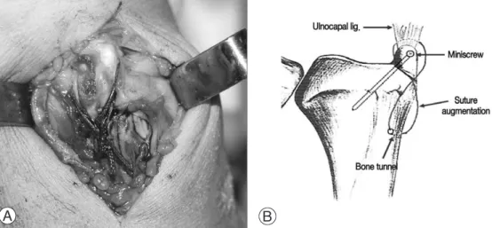
전신 마취 하에서 환자의 환측 상완부에 압박대를 착용시킨 상태에서 수술을 시행하였다. 원위 요골 골 절에 대하여 요수근 굴건 건초를 통한 수장측 도달법 을 이용하여 관혈적 정복 후 수장측 잠김나사 금속판 (volar locking plate)을 이용하여 내고정한 후 척골 경상돌기 골절에 대한 수술을 진행하였다. 척골 경상 돌기 척측면을 따라 3 cm의 종절개를 한 후에 척수근 신근과 척수근 굴근 사이를 박리하여 골절 부위를 노 출시켰다. 척골 경상돌기 골편을 정복한 후 척수근 인 대 안쪽으로 통과시킨 2가닥의 굵은 흡수성 폴리글락 틴(1-0 Vicryl) 봉합사를 척골 경상돌기 위로 사선방 향으로 교차시키고 근위부로 견인하여 골편의 정복을 유지한 채로 경상돌기 끝부분에서 사선방향으로 근위 요골쪽으로 드릴링하고 직경 1.2 mm, 길이 18 mm 의 소형 나사못을 이용하여 내고정을 하였다. 봉합사
일측을 척골 경부의 골터널을 통과시켜‘8’자 형태로 단단하게 묶어 나사못 내고정을 보강하였다(Fig. 1).
상완부의 압박대를 풀고 지혈 후 척측 심부 건막과 남 은 창상을 봉합하였다.
3. 수술 후 관리
수술 후 3~4주간 단상지 석고 부목을 이용하여 고 정하고 가골 형성이 진행됨에 따라 부목을 제거하여 관절 운동을 허용하였다.
4. 결과 평가
결과 평가는 방사선학적 골유합과 척측 동통 유무 및 관절 운동 범위를 평가하였으며 변형된 Green과 O’Brien의 방법14을 이용하여 기능적 결과를 평가하였 다. 추시 방사선 사진상 골절선을 가로지르는 골소주 의 연결이 보이면 골유합이 이루어진 것으로 판단하였 다. 평균 추시 기간은 13.7개월이었다.
결 과
전체 14명의 환자 중 10명의 환자에서 평균 8주 (5~11주)에 방사선학적 골유합을 얻을 수 있었다 (Fig. 2). 4례에서는 골유합 시기가 수술 후 5개월에 서 11개월 사이로 지연유합을 보였다. 합병증으로 1례 에서 나사못의 이완 소견을 보였으나 추가적인 처치 없이 수술 후 9개월에 골유합을 얻었다. 3례에서 경도 의 척측 동통을 호소하였으나 모든 환자에서 기능적 범위의 회외전과 회내전이 가능하였다(Fig. 3). 최종 추시상 변형된 Green과 O’Brien의 방법을 이용하여 평가한 기능적 결과는 매우 우수가 8례, 우수가 4례,
Fig. 1. A clinical photograph and a diagram of miniscrew fixation with figure of 8 tension-band suture augmentation.
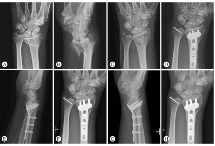
Fig. 2. A case (No 9) of intra-articular distal radius fracture with ulnar styloid avulsion. Initial AP (A) & Lat (B) radiographs. Post- reduction radiograph revealed unreduced ulnar styloid (C). Miniscrew fixation was augmented with tension-band suture (D, E). Bone union was obtained at 5 weeks after operation (F). At last follow up of 14 months (G, H), the patient had painless full range of wrist and forearm motions. The grip strength was 72% of normal side. The final result was assessed as ‘good’.
Fig. 3. Fifty-one year old lady (No 5) with AO C2 type distal radius fracture with ulnar styloid avulsion (A). Postoperative radi- ograph (B). Bony union was delayed until 8 months (C). At last follow up, radiograph revealed bony union (D). Near full range of motions were obtained (E, H).
양호가 2례였다(Table 1).
고 찰
원위 요골 골절과 동반된 척골 경상돌기 골절의 형 태나 골유합 여부와 기능적 결과와의 관련성에 대하여 일부 회의적인 보고들이 있다15-18. 척골 경상돌기 근위 부와 척골두 와부위에 삼각 섬유연골 복합체가 부착되
는데10,12,19, 이들 연구에서는 척골 경상돌기 골절을 해
부학적 위치에 따라서 구별하지 않고 골절 유무만을 고려하거나, 단순 방사선 사진상 나타나지 않는 삼각 섬유연골 복합체 등 연부조직 손상을 고려하지 않고 있다. 한편 수근 관절부에 대한 한 두장의 단순 방사 선 사진만으로 경상돌기 기저부 골절이 삼각 섬유연골 복합체 심부 부착부를 포함하는지 여부를 정확하게 평 가하기 어려울 수 있는데20, 수근 관절 전후방 단순 방 사선 사진을 정확한 중립위에서 촬영하지 않으면 경상 돌기와 척골두의 음영이 중첩되어 나타나기 쉽고 척골 두 원위단 전후면의 윤곽이 서로 일치하지 않을 경우 정확한 골절 부위를 판단하기 어렵다20,21. 이러한 골절 분류상의 어려움이나 동반된 연부조직 손상에 대한 부 정확한 평가가 척골 경상돌기 골절 또는 불유합과 임 상적 결과와의 상관성을 평가하는데 있어 제한 요인으 로 작용한 것으로 보인다21,22.
척골 경상돌기 골절이나 동반된 삼각 섬유인대 복합 체의 손상과 원위 요척 관절의 불안정성이 관련이 있 다는 것을 보고하는 연구들이 많으며8,10,11,13,16,19,23
일부
연구에서는 특히 척골 경상돌기 기저부 견열 골절이나 불유합이 수근 관절부 손상에서 임상 결과를 결정하는 데 중요한 역할을 하는 것을 보여 주고 있다6,11,12,24,25
. 이는 장기 추시 결과 척측 동통을 호소하는 많은 환자 에서 원위 요척 관절의 불안정성 및 척골 경상돌기 기 저부 골절이 동반된 것으로도 확인된다8,10,11.
척골 경상돌기 기저부 골절이라도 척골두 와의 삼각 섬유연골 복합체 심부 분지 부착부위가 포함되지 않았 을 경우 원위 요척 관절의 안정성이 남아 있어 수술을 통한 내고정을 요하지 않을 수 있다. 따라서 급성기에 불안정성 원위 요골 골절에 대한 내고정 후 도수검사 로 원위 요척 관절의 안정성을 평가하는 것은 비록 마 취 하에서라 하더라도 심한 부종 등으로 용이하지 않 지만, 방사선 사진을 면밀히 관찰하고 필요할 경우 CT 등 정밀검사를 추가로 시행하며 마취 하에서 원위 요척 관절에 대한 정확한 도수검사를 통해 불안정성 여부를 구분하려는 노력은 중요하다. 원위 요골 골절 에 대한 관혈적 정복술 및 내고정술 후 척골 경상돌기 골편이 자연 정복 되기도 하는데 자연 정복된 척골 경 상돌기의 골유합을 얻기 위해서는 6주 이상의 석고 부 목 고정이 필요하므로 방사선 검사 및 수술 중 요골 고정술 후 시행한 원위 요척 관절 불안정성에 대한 도 수검사에서 뚜렷한 불안정성이 있거나 골편 불유합 시 불안정성이 남을 것으로 예상되는 경우 골편의 정복 여부에 관계없이 내고정을 시행하는 것이 바람직하다.
척골 경상돌기 골절에 대한 수술적 방법에는 K-강선 과 장력대를 이용한 방법, 지연나사를 이용한 방법, Table 1. The summary of clinical results
Functional result*
Case No Age Sex F/U Union time Pronation/
Pain Functional Range of Grip Total Final (mo)� (wk)� supination
status motion strength score result
11 56 F 10 8 90/901 25 25 25 15 190 Excellent
12 66 F 12 11 m 90/901 25 25 25 15 190 Excellent
13 31 M 11 11 70/901 25 25 25 15 190 Excellent
14 51 M 18 5 90/100 25 25 25 15 190 Excellent
15 51 F 15 18 m 85/901 25 25 25 15 190 Excellent
16 69 F 13 6 50/901 25 25 25 15 190 Excellent
17 46 F 10 6 80/801 25 25 25 10 185 Good
18 45 F 14 8 90/901 20 25 25 15 185 Good
19 45 M 14 5 90/901 25 25 25 10 185 Good
10 62 F 18 15 m 90/901 25 25 25 25 100 Excellent
11 53 F 18 19 m 80/851 20 20 15 20 165 Fair
12 45 F 14 8 60/901 25 25 15 25 180 Good
13 65 F 18 5 70/901 20 25 15 25 175 Fair
14 63 F 16 8 80/851 25 25 25 15 190 Excellent
AV±SD 53 13 7±1.9** 80/891 24±1.9 25±1.3 23±4.3 16±5.04 86±10
*by Green and O’Brien’s criteria, **Except for 4 cases of delayed union, �mo: months, wk: weeks
고정나사 봉합(suture anchor)을 이용한 방법, 전완 부를 회외전 위치로 요척골을 외고정하여 간접적인 고 정효과를 이용하는 방법 등이 있다1,9. 일반적인 압박 나사를 이용하기에는 척골 경상돌기 골편의 크기가 작 아 어려움이 있으며 가장 흔히 사용되는 K-강선과 장 력대를 이용한 내고정술은 작은 경상돌기 골편에 2개 의 K-강선을 삽입하기가 기술적으로 쉽지 않으며, 작 은 골편에 무리해서 2개의 K-강선을 삽입하려 시도할 때, 때로는 골편의 분쇄를 초래하기도 한다. 또한 K- 강선 끝단에 의한 연부조직 자극이나 강선의 이완 등 합병증이 올 수 있고 이로 인하여 내고정물의 조기 제 거가 필요할 수도 있다9. 최근 보고된 외고정기를 이용 하여 회외전위로 고정하는 방법은 간접고정의 방법으 로 장기간의 회외전 위에서의 고정으로 회내전 제한이 남기 쉽고 핀 주위 감염증이 발생할 수 있다는 단점이 있다9. 저자들이 사용한 방법은 하나의 소형 나사못을 이용하여 척골 경상돌기를 비교적 쉽게 고정하고, 고 정의 이완을 방지하기 위하여 긴장대 봉합을 부가하여 추가적인 안정성을 부여함으로써 조기에 관절 운동을 허용할 수 있으며 연부조직 자극이 없어 내고정물을 제거하지 않아도 되는 장점이 있다. 저자들은 불안정 성 원위 요골 골절에 동반된 척골 경상돌기 기저부 골 절에 대하여 장력대 봉합 방법을 부가한 소형 나사못 고정술로 치료한 증례들에서 빠른 골유합과 우수한 기 능적 결과를 얻을 수 있었다.
결 론
불안정성 원위 요골 골절과 동반된 척골 경상돌기 기저부 골절은 잠재적인 불안정성을 갖기 때문에 수술 적인 치료가 필요하며, 장력대 봉합 방법을 부가한 소 형 나사못 고정술은 술식이 비교적 쉽고, 빠른 골유합 과 우수한 기능적 결과를 보여 임상적으로 유용한 술 식으로 생각되었다.
참고문헌
01) Geissler WB, Fernandez DL, Lamey DM. Distal radioul- nar joint injuries associated with fractures of the distal radius. Clin Orthop. 1996;327:135-46.
02) Jupiter JB, Fernandez DL. Complications following distal radial fractures. Instr Course Lect. 2002;51:203-19.
03) Lindau T, Hagberg L, Adlercreutz C, Jonsson K, Aspenberg P. Distal radioulnar instability is an indepen- dent worsening factor in distal radius fractures. Clin Orthop. 2000;376:229-35.
04) Lindau T, Runnquist K, Aspenberg P. Patients with laxity
of the distal radioulnar joint after distal radial fractures have impaired function, but no loss of strength. Acta Orthop Scand. 2000;73:151-6.
05) Lindau T. Treatment of injuries to the ulnar side of the wrist occurring with distal radius fractures. Hand Clin.
2005;21:417-25.
06) Nakamura R, Horii E, Imaeda T, Nakao E, Shionoya K, Kato H. Ulnar styloid malunion with dislocation of the distal radioulnar joint. J Hand Surg Br. 1998;23:173-5.
07) May MM, Lawton JN, Blazar PE. Ulnar styloid fractures associated with distal radius fractures: incidence and implications for distal radioulnar joint instability. J Hand Surg Am. 2002;27:965-71.
08) Stoffelen D, De Smet L, Broos P. The importance of the distal radioulnar joint in distal radial fractures. J Hand Surg Br. 1998;23:507-11.
09) Ruch DS, Lumsden BC, Papadonikolakis A. Distal radius fractures: A comparison of tension-band wiring versus ulnar outrigger external fixation for the management of distal radioulnar instability. J Hand Surg Am. 2005;30:
969-77.
10) af Ekenstam F, Hagert CG. Anatomical studies on the geometry and stability of the distal radioulnar joint. Scand J Plast Reconstr Surg. 1985;19:17-25.
11) Hauck RM, Skahen J 3rd, Palmer AK. Classification and treatment of ulnar styloid nonunion. J Hand Surg Am.
1996;21:418-22.
12) Mikic ZD. Treatment of acute injuries of the triangular fibrocartilage complex associated with distal radioulnar joint instability. J Hand Surg Am. 1995;20:319-23.
13) Tsukazaki T, Iwasaki K. Ulnar wrist pain after Colles’
fracture. 109 fractures followed for 4 years Acta Orthop Scand. 1993;64:462-4.
14) Green DP, O’Brien ET. Open reduction of carpal disloca- tion: Indications and operative techniques. J Hand Surg Am. 1978;3:250-65.
15) Catalano LW 3rd, Cole RJ, Gelberman RH, Evanoff BA, Gilula RA, Borrelli J Jr. Displaced intra-articular fractures of the distal aspect of the radius. Long-term results in young adults after open reduction and internal fixation. J Bone Joint Surg Am. 1997;79:1290-302.
16) Lindau T, Arner M, Hagberg L. Intraarticular lesions in distal fractures of the radius in young adults. A descriptive arthroscopic study in 50 patients. J Hand Surg Br.
1997;22:638-43.
17) Lindau T, Adlercreutz C, Aspenberg P. Peripheral tears of the triangular fibrocartilage complex cause distal radioul-
nar joint instability after distal radial fractures. J Hand Surg Am. 2000;25:464-8.
18) Richards RS, Bennett JD, Roth JH, Milne K. Arthroscopic diagnosis of intra-articular soft tissue injuries associated with distal radial fractures. J Hand Surg Am. 1997;22:772-6.
19) Melone CP, Nathan R. Traumatic disruption of the trian- gular fibrocartilage complex. Clin Orthop. 1992;275:
65-73.
20) Ishii S, Palmer AK, Werner FW, Short WH, Fortino MD.
An anatomic study of the ligamentous structure of the tri- angular fibrocartilage complex. J Hand Surg Am. 1998;23:
977-85.
21) Yanagida H, Ishii S, Short WH, Werner FW, Weiner MM, Masaoka S. Radiologic evaluation of the ulnar styloid. J Hand Surg Am. 2002;27:49-56.
22) Flinkkila T, Raatikainen T, Hamalainen M. AO and
Frykman's classifications of Colles' fracture. No prognos- tic value in 652 patients evaluated after 5 years. Acta orthop Scand. 1998;69:77-81.
23) Kaukonen JP, Karaharju EO, Porras M, L u¨thje P, Jakobsson A. Functional recovery after fractures of the distal forearm. Analysis of radiographic and other factors affecting the outcome. Ann Chir Gynaecol. 1988;77:27- 31.
24) Oskarsson GV, Aaser P, Hjall A. Do we underestimate the predictive value of the ulnar styloid affection in Colles fractures? Arch Orthop Trauma Surg. 1997;116:341-4.
25) Shaw JA, Bruno A, Paul EM. Ulnar styloid fixation in the treatment of posttraumatic instability of the radioulnar joint: a biomechanical study with clinical correlation. J Hand Surg Am. 1990;15:712-20.