영남지역 소아 정맥혈전색전증의 역학 및 위험인자에 대한 다기관연구
이소람1ㆍ윤종혁1ㆍ임재영1ㆍ최희원2ㆍ이재민3ㆍ서진경4ㆍ김지윤4ㆍ양유진5ㆍ박경미5 임영탁5ㆍ박지경6ㆍ최은미7ㆍ심예지7ㆍ김흥식7ㆍ박상규8ㆍ공섬김9ㆍ최은진10ㆍ박은실1,11
1경상대학교 의과대학 소아과학교실, 2동아대학교 의과대학 소아과학교실, 3영남대학교 의과대학 소아과학교실,
4경북대학교 의과대학 소아과학교실, 5부산대학교 어린이병원 소아과학교실, 6인제대학교 부산백병원 소아과학교실,
7계명대학교 의과대학 소아과학교실, 8울산대학교 의과대학 소아과학교실, 9고신대학교 의과대학 소아과학교실,
10대구가톨릭대학교 의과대학 소아과학교실, 11경상대학교 의과대학 건강과학연구원
Childhood Venous Thromboembolism in Yeungnam Region in Korea: Multicenter Study
Soram Lee, M.D.1, Jong Hyuk Youn, M.D.1, Jae Young Lim, M.D.1, Hee Won Chueh, M.D.2,
Jae Min Lee, M.D.3, Jin Kyung Suh, M.D.4, Ji Yoon Kim, M.D.4, Eu Jeen Yang, M.D.5, Kyung Mi Park, M.D.5, Young Tak Lim, M.D.5, Jikyoung Park, M.D.6, Eun Mi Choi, M.D.7, Ye Jee Shim, M.D.7, Heung Sik Kim, M.D.7,
Sang Kyu Park, M.D.8, Seom Gim Kong, M.D.9, Eun Jin Choi, M.D.10 and Eun Sil Park, M.D.1,11
1Department of Pediatrics, Gyeongsang National University College of Medicine, Jinju, 2Department of Pediatrics, Dong-A University College of Medicine, Busan, 3Department of Pediatrics, Yeungnam University College of Medicine, Daegu, 4Department of Pediatrics, School of
Medicine, Kyungpook National University, Daegu, 5Department of Pediatrics, Pusan National University Children’s Hospital, Yangsan,
6Department of Pediatrics, Inje University Busan Paik Hospital, Busan, 7Department of Pediatrics, Keimyung University School of Medicine, Daegu, 8Department of Pediatrics, Ulsan University College of Medicine, Ulsan, 9Department of Pediatrics, Kosin University College of Medicine, Busan, 10Department of Pediatrics, College of Medicine, Daegu Catholic University, Daegu, 11Health Science Institute, Gyeongsang National
University College of Medicine, Jinju, Korea
Background: Venous thromboembolism (VTE) is rare in pediatric patients compared to adults, but it’s incidence is gradually increasing. The purpose of this study was to analyze the incidence, risk factors, and prognosis of pediatric patients with VTE in Korea.
Methods: Between January 2000 and July 2017, 249,312 medical records of the patients older than 1 year who were hospitalized in the department of pediatrics of 10 university hospitals in Yeungnam region were retrospectively reviewed.
Results: The overall incidence of VTE was 4.9 per 10,000 admissions. Of the total 123 patients, 80 (65.0%) were male and the median age was 10.8 years (range, 1.0-23.5 years). Magnetic resonance imaging was performed most frequently to confirm the diag- nosis of VTE (43.1%). Thrombosis occurred in the cerebral vessels (46.3%), lower ex- tremities (23.8%), pulmonary (19.5%), abdomen (9.8%), and upper extremities (4.1%).
One hundred and six patients had underlying causes such as cancer (27.6%), infection (26.8%), intravenous catheter insertion (17.9%), and surgery (14.6%). Protein C was eval- uated in 39 patients (31.7%), protein S in 40 (32.5%), antithrombin (AT) III in 52 (42.3%), and homocysteine in 21 (17.1%). Among them, one patient with a family history of AT III deficiency had SERPINC gene mutation. Seventy-seven patients (62.6%) started anti- coagulation treatment. Most (52.0%) were treated for more than 90 days.
Conclusion: Healthcare providers must be aware of the potential for VTE development in childhood. In the near future, a nationwide survey should be investigated to de- termine the incidence rate and the trends in VTE among Korean children.
pISSN 2233-5250 / eISSN 2233-4580 https://doi.org/10.15264/cpho.2018.25.1.43 Clin Pediatr Hematol Oncol 2018;25:43∼49
Received on March 26, 2018 Revised on April 3, 2018 Accepted on April 9, 2018
Corresponding Author: Eun Sil Park Department of Pediatrics, Health Science Institute, Gyeongsang National University College of Medicine, Jinju-daero 816 beon-gil, Jinju 52727, Korea
Tel: +82-55-750-8829 Fax: +82-55-752-9339 E-mail: [email protected]
ORCID ID: orcid.org/0000-0001-9344-7191
Key Words: Venous thromboembolism, Pediatric patients, Epidemiology
Introduction
The incidence of venous thromboembolism (VTE) is known to be relatively low in childhood; the reported in- cidence rate ranges from 0.7 to 4.9 per 100,000 per- son-years [1,2], while total incidence of Korea population is 13.8 per 100,000 person [3]. However, an increasing in- cidence of VTE in childhood was reported in the United States and Canada, especially in the tertiary care setting [1,4]. General population data from Denmark revealed that the population incidence is relatively stable (2.09 per 100,000 person-years) but with an upward trend showing an annual increase of 9.6% from 2001-2006 [5]. Pediatric VTE has a significant impact on both acute and chronic health outcomes, including increased risk of mortality, re- currence of VTE, and post-thrombotic syndrome [1,6-8].
According to a Canadian study, the mortality rate directly attributable to VTE was 2.2% and morbidity including re- current thrombosis (8.1%) and postphlebitic syndrome (12.4%) was substantial. The increasing incidence of child- hood VTE is postulated by experts to be a result of ad- vanced tertiary healthcare resulting in improved survival of critically ill children at the cost of VTE. The evolution of diagnostic techniques and image modality also contributed to the increased incidence rate.
In 2016, Choi et al. [9] reported that the incidence rate of childhood VTE at a single center was 3.27 per 10,000 admissions; the authors also reported risk factors, diagnosis, and treatment data, but there is still lack of a nationwide epidemiologic survey on VTE in children. To the best of our knowledge, this is the first multicenter study investigat- ing childhood VTE in Korea. The primary aim of this study was to evaluate the incidence rate and outcome of child- hood VTE on a large scale, including risk factors and treatment.
Materials and Methods
Medical records of the patients hospitalized in the de- partment of pediatrics of 10 university hospitals in the Yeungnam region were retrospectively reviewed by pedia-
tricians from January 2000 to July 2017. Patients were se- lected by diagnostic codes (based on the International Classification of Diseases, ICD, Tenth Edition) and radio- logic findings. ICD-10 codes were I80.2 and I80.3 for deep vein thrombosis (DVT); I26, I26.0, and I26.9 for pulmonary embolism (PE); D73, I81.1, I82.0, I82.2, I82.3, and K55.0 for intraabdominal thrombosis; G08.0 and I67.6 for cerebral vein thrombosis; and I82.8 and I82.9 for upper extremity DVT. Patients were also included in the study if there was any mention of thrombosis in the radiological reports.
Patients younger than 1 year old were excluded because it was mostly catheter-related thrombosis in the neonatal in- tensive care unit. Demographic characteristics including sex and age at diagnosis, underlying disease, and clinical risk factors were collected. Results of thrombophilic testings and adjusted treatment data at the time of diagnosis were also collected.
Results
Of the total 249,312 hospitalizations during the study pe- riod, 142 medical records were reviewed retrospectively based on coding and radiologic data, among which with moyamoya disease or hemorrhagic cerebral infarction were excluded. Finally, 123 admissions were included and no patients were duplicated. The total incidence of VTE was 4.9 per 10,000 admissions (0.049%).
1) Characteristics of patients
Of the 123 patients, 80 (65.0%) were male and 43 (35.0%) were female. The median age at diagnosis was 10.8 years (range, 1.0-23.5 years), while that of the male was 11.7 years and the female was 9.2 years. Fig. 1 shows age distribution of the patients with 1-year interval. In pre- vious studies, a bimodal peak incidence at under 1 year of age and during adolescence was reported. Because the current study excluded patients under 1 year old, the age distribution showed a relatively higher frequency among teenagers. VTE in cerebral vessels was the most common diagnosis (46.3%), followed by the lower extremities and pulmonary vessels (Table 1).
Table 2. Investigations performed to confirm the diagnosis of VTE
N=123 (%)
MRI 53 (43.1)
CT 41 (33.3)
Ultrasonography 17 (13.8)
Venography or angiography 13 (10.6)
Echocardiography 5 (4.1)
V/Q scan 1 (0.8)
Othera) 1 (0.8)
MRI, magnetic resonance imaging; CT, computed tomography;
V/Q, ventilation/perfusion.
a)Other: A 15.4-year-old male with mental change had under- lying risk of acute myeloid leukemia and systemic lupus erythematous. At the time of visiting emergency room, cardiopulmonary resuscitation was performed but he was expired. Cerebral and pulmonary embolism were diagnosed by clinical and neurologic exam.
Table 1. Characteristics of the patients
N
Total VTE patients 123
Male/Female 80/43
Age at diagnosis, median (range), year 10.8 (1.0-23.5) Location of VTE (%)
Pulmonary 24 (19.5)
Abdominal 14 (11.4)
Cerebral 57 (46.3)
Upper extremities 5 (4.1)
Lower extremities 29 (23.6)
Others 8 (6.5)
VTE, venous thromboembolism.
Fig. 1. Age distribution of the patients with 1 year interval.
Table 3. Underlying clinical conditions (risk factors) N=123 (%)
Cancer 34 (27.6)
Infection 33 (26.8)
Catheter 22 (17.9)
Surgery 18 (14.6)
None 17 (13.8)
Congenital heart disease 16 (13.0)
Dehydration 9 (7.3)
Immobilization 4 (3.3)
Medication 2 (1.6)
Trauma 2 (1.6)
Others 36 (29.3)
2) Diagnosis
Magnetic resonance imaging (MRI) was performed most frequently to confirm the diagnosis (43.1%), followed by computed tomography (CT) (33.3%) and ultrasonography (13.8%) (Table 2). Cerebral VTE was diagnosed mainly by MRI (80.7%); PE, CT (75.0%); VTE of lower limb, ultra- sonography (48.3%).
3) Underlying clinical conditions (risk factors)
One hundred six patients (86.2%) had risk factors such as cancer (34 patients, 27.6%), infection (33, 26.8%), intra- venous catheter insertion (22, 17.9%), surgery (18, 14.6%), and congenital heart disease or prosthetic valve (16, 13.0%) (Table 3). Other diagnoses included vascular disease, hem- atologic disease, and systemic diseases such as systemic lu-
pus erythematous, nephrotic syndrome, and others. Of the 34 patients, diagnosed with cancer, acute lymphoblastic lymphoma was present in 18 (52.9%) and solid tumor in 9 (26.5%) (Fig. 2). Fifty-seven patients (46.3%) had two or more risk factors.
4) Congenital thrombophilia
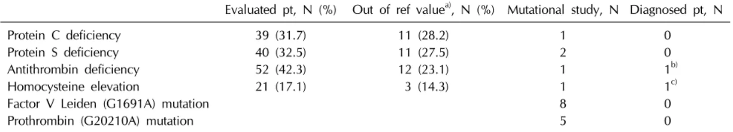
Protein C level was evaluated in 39 patients (31.7%), pro- tein S in 40 (32.5%), antithrombin (AT) III in 52 (42.3%), and homocysteine in 21 (17.1%) (Table 4). Among them, 11 out of 39 (28.2%), 11 out of 40 (27.5%), 12 out of 52 (23.1%), and 3 out of 21 (14.3%) showed values below or above the reference range. However, only 2 patients under- went mutational analysis of related genes.
Table 4. Congenital thrombophilia
Evaluated pt, N (%) Out of ref valuea), N (%) Mutational study, N Diagnosed pt, N
Protein C deficiency 39 (31.7) 11 (28.2) 1 0
Protein S deficiency 40 (32.5) 11 (27.5) 2 0
Antithrombin deficiency 52 (42.3) 12 (23.1) 1 1b)
Homocysteine elevation 21 (17.1) 3 (14.3) 1 1c)
Factor V Leiden (G1691A) mutation 8 0
Prothrombin (G20210A) mutation 5 0
a)Reference value of Protein C defined as 70-120%, Protein S 60-150%, Antithrombin III 80-120%, Homocysteine 4.2-15.3 mol/L.
b)SERPINC1 gene mutation.
c)MTHFR (C677T) heterozygous variant.
Fig. 2. Numbers of the patients with VTE according to the diagnosis of malignancies.
Table 5. Congenital thrombophilia without any risk factor (N=17)
Evaluated pt, N (%) Out of ref valuea), N (%) Mutational study, N Diagnosed pt, N
Protein C deficiency 11 (64.7) 0 0 0
Protein S deficiency 11 (64.7) 4 (36.4) 0 0
Antithrombin deficiency 9 (52.9) 1 (11.1) 0 0
Homocysteine elevation 8 (47.1) 1 (12.5) 1 1b)
Factor V Leiden (G1691A) mutation 1 0
Prothrombin (G20210A) mutation 0 0
a)Reference value of Protein C defined as 70-120%, Protein S 60-150%, Antithrombin III 80-120%, Homocysteine 4.2-15.3 mol/L.
b)MTHFR (C677T) heterozygous variant.
Among all patients, one patient with a family history of AT III deficiency had SERPINC gene mutation and one was diagnosed with MTHFR heterozygous variant. Factor V Leiden and prothrombin mutations were evaluated in 8 (15.4%) and 5 (4.1%) patients, respectively, and none of the patients had mutation.
When we analyzed the 17 patients without risk factors for VTE, the frequency of evaluation for congenital thrombo-
philia was higher than that of all patients (Table 5). Only the patient with MTHFR mutation underwent mutational analysis.
5) Treatment and outcomes
Seventy-seven patients (62.6%) started anticoagulation treatment. Twenty-six patients (33.8%) were treated with low molecular weight heparin and 19 (15.4%) were treated with conventional heparin (Table 6). The percentage of pa- tients who did not receive any treatment was high (37.4%).
Most patients (N=40, 52.0%) were treated for more than 90 days, and of the 49 patients who continued treatment, 16 (32.7%) were treated with warfarin.
Twenty-one patients (17.1%) had complications during follow-up, and 14 of them died due to septic shock (5 pa- tients); cancer related complication (4); pulmonary embo- lism (2); heart failure (1); gastrointestinal bleeding (1); and unknown cause (1).
Discussion
In prospective pediatric registries in North America and
Table 6. Treatment
N=123 (%) Medication
Conventional heparin 19 (15.4)
LMWH 26 (21.1)
Warfarin 13 (10.6)
Aspirin 14 (11.4)
Clopidogrel 7 (5.7)
Rivaroxaban 1 (0.8)
Urokinase 2 (1.6)
IVC filter 1 (0.8)
Surgery (thrombectomy) 2 (1.6)
Unknown 6 (4.9)
No treatment 46 (37.4)
LMWH, low molecular weight heparin; IVC, inferior vena cava.
Europe, the annual incidence of VTE was estimated to be 0.7 to 1.4 per 100,000 children, 5.3 per 10,000 hospital ad- mission among children, and 24 per 10,000 admissions of neonates to neonatal intensive care units [2,10,11].
Moreover, a dramatic increase in the US hospital-based pe- diatric VTE incidence rate (188 per 100,000 discharges) was reported in the 2000s [4,7]. Our retrospective multi-center cohort study revealed that the incidence was 4.9 per 10,000 admissions, a rate similar to previous reports. However, our data have several limitations. First, this study included data from multiple centers during extended time period, thus various diagnostic techniques and laboratory methods that differ in accuracy and consistency were included. Second, selection bias by researchers was inevitable in our retro- spective analysis as many asymptomatic patients with VTE might be omitted.
One difference between adults and children with VTE was that the majority of cases (over 70%) in children were associated with clinical risk factors including Inherited thrombophilia (IT), whereas in adults, aging is the domi- nant risk factor for VTE (population attributable risk >90%) [12]. There are many acquired and transient conditions that lead to a prothrombotic state including sepsis, cancer, con- genital heart disease, central venous catheter placement, surgery, strict immobilization, pregnancy, oral contra- ceptives, and persistent antiphospholipid antibody. Activated protein C resistance (designated as factor V Leiden) in the most common hereditary abnormalities predisposing to VTE
[13,14]. The factor II variant G20210A is another common gene mutation; other inherited risk factors are disorders of AT III, protein C, protein S, and dysfibrinogenemia. The relative risk of IT leading to VTE in children was evaluated in a previous meta-analysis, which showed the odds ratio (OR) of AT III deficiency to be 9.4 (95% confidence interval [CI] 3.4-26.66), protein C deficiency 7.7 (95% CI 4.4-13.4), protein S deficiency 5.8 (95% CI 3.0-11.0), factor V variant G1691A 3.6 (95% CI 3.8-4.8), and factor II variant G20210A 2.6 (95% CI 1.6-4.4), which is similar to data in adults [12,13,15]. Although genetic variants in factor II (G20210A) and factor V (G1691A) show different patterns between eth- nic groups, there is limited data concerning IT in childhood.
The rate of recurrent VTE was suggested to be 3%
among neonates and 21% among children without idio- pathic VTE based on follow-up reports [2,15,16]. The most important determinant of recurrence is the presence of tran- sient clinical risk factors during the time of the first episode [17]. After an unprovoked first episode of VTE, the risk of recurrence was 10% during the first year after cessation of anticoagulant and 5% per year thereafter. In terms of IT, the risk of recurrent VTE is controversial. Some studies re- ported that children with AT deficiency had a 6.5-fold high- er risk of recurrence compared with patients with non- thrombophilic VTE [18]. Numerous case control studies of patients with VTE due to a specific thrombophilia and pa- tients with nonthrombophilic VTE as controls showed rela- tive risks of 1.5 to 2.0 for most IT [12,13,19-21]. Because there is a lack of evidence that positive thrombophilia test results have an effect on patient management, thrombo- philia screening in children with VTE and among thrombo- sis-prone families is still debated [22]. Although knowledge of IT may be useful to promote awareness of the potential need for prophylaxis in high risk situations, the potential harm of identifying a non-contributory problem leading to inappropriate management also exists [23]. To date, throm- bophilia testing has been recommended in a limited num- ber of patients with VTE, particularly if they are young (<50 years old), especially in association with weak pro- voking factors (minor surgery, immobility, unprovoked VTE, or combination oral anticoagulants), a positive family
history for disease, recurrent VTE events, or thrombosis in unusual sites such as the splanchnic or cerebral veins [24].
The existence of triggering factors for VTE is an im- portant determinant of the duration of therapy; usually, 3 months of anticoagulant treatment is sufficient for patients with a first episode of VTE secondary to a reversible risk factor [25]. For those with an unprovoked (idiopathic) event and those with recurrent events, indefinite anticoagulation therapy should be considered [26]. Determining which pa- tients will be recurrent remains difficult, and the role of the presence or absence of IT in guiding decision making about the duration of therapy remains controversial and in- completely proven.
The dramatic rise in the incidence of pediatric VTE in tertiary care pediatric centers was previously recognized and is attributed to intensive medical intervention at tertiary care centers and improved survival of critically ill patients.
Also, the development of diagnostic technology contributed to the increased incidence by early detection of VTE. Due to recurrent VTE and post-thrombotic syndrome (PTS), the risk of major morbidities requires careful follow-up after completion of planned anticoagulant therapy. At follow-up visits, detailed evaluation of the patient’s history and phys- ical examination can focus on the detection of recurrent VTE, and the clinical scoring system for the diagnosis of PTS should be adjusted.
In conclusion, close attention should be paid to the de- velopment of VTE in childhood, along with the develop- ment of improved medical care and a deliberate and in- dividualized approach for the detection of IT and proper follow up. In the near future, a nationwide survey should be investigated to identify the incidence rate and trends in the VTE incidence among Korean children.
References
1. Andrew M, David M, Adams M, et al. Venous thromboembolic complications (VTE) in children: first analyses of the Canadian Registry of VTE. Blood 1994;83:1251-7.
2. van Ommen CH, Heijboer H, Büller HR, Hirasing RA, Heijmans HS, Peters M. Venous thromboembolism in child- hood: a prospective two-year registry in the Netherlands. J Pediatr 2001;139:676-81.
3. Jang MJ, Bang SM, Oh D. Incidence of venous thromboembo- lism in Korea: from the Health Insurance Review and Assessment Service Database. J Thromb Haemost 2011;9:
85-91.
4. Raffini L, Huang YS, Witmer C, Feudtner C. Dramatic increase in venous thromboembolism in children's hospitals in the United States from 2001 to 2007. Pediatrics 2009;124:1001-8.
5. Tuckuviene R, Christensen AL, Helgestad J, Johnsen SP, Kristensen SR. Pediatric venous and arterial noncerebral thromboembolism in Denmark: a nationwide pop- ulation-based study. J Pediatr 2011;159:663-9.
6. Creary S, Heiny M, Croop J, et al. Clinical course of post- thrombotic syndrome in children with history of venous thromboembolism. Blood Coagul Fibrinolysis 2012;23:39-44.
7. Setty BA, O'Brien SH, Kerlin BA. Pediatric venous throm- boembolism in the United States: a tertiary care complication of chronic diseases. Pediatr Blood Cancer 2012;59:258-64.
8. Gokce M, Altan I, Unal S, et al. Recurrent pediatric thrombo- sis: the effect of underlying and/or coexisting factors. Blood Coagul Fibrinolysis 2012;23:434-9.
9. Choi HS, Choi CW, Kim HM, Park HW. Venous thromboemb- olism in pediatric patients: a single institution experience in Korea. Blood Res 2016;51:164-70.
10. Schmidt B, Andrew M. Neonatal thrombosis: report of a pro- spective Canadian and international registry. Pediatrics 1995;96:939-43.
11. Monagle P, Adams M, Mahoney M, et al. Outcome of pediatric thromboembolic disease: a report from the Canadian Childhood Thrombophilia Registry. Pediatr Res 2000;47:763-6.
12. Young G, Albisetti M, Bonduel M, et al. Impact of inherited thrombophilia on venous thromboembolism in children: a sys- tematic review and meta-analysis of observational studies.
Circulation 2008;118:1373-82.
13. Kearon C, Julian JA, Kovacs MJ, et al. Influence of thrombo- philia on risk of recurrent venous thromboembolism while on warfarin: results from a randomized trial. Blood 2008;112:
4432-6.
14. Choi HS. Venous thromboembolism in children. Clin Pediatr Hematol Oncol 2017;24:1-10.
15. Nowak-Göttl U, Junker R, Kreuz W, et al. Risk of recurrent venous thrombosis in children with combined prothrombotic risk factors. Blood 2001;97:858-62.
16. Goldenberg NA, Knapp-Clevenger R, Manco-Johnson MJ;
Mountain States Regional Thrombophilia Group. Elevated plasma factor VIII and D-dimer levels as predictors of poor outcomes of thrombosis in children. N Engl J Med 2004;351:1081-8.
17. de Jong PG, Coppens M, Middeldorp S. Duration of anti- coagulant therapy for venous thromboembolism: balancing benefits and harms on the long term. Br J Haematol 2012;158:433-41.
18. Limperger V, Kenet G, Goldenberg NA, et al. Impact of
high-risk thrombophilia status on recurrence among children with a first non-central-venous-catheter-associated VTE: an observational multicentre cohort study. Br J Haematol 2016;175:133-40.
19. Baglin T, Luddington R, Brown K, Baglin C. Incidence of re- current venous thromboembolism in relation to clinical and thrombophilic risk factors: prospective cohort study. Lancet 2003;362:523-6.
20. Marchiori A, Mosena L, Prins MH, Prandoni P. The risk of recurrent venous thromboembolism among heterozygous car- riers of factor V Leiden or prothrombin G20210A mutation.
A systematic review of prospective studies. Haematologica 2007;92:1107-14.
21. Margaglione M, D'Andrea G, Colaizzo D, et al. Coexistence of factor V leiden and factor II A20210 mutations and re- current venous thromboembolism. Thromb Haemost 1999;82:
1583-7.
22. Klaassen ILM, van Els AL, van de Wetering MD, van Ommen CH. Increasing incidence and recurrence rate of venous thromboembolism in paediatric oncology patients in one sin- gle centre over 25 years. Thromb Haemost 2017;117:2156-62.
23. Kenet G, Limperger V, Shneyder M, Nowak-Göttl U. Risk fac- tors for symptomatic venous and arterial thromboembolism in newborns, children and adolescents-What did we learn within the last 20 years? Blood Cells Mol Dis 2017;67:18-22.
24. Connors JM. Thrombophilia testing and venous thrombosis.
N Engl J Med 2017;377:1177-87.
25. Kerlin BA. Current and future management of pediatric ve- nous thromboembolism. Am J Hematol 2012;87:S68-74.
26. Monagle P, Chan AKC, Goldenberg NA, et al. Antithrombotic therapy in neonates and children: antithrombotic therapy and prevention of thrombosis, 9th ed: American College of Chest Physicians Evidence-Based Clinical Practice Guidelines. Chest 2012;141:e737S-801S.