Lactobacillus rhamnosus 파쇄물의 항산화 및 미백효과
최우석1, 권희석2, 임혜원3, 노라환4, 이현용4*
1강원대학교생물의소재공학과
2한국코스모화장품
3세바바이오텍
4서원대학교식품공학과
Received : February 18, 2013 / Accepted : April 3, 2013
서 론
의학기술이발전되고현대사회의생활수준이점점향상됨 에따라인간의평균수명도증가되면서사람들의관심은건 강하게오래살며아름답게늙어가는것으로집중되고있다. 이 러한시대의관심에부응하여기능성식품및화장품분야 의시장규모가확대되고, 특히천연물질을활용한분야의연 구가급속히진행되고있다[3].
천연물질중많이사용되는프로바이오틱스는체내에들 어가서건강에도움을주는살아있는균을지칭하는데, 현재 프로바이오틱스로 사용되는 것은 Bifidobacterium 속,
Enterococcus 속, Lactobacillus 속, Clostridium butyricum, Lactobacillus sporogenes, Bacillus subtilis 및 Bacillus polyfermenticus 등이있으며이들의면역조절및항암기 능에대한연구들이보고되고있다[6, 17].
프로바이오틱스로사용되는유산균(Lactobacillus)은치즈, 발효유및유산균음료등의발효유제품을비롯하여각종 발효식품의제조에광범위하게응용되고있는산업적으로 중요한미생물이다. 예로부터오랫동안식품의발효와장질 환치료및예방의정장효과를목적으로유산균이사용되 어발전을거듭하여왔다. 이러한유산균을이용한발효물은 요구르트나치즈와같은형태로제품화가되거나, 김치및 청국장같은산업에크게영향을미치고있다. 하지만일반 유산균을이용하여제품을발효할경우유산균종류에따라 발효수율및생리활성물질의생산수율이달라질수있으 며, 식품및화장품의원료로써활용되어지는등유산균종 Whitening Effects of Lactobacillus rhamnosus Associated with Its Antioxidative Activities. Choi, Woo Seok1, Hee- Souk Kwon2,Hye Won Lim3, Ra Whan No4, and Hyeon Yong Lee4*. 1Department of Medical Biomaterials Engineering, Kangwon National University, Chuncheon 200-701, Korea, 2Hankook Cosmo Cosmetics Co., Bucheon 421-808, Korea,
3Shebah Biotech Co., Chuncheon Bioindustry Foundation Hi-Tech Venture Town, Chuncheon 200-161, Korea, 4Department of Food Science and Engineering, Seowon University, Chungju 361-742, Korea
This study was performed to investigate the whitening effects of Lactobacillus rhamnosus in addition to its antioxidative activi- ties. The cytotoxicity of the Lactobacillus rhamnosus was 7.6% at 10.0% (v/v) concentration. Its cytotoxicity was lower than 3.2% of Lactobacillus casei when adding the same concentration. Lactobacillus rhamnosus exhibited high antioxidative activi- ties at 14.9% of DPPH radical scavenging activity, and a lower reducing power was measured. Lactobacillus casei exhibited relatively lower antioxidative activities at 13.4%. The tyrosinase inhibition activity of Lactobacillus rhamnosus was observed at 31.3% when adding 10.0% (v/v), as compared to 17.7% for Lactobacillus casei. Lactobacillus rhamnosus demonstrated strong inhibition activity for melanin synthesis at 58.6% when adding 10.0% (v/v), while Lactobacillus casei increased to 80.6%. It was also observed that the high antioxidative activities of Lactobacillus rhamnosus were strongly correlated to whitening activities, due to the inhibition of both tyrosinase and melanin synthesis. These results support the expanded use of lactic acid bacteria as a functional bioresources in the cosmetics industry.
Keywords: Lactic aicd bacteria, Lactobacillus rhamnosus, antioxidative activity, whitening effect
*Corresponding author
Tel: +82-43-299-8471, Fax: +82-43-299-8471 E-mail: [email protected]
© 2013, The Korean Society for Microbiology and Biotechnology
류에따라발효에따른활성이다르기때문에발효능력이뛰 어난신규유산균의확보는경제적으로매우큰효과를가
질수있다[18, 32]. 또한유산균의자신을보호하는항산화
메커니즘이발견[1, 15, 27] 됨에따라유산균의항산화효과 에대한연구가시작되고있다.
특히 Latobacillus 균은장내정상세균으로면역증강, 항 바이러스, 항균효과를나타내는것으로보고된바있으며[8,
21]. 국내외연구진들에의하여피부장벽을강화하는효능
및미백, 보습효과가뛰어나다는연구결과가보고된바있
다[5, 26]. 유산균에의한효능들은서로밀접하게영향을가지
고있다고사료되어, 본연구진은 Lactobacillus rhamnosus의 항산화및미백활성의연관성을확인하여기능성화장품으 로써의발전을위한연구를꾀하였다.
피부는자외선의영향을받기쉬운기관이다. 자외선의영 향으로피부에활성산소가생성되고이에따라생성된활성 산소종은피부세포를손상시키며손상받은세포는식세포 에의해제거된다. 활성산소는매우불안정한상태이므로, 강한반응성을갖고, 세포내매우강한독성을갖게하여그 중 O2·은 thiol, ascorbate, tocopherol 그리고 catecholamine 을산화시켜 (Fe-s)4 cluster 함유단백질을공격하기도한다
[10]. 활성산소가노화및각종질병의직접적이며, 근본적인
원인으로작용함에따라과량의활성산소가존재하면체내 의항산화물질을고갈시키면서멜라닌생성반응을증가시 킨다. 이는곧각질형성세포에작용하여사이토카인을분비 시켜직·간접적으로멜라닌세포의증식과합성을증가시켜
피부의색소침착및기미형성이증가된다[31]. 추가로피부
는자외선을받아도피부에서색소침착이증가되는데, 이와 같이색소침전이증가하는것은자외선흡수에의한피부세 포의손상을억제할목적으로멜라닌생성이증가된결과이 다. 자외선에의한광노화는깊은주름살과잔주름살생성, 색소탈색, 피부건조, 탄력성감소및색소침착등의변화
가나타난다[17, 22]. 결국자외선에의한피부의노화가진
행되면멜라닌증식이항시적으로되고그결과기미가생
성된다[28]. 이멜라닌생성에있어서가장중요한역할을하
는효소가바로 tyrosinase이며 melanosome 내에 tyrosine 을산화시켜 3, 4-dihydroxyphenylalanine (DOPA)를만드 는 tyrosine hydroxylase로 DOPA를산화시켜 dopachrome 을만드는 DOPA oxidase로작용하여최종적으로 melanin
polymer를합성하게된다. 따라서피부미백제의개발에있
어서 tyrosinase활성억제실험은유용한일차평가법으로
인정되고있다[23, 25].
향장소재로써의항산화및미백활성은멜라닌세포에직 접적으로작용되는활성산소와관련되며, 멜라닌세포생성 경로내의생성인자억제및활성산소의증가를억제하면미
백활성이증가한다[2, 30]. 더나아가각종질병및노화의
직접적으로관련된활성산소를제거하는것이멜라닌색소 형성억제에효과적임을알수있다. 이에따라항산화및미 백활성을높일수있는소재를찾으려는연구가활발히진 행되고있다[4, 19].
본연구진은 lactic acid bacteria인 Lactobacillus rhamnosus 의세포독성실험및항산화활성실험을바탕으로하여유 산균파쇄물이활성산소를억제할수있는능력이있는지확 임함과동시에미백기능성향장소재로써가치가있는지여 부를 확인할 수 있는 tyrosinase inhibition activity와 melanogenesis activity를진행하였다.
재료 및 방법
균주 확보 및 배양조건
본 연구에서 사용된 유산균 Lactobacillus rhamnosus
(KCCM40069)와보편적으로 사용되는프로바이오틱스인
Lactobacillus casei (KCCM12452)를 초기균수 105 CFU/
ml로 37oC에서 24시간동안 1 L MRS broth에서각각혐기 배양을한후, 항산화활성및미백활성을측정하기위하여 균주와볼밀용볼(직경 1 mm)을볼밀(HK-BA01, SAMHAW
CERAMIC COMPANY, KOREA)에넣어하루동안파쇄시
킨후파쇄물을 0.45µm 필터에여과한후여과물을가지고
실험을진행하였다.
세포주 및 세포 생육 배지
실험에 이용된 세포주로 human fibroblast CCD-986sk (KCLB 21947, KOREA), melanoma cell B16F10 (KCLB
80008, KOREA)를사용하였으며, 세포배양에필요한배지
로 RPMI 1640 (GIBCO, USA)을사용하였고그밖의세포 배양에 필요한 시약으로 hepes buffer (SIGMA, USA)와 fetal bovine serum (GIBCO, USA), gentamycin sulfate (SIGMA, USA), trypsin-EDTA (SIGMA, USA)를사용하였 다. 실험에사용된세포는 RPMI 1640 (GIBCO, USA) 배 지 90%에 FBS를 10%로적용시켜 배양하여실험에이용 하였다.
세포 독성 측정
Mosmann 방법을 변형시켜세포 생존율을측정하였다.
CCD-986sk 세포를 96 well plate에 1.5×10⁴ cells/well 농 도로접종한후, 80% 정도의배양시점에서각 well에시료 를투여하고 CO2 incubator에서 24시간동안 배양하였다. 5µg/ml 농도의 MTT 용액을각 well에첨가하고 4시간후 상층액을제거하고, 10 µl의 acid-isopropanol (0.04 N HCl in isopropanol)을 첨가한 후 microplate reader (Tecan, USA)로 565 nm의파장에서흡광도를측정하였다.
DPPH 자유라디칼 소거활성 측정
DPPH (α,α-diphenyl-β-picrylhydrazyl) radical에 대한 소거활성은 Dietz 등 [6]의방법을약간변형하여실험하였 다. 96-well plate에용매를 ethanol로하여제조한 0.1 mM DPPH용액 200 µl와 2.5%, 5.0%, 10.0% (v/v)의농도로조 절된 Lactobacillus rhamnosus와 Lactobacillus casei, postive control인 ascorbic acid 80µl을 각각 혼합하여 25oC에서 20분동안암실에방치한후 525 nm에서흡광도 측정하였다. 측정값은 아래의 DPPH radical scavenging activity (%)로계산하였다.
DPPH radical scavenging activity (%)
=
환원력 측정
환원력은 sample 및 postive control를각각 2.5%, 5.0%, 10.0% (v/v)의 농도 2.5 ml에 200 mM sodium phosphate buffer (SIGMA, USA) 2.5 ml와 1% potassium ferricyanide (HAYASHI PURE CHEMICAL, JAPAN) 2.5 ml을혼합시 키고, 50oC에서 20분간 incubation시킨 다음 2.5 ml trichloroacetic acid (SIGMA, USA) (10%, w/v)를첨가하여 3500 rpm에서 10분간원심분리하였다. 상층액 5 ml에탈 이온수 5 ml와 1% ferric chloride (SIGMA, USA) 1 ml를 첨가시킨 후 UV-vis Spectrophotometer (852A Diode Array Spectrophotometer, Hewlett Packard)를 이용하여 700 nm에서흡광도를측정하였다[20].
Tyrosinase 억제 효과 탐색
dopachrome법을이용하여 tyrosinase 억제효과를측정하 였다[24]. mushroom tyrosinase-150 unit 150 µl와 2.5 mM L-tyrosine 225 µl, 0.4 M hepes buffer (pH 6.8) 225 µl, ethanol 용액또는 1 mg/ml 농도의시료 300 µl를혼합한후 배양전, 15분간배양후의각각의흡광도를 475 nm의파장 에서측정하였다.
Tyrosinase Inhibition (%) = (D−C)−(B−A) / (D−C)×100
A와 B는각각시료가첨가된용액의배양전과배양후의
흡광도이며, C와 D는각각시료를넣지않은용액의배양전 과배양후의흡광도이다. 이들 tyrosinase 억제효과는 100 으로나타날때완전한억제를의미하며, 0일때전혀억제 를하지못하는것을의미한다.
Melanoma cell를 이용한 멜라닌 생성량 측정
멜라닌생성량측정을위해 melanoma cell B16F10을각 각의 시료와 positive control인 arbutin을 2.5%, 5.0%,
10.0% (v/v)의 농도로 처리하여 미백활성을 측정하였다.
Melanoma cell B16F10을 96-well plate에 1.5×10⁴ cells/
well 농도로접종한후시료를 3일간처리하였다. 3일후에 각 well의배지를제거한후에 PBS로세척하고, 각 well 당 1 ml의 1 N NaOH를가한다음교반시켜멜라닌성분이용 출되도록하였다. 용출된멜라닌의함량을측정하기위하여 microplate reader를이용하여 400 nm에서흡광도를측정 하였다[9].
통계처리
데이터의통계처리는모두 3회반복으로행해졌으며, 실 험값의통계는 SAS (Statistical Analysis System) 프로그램 을사용하여평균을구하였으며, 각처리구간의최소유의차 (p < 0.05) 수준에서통계처리하였다.
결과 및 고찰
세포 독성 측정
Lactobacillus rhamnosus의파쇄물의 human fibroblast
cell인 CCD-986sk에대한세포독성을알아보기위하여본
실험을진행하였다. Lactobacillus rhamnosus가장내일반 유산균과비교하여피부세포에서어떠한차이를나타내는지 비교및관찰하기위하여대조군으로써 Lactobacillus casei 파쇄물을사용하였다. 각 sample은 2.5%, 5.0%, 10.0% (v/v) 농도로처리하였으며, 그결과농도의존적으로세포독성이
증가되는 것을 확인할 수 있었다. 10.0% (v/v)의 농도의
Lactobacillus rhamnosus 파쇄물의경우 7.6%의세포독성 을나타내어, 같은농도의 Lactobacillus casei 파쇄물의세 포독성인 9.8%보다낮은독성을가진다는것을 Fig. 1을통 해서알수있다. 따라서보편적으로사용되는프로바이오틱 control O.D. - sample O.D.
control O.D.
--- 100×
Fig. 1. Cytotoxicity of Lactobacillus rhamnosus and Lacto- bacillus casei.
*Mean values± SD from triplicate separated experiments are shown. Mean with difference letter (A-B) within same concen- tration are significantly different at p < 0.05 and mean with difference letter (a-b) within same sample are significantly different at p < 0.05.
스인 Lactobacillus casei보다 Lactobacillus rhamnosus 파
쇄물이 fibroblast에주는자극이현저하게낮은것을확인
할수있다. 상기프로바이오틱스와비교하여도현저히낮은 세포독성을나타내는 Lactobacillus rhamnosus가추후식품 이나향장원료로서큰문제점없이사용될수있을것이라 고사료된다.
DPPH 자유라디칼 소거활성 측정
DPPH는안정된자유라디칼로 517 nm 근처에서최대로
흡수하며, 수소또는전자를받으면 517 nm 근처에서흡광
도가감소한다. 따라서, 항산화활성을판단할수있는 DPPH 자유라디칼소거활성측정을통해라디칼을환원시키는능
력이크면항산화활성이크다고기대할수있다[14]. 이러
한원리로측정된 DPPH 자유라디칼소거활성결과를 Table
1에 나타내었다. 실험군으로 Lactobacillus rhamnosus와 Lactobacillus casei 각각의파쇄물을이용하였으며, postive control로써 ascorbic acid를사용하였다.
Lactobacillus rhamnosus와 Lactobacillus casei 모두
DPPH 자유라디칼소거능이농도의존적으로증가하였으며,
각각 10.0%(v/v) 농도에서 14.9%, 13.2%의 DPPH 자유라디 칼소거활성을나타냈다. 본실험의결과는 postive control 인 ascorbic acid과증숙더덕추출물 1.25 mg/ml의농도에서 41.58%의항산화활성의결과[29]보다작은값이지만, 농도
의증가에따른 DPPH 자유라디칼소거활성증가를확인할
수있다.
활성산소가감소할경우체내의항산화물질을고갈시키것 을막는동시에, 각질형성세포에작용하여사이토카인의분 비를억제시킴으로써멜라닌세포에영향을미치게된다[31].
따라서활성산소의감소는직·간접적으로멜라닌세포생성 경로및인자들에영향을미치게되어멜라닌생성작용들 도억제가된다. 결국항산화활성의증가에따른추가적인 미백활성에도영향을미칠것이라고사료된다.
환원력 측정 분석
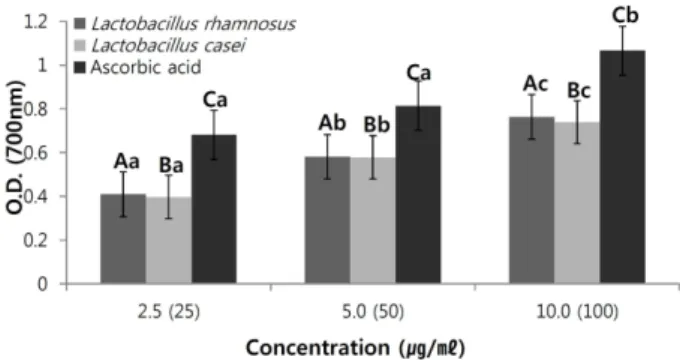
환원력은활성산소에전자를제공할수있는활성수소의 수치를측정한것으로흡광값이높으면활성산소제거능력이 높음을나타낸다. Lactobacillus rhamnosus와 Lactobacillus casei 파쇄물을각각 2.5%, 5.0%, 10.0% (v/v)의농도로사용 하였으며 ascorbic acid는 positive control로사용하였다. 실
험결과를보면각 sample의파쇄물농도가점차적으로증
가함에따라농도의존적으로환원력이증가하는경향을볼 수있었으며, positive control인 ascorbic acid의 O.D. 값이 1.0635로 가장 강한 환원력을 나타내었고, Lactobacillus rhamnosus가 0.7636, Lactobacillus casei가 0.7391의 O.D. 값 이측정되었다(Fig. 2). 위의결과로 Lactobacillus rhamnosus의 환원력은초고압공정을거친지치의추출물[13]보다작은 값이지만, 일반적인장내유산균인 Lactobacillus casei보다
높은항산화활성을가짐을확인할수있다. DPPH 자유라
디칼소거활성실험과마찬가지로항산화활성의증가는멜 라닌색소형성의신호전달경로에영향을미침으로써멜라 닌 색소 형성억제에 효과적으로 알려져 있다[31]. 따라서 Lactobacillus rhamnosus 파쇄물이체내의활성산소를감 소시키므로항산화활성이증가됨에따라활성산소에영향 을받는멜라닌생성기작에도영향을미치게되어피부미백 효능이동반될것이라사료되기에다음과같은미백효능실 험을진행하였다.
Tyrosinase 억제 효과 탐색
Tyrosinase는피부의멜라닌색소의생성을촉진하는효
소로 tyrosinase의저해활성을측정함으로써각시료들의미
백효과를알아볼수있다.
Lactobacillus rhamnosus와 Lactobacillus casei 파쇄물 Table 1. Free radical scavenging activity of the Lactobacillus
rhamnosus and Lactobacillus casei.
Sample (10%, v/v) The DPPH radical scavenging activity (%)*
Lactobacillus rhamnosus 14.91%± 0.48Aa Lactobacillus casei 13.21%± 0.26Aa
Ascorbic acid 56.56%± 0.18Bb
*Mean values± SD from triplicate separated experiments are shown.
Mean with difference letter (A-C) within same concentration are significantly different at p < 0.05 and mean with differ- ence letter (a-b) within same sample are significantly differ- ent at p < 0.05.
Fig. 2. Reducing power of Lactobacillus rhamnosus and Lactobacillus casei.
*Mean values± SD from triplicate separated experiments are shown. Mean with difference letter (A-C) within same concen- tration are significantly different at p < 0.05 and mean with difference letter (a-c) within same sample are significantly different at p < 0.05.
을각각 2.5%, 5.0%, 10.0% (v/v)의농도로처리하고, positive control로써 hydroquinon을 25, 50, 100 µg/ml 농도로투여 하여 tyrosinase 저해활성을 측정하였다(Fig. 3). Positive control인 hydroquinone의경우가장 높은농도인 100 µg/
ml에서 40.65%의미백활성을나타내었으며, Lactobacillus rhamnosus가 2.5%, 5.0%, 10.0% (v/v)의농도에서각각 20.6%, 26.8%, 31.1%의 tyrosinase 저해 활성을 나타내었으며, Lactobacilllus casei도같은 농도에서각각 11.6%, 13.2%, 17.7%의 tyrosinase 저해활성을나타내었다. Lactobacillus rhamnosus가 Lactobacillus casei와비교하여약 2배에가까 운높은미백활성을보였다. Kim의연구에서는 1 mg/ml 농
도의녹차추출물이 38.64%의미백활성을나타낸것과비교
하면 Lactobacillus rhamnosus 파쇄물만으로도천연추출 물만큼의미백활성을나타낸다는것을확인할수있었다. 위의 결과로 Lactobacillus rhamnosus 파쇄물이 Lactobacillus casei 파쇄물의약 2배이상의 tyrosinase 저해활성을나타냄에따 라, 멜라닌생성에관여하는효소인 tyrosinase를저해하여 멜라닌생성을방해해미백효과를나타낼것으로생각되며, 추후미백기능을가지는향장소재로써효과적으로이용될 수있을것으로사료된다.
Melanoma cell을 이용한 멜라닌 생성량 측정
Melanoma 세포를이용하여시료투여시멜라닌생합성
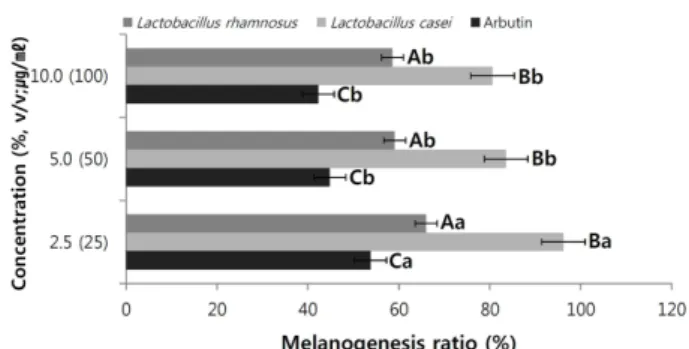
과세포생존율에미치는영향을관찰하기위하여농도별로 처리하고 24시간후, 멜라닌생성량을측정하였다(Fig. 4).
실험 결과, Lactobacillus rhamnosus의 경우 2.5%, 5.0%, 10.0% (v/v) 농도에서각각 66%, 59% 58.6%의생성률을보 였으며, Lactobacillus casei 파쇄물은같은농도에서 96.2%, 83.6%, 80.6%의 생성률을 보였다. positive control인 hydroquinone의 경우 25, 50, 100 µg/ml의농도에서각각
53.8%, 46.2%, 40.9%의낮은생성률을보였다. 위의세포독 성결과에서 Lactobacillus casei가가장높은세포독성을가 지는데에도불구하고 Lactobacillus rhamnosus 파쇄물의 멜라닌생성량이낮은결과로보아, 멜라닌의생성을억제하 는활성물질이함유되어있을것이라사료된다. 미백효능이 뛰어나다고알려진고로쇠수액의경우 100µg/ml의농도에
서 35% 가량의멜라닌생성저해율을나타낸것과비교하여
도 Lactobacillus rhamnosus 파쇄물의미백활성이 월등히 우수한것이확인되었다[12]. 또한초고압추출공정에의한 당귀추출물의미백및자외선차단효과연구에서보고된
초고압처리된당귀의멜라닌생성량이 82.4%인것과비교
하면 Lactobacillus rhamnosus 파쇄물의미백활성이높다는 것을유추할수있다[11]. Positive control인 hydroquinone의 미백활성이 Lactobacillus rhamnosus에비해약 15% 정도 높지만, 대표적인미백제로알려진 hydroquinone은높은미 백활성과함께독성을가지고있는합성물질로현재많은연 구진들이이러한합성미백제를대체할수있는천연물질들 에대해연구중에있다. 위의결과를토대로독성이거의없 는 Lactobacillus rhamnosus가합성미백제인 hydroquinone의 미백활성보다낮지만피부적합성이높아합성미백제및기 타천연물을대체할수있는긍정적인향장소재로다가갈수 있으며, 멜라닌생성은줄이면서세포독성은낮아야하는향 장미백제로서 Lactobacillus rhamnosus의활용가능성이매 우높을것으로사료된다.
요 약
유산균은국내외연구진들에의하여피부장벽을강화하는 효능및미백, 보습효과가뛰어나다는연구결과가보고된 바 있다[5, 26]. Lactic acid인 Lactobacillus rhamnosus는 Fig. 3. Tyrosinase inhibition activity of Lactobacillus rhamnosus
and Lactobacillus casei.
*Mean values± SD from triplicate separated experiments are shown. Mean with difference letter (A-C) within same concen- tration are significantly different at p < 0.05 and mean with difference letter (a-b) within same sample are significantly different at p < 0.05.
Fig. 4. Melanogenesis ratio of Lactobacillus rhamnosus and Lactobacillus casei.
*Mean values± SD from triplicate separated experiments are shown. Mean with difference letter (A-C) within same concentration are significantly different at p < 0.05 and mean with difference letter (a-b) within same sample are significantly different at p < 0.05.
보편적으로많이사용하는프로바이오틱스인 Lactobacillus casei보다낮게측정되는세포독성결과와높은 tyrosinase 억제활성을바탕으로멜라닌생성을줄임에따라미백활성을 가진다. 또한 Lactobacillus rhamnosus 파쇄물은 melanoma cell의멜라닌생성저해능이 Lactobacillus casei보다월등 히높아, 높은미백향장활성을가지는것을확인할수있
다(Table 2). 이는곧천연물질로써기존에사용하던합성미
백제보다피부에독성을주지않고, 부작용없이피부에적 합한향장소재로판단되며, 현대의새로운미백향장소재로 서활용가능성이매우높을것으로사료된다.
더나아가항산화활성은멜라닌세포형성억제에직접 적으로영향을주며, 주름및노화등의출발점이되어, 미 백활성도증가됨을확인할수있다. 따라서, 기능성향장소 재로써가치가높은 Lactobacillus rhamnosus는합성물질 및기타천연물을대체할수있는항산화및미백기능성향 장소재로다가갈수있으며, 향장소재로써선택의폭을늘릴 수있을것으로사료된다.
Acknowledgements
This study was supported by a grant of the Korea Healthcare technology R&D Project, Ministry of Health & Welfare, Republic of Korea (Grant NO. : A103017).
References
1. Ahoutupa, M., M. Saxelin, and R. Korpela. 1996. Antioxidative properties of Lactobacillus GG. Ntr. Today (suppl.). 31: 518- 528.
2. Bennett, P. B., R. E. Marquis, and I. Demchenko. 1998. High pressure biology and medicine. University of Rochester Press. New York.
3. Cho, W. G. 2007. Comparision of drug delivery using hairless and pig skin. J. Korean Oil Chem. Soc. 24: 410-415.
4. Dean, R. T. and M. J. Gieseg Davises. 1993. Reactive spe- cies and their accumulation on radical damaged proteins.
Trends Biochem. Sci. 18: 437-441.
5. Di Mazio, L., B. Cinque, C. De Simone, and M. G. Cifone.
1999. Effect of the lactic acid bacterium Streptococcus ther- mophilus on ceramide levels in human keratinocytes in vitro and stratum corneum in vivo. J. Invest. Dermatol. 113: 98-106.
6. Dietz, B. M., Y. H. Kang, G. Liu, A. L. Eggler, P. Yao, L. R.
Chadwick, G. F. Pauli, N. R. Farnsworth, A. D. Mesecar, and R. B. van Breemen. 2005. Xanthohumol isolated from Humu- lus Lupulus inhibits menadione-induced DNA damage through induction of quinone reductase. Chem. Res. Toxicol.
18: 1296-1305.
7. Furrie Elizabeth. Probiotics and allergy. 2005. P. Nutr. Soc. 64:
465-469.
8. Gibson, G. R. and M. B. Roberfroid. 1995. Dietary modulation of human colonic microbiota: Introducing the concept of probi- otics. J. Nutr. 125: 1401-1412.
9. Kim, C. H., M. C. Kwon, J. G. Han, C. S. Na, H. G. Kwak, G. P.
Choi, U. Y. Park, and H. Y. Lee. 2008. Skin-whitening and UV- protective effects of Angelica gigas Nakai ectracts on ultra high pressure extraction process. Korean J. Med. Crop Sci.
26: 110-116.
10. Kim, H. Y. and J. S. Ham. 2003. Antioxidative ability of lactic acid bacteria. Korean J. Food Sci. Ani. Res. 23: 186-192.
11. Kim, I. C. 2008. Antioxidative property and whitening effect of the polygoni multiflori radix, polygonati rhizoma and ephedrae herba. J. Korean Oil Chem. Soc. 25: 533-538.
12. Kim, J. S., Y. C. Seo, W. Y. Choi, H. S. Kim, B. H. Kim, D. H.
Shin, C. S. Yoon, H. W. Lim, J. H. Ahn, and H. Y. Lee. 2011.
Enhancement of antioxidant activities and whitening effect of acer mono sap through nano encapsulation processes.
Korean J. Med. Crop Sci. 19: 191-197.
13. Kim, J. S., M. H. Jeong, W. Y. Choi, Y. C. Seo, C. J. Ma, J. H.
Ahn, N. S. Kim, B. Hwang, J. S. Cho, and H. Y. Lee. 2011.
Enhancement of whitening effects of Lithospermum eryth- rorhizon extracts by ultra high pressure. Korean J. Med. Crop Sci. 19: 97-102.
14. Kim, S. S., M. H. Jeong, Y. C. Seo, J. S. Kim, N. S. Kim, J. H.
Ahn, B. Hwang, and H. Y. Lee. 2010. Comparison of antioxi- dant activity by high pressure extraction of Codonopsis lan- ceolata from different production areas. Korean J. Med. Crop Table 2. Comparison of cosmeceutical activities of Lactobacillus rhamnosus and Lactobacillus casei.
Concentration (%, v/v)
Lactobacillus rhamnosus Lactobacillus casei
2.5 5.0 10.0 2.5 5.0 10.0
Cytotoxicity (MTT) 2.7 4.6 7.6 6.1 7.2 9.8
Tyrosinase inhibition activity 20.6 26.8 31.3 11.6 13.2 17.7
Melanogenesis activity 66 591 58.6 96.2 83.6 80.6
Radical scavenging activity (DPPH) 13.7 14.4 14.9 12.2 12.7 13.4
Reducing power (O.D.) 0.410 0.581 0.763 0.397 0.578 0.739
*Mean values± SD from triplicate separated experiments are shown.
Sci. 18: 248-245.
15. Korpela, R., K. Peuhkuri, T. Lahteenmaki, E. Sievi, M. Saxelin, and H. Vapaatalo. 1997. Lactobacillus rhamnosus GG shows antioxidative properties in vascular endothelial cell culture.
Milchwissenschaft. 52: 503-505.
16. Kopp-Hellihan Lori. 2001. Prophylactic and therapeutic uses of probiotics. J. Am. Dietetic Assoc. 101: 229-241.
17. Lim, A. K., Y. J. Jung, J. S. Kim, Y. H. Kim, J. H. Kwak, J. H.
Hong, H. Y. Kim, and D. I. Kim. 2010. Skin UVB photo aging effect from extract of fermented Reynoutria elliptica. J Korean Soc Food Sci Nutr. 39: 369-375.
18. Lim, S. W., H. C. Ryoo, and S. H. Lee. 2002. Understanding of skin aging and its prevention and care. The J. Skin Barrier Res. 4: 71-80.
19. Macrae, R. G., R. K. Robinson, and M. J. Sadler. 1993. Ency- clopedia of food science. Food Technol. Nutr. 1: 607-171.
20. Oyaizu M. 1986. Studies on products of browning reaction:
antioxidative activities of products of browning reaction pre- pared from glucosamine. Jpn. J. Nutr. 44: 307-315.
21. Park, H. S., H. S. Lee, and T. B. Uhm. 1998. Selection of microorganisms for probiotics and their characterization. J.
Korean Soc. Food Sci. Nutr. 27: 433-440.
22. Park, J. M., J. Y. Lee, T. S. Park, S. J. Hyun, H. H. Kim, Y. J.
Cho, O. J. Kwon, A. R. Son, D. S. Kim, and B. J. An. 2008. A study on the cosmeceutical activities of prunus sargentii R.. J.
Korean Soc. Appl. Biol. Chem. 51: 70-78.
23. Pavel, S. and F. A. Muskiet. 1980. Euemlanin (precursor) metabolites as markers for pigmented malignant melanoma, a preliminaryreport. Cancer Detect Prev. 6: 311-318.
24. Pomerantz, S. H. 1963. Seperation, purification and proper- ties of two tyrosinases from hamster melanoma. J. Biol.
Chem. 238: 2351-2357.
25. Prota, G. 1990. Recent advances in the chemistry of melano- genesis in mammals. J. Invest. Dermatol. 75: 122-128.
26. Prince, T., A. J. McBain, and C. A. O’Neill. 2012. Lactobacillus reuteri protects epidermal keratinocytes from Staphylococcus aureus-induced cell death by competitive exclusion. Appl.
Environ. Microbiol. 78: 5119-5126.
27. Sanders, J. W., K. J. Leehouts, A. J. Haandrikman, G. Ven- ema, and J. Kok. 1995. Stress response in Lactococcus lac- tis:cloning, expression analysis and mutation of the lactococcal superoxide dismutase gene. J. Bacteriol. 177: 5254-5260.
28. Shin, J. Y. 2001. Screening of natural products that have activ- ities against skin-aging. Korea J. Food Nutr. 14: 568-572.
29. Song, C. H., Y. C. Seo, W. Y. Choi, C. G. Lee, D. U. Kim, J. Y.
Chung, H. C. Chung, D. S. Park, C. J. Ma, and H. Y. Lee.
2012. Enhancement of antioxidative activity of codonopsis lanceolata by stepwise steaming process. Korean J. Med.
Crop Sci. 20: 238-244.
30. Tobin, D. and A. J. Thody. 1994. The superoxide anion may mediate short but not long term effects of ultraviolet radiation on melanogenesis. J. Am. Acad. Dermatol. 3: 99-105.
31. Yamakoshi, J., F. Otsuka, A. Sano, S. Tokutake, M. Saito, M.
Kikuchi, and Y. Kubota. 2003. Lightening effect on ultraviolet- induced pigmentation of guinea pig skin by oral administration of a proanthocyanidin-rich extract from grape seeds. Pig Cell Res. 16: 629-638.
32. Yoon, T. J., W. S. Yang, S. M. Park, H. Y. Jung, A. N. Lee, J. H.
Jung, T. B. Kang, Y. C. Yoo, and J. B. Kim. 2009. In vivo toxic- ity and immunoadjuvant activity of Korean mistletoe (Viscum album coloratum) extract fermented with Lactobacillus.
Korean J. Food Sci. Technol. 41: 560-565.