Carcinoid tumors of the testes are very rare. Since Simon et al. reported the first case of primary testicular carcinoid tumor, about 70 carcinoid tumor cases have been record in the literature. However, no reports on the imaging findings of primary testicular carcinoid tu- mor have been published in the Korean literature.
We report a case of primary testicular carcinoid tumor along with a description of the ultrasonographic find- ings.
Case Report
A 50-year-old man presented with left scrotal swelling which he had been suffering from for three months. The patient denied having any other symptoms. He was re- ferred to our hospital by a local clinic for further evalua- tion. Physical examination revealed a palpable non-ten- der hard mass in the left testis. Scrotal ultrasonography showed an isoechoic solid mass with a peripheral hy-
poechoic rim and small anechoic cystic areas in the left testis, but no calcification was seen (Fig. 1A). Increased flow signals surrounding the intratesticular mass were seen on the Doppler image (Fig. 1B). The right testis was normal in both the physical examination and ultra- sonography. We suspected a non-seminomatous germ cell tumor of the left testis and, although secondary in- tratesticular cancer is uncommon, the presence of a sec- ondary malignance could not be excluded.
The levels of serum testicular tumor markers such as α-fetoprotein, β-human chorionic gonadotrophin and lactate dehydrogenase were normal. However, the level of urine 5-hydroxy-indole acetic acid was not checked.
Computed tomography scans of the chest, abdomen and pelvis were unremarkable. The patient underwent left orchiectomy.
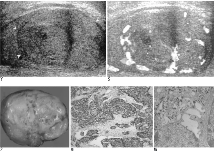
The gross specimen consisted of an ovoid, solid, whitish-yellow mass, measuring 3.2×2.4×2.0 cm re- placed by tumor except for the peripheral viable area (Fig. 1C). The nodular surface showed small cysts in the upper portion and spotted hemorrhage. Immunochemi- cally, the tumor cells were positive for cytokeratin, NSE and chromogranin (Fig. 1D). However, PLAP immunos- taining was negative. The electron microscopic study showed numerous dense-core neurosecretory granules in the cytoplasm of the tumor cells (Fig. 1E). The level of 5-hydroxy-indoleacetic acid in the urine collected fol-
J Korean Radiol Soc 2005;53:33-35
─ 33 ─
Primary Carcinoid Tumor of the Testis: Case Report1
Jeung Hee Moon, M.D. , Dae Young Yoon, M.D., Chul Soon Choi, M.D., Eun Joo Yoon, M.D., Sang Joon Park, M.D.,Young Lan Seo, M.D., Im Kyung Hwang, M.D., Yu-Jin Lee, M.D., Sung Yong Kim, M.D.2
Primary testicular carcinoid tumors are extremely rare and account for less than 1%
of all testicular tumors. We recently experienced a case of primary carcinoid tumor of the testis in a 50-year-old man who presented with scrotal swelling. The ultrasono- graphic findings were evaluated and compared with those of previous reports.
Index words :Carcinoid Testicular tumor Ultrasound (US)
1Department of Radiology, Hallym University College of Medicine, Kangdong Sacred Heart Hospital
2Department of Urology, Hallym University College of Medicine, Kangdong Sacred Heart Hospital
Received August 3, 2004 ; Accepted June 9, 2005
Address reprint requests to : Jeung Hee Moon, M.D., Department of Radiology, Kang-Dong Sacred Heart Hospital, 442 Gil-dong, Kangdong- gu, Seoul 134-701, Republic of Korea.
Tel. 82-2-2224-2312 Fax. 82-2- 448 -7370 E-mail: [email protected]
lowing the operation was within the normal range.
Discussion
Primary testicular carcinoid tumors are extremely rare, and most patients with this disease present in their fifth decade of life (1). The origin of carcinoid tumors is argentaffin or Kulchitsky cells, which are usually locat- ed within the crypts of Lieberkuhn (2).
Testicular carcinoid tumors have been divided into three subgroups, viz. primary testicular carcinoid tu- mors, carcinoid tumors associated with teratoma and carcinoid tumors metastatic to the testis. Even though the latter two groups are relatively unusual, the most important differential diagnosis is metastatic carcinoid tumor, and the differentiation between primary and
metastatic carcinoid tumor is difficult, both clinically and morphologically (3). To establish the diagnosis of a primary carcinoid tumor, the existence of primary le- sions elsewhere in the body must be excluded (4). The majority of carcinoid tumors originate from the gastroin- testinal tract, especially the appendix and ileocecal re- gion. CT of the abdomen and pelvis should be per- formed to exclude the existence of primary tumors in this region and lymphatic spread. The second most com- mon origin of carcinoid tumors is the lung; therefore, a plain radiograph of the chest or chest CT is helpful to rule out the presence of bronchial carcinoid tumor (1, 5, 6).
A painless mass and prominent testicular enlargement are common clinical findings. Other symptoms includ- ing carcinoid syndrome are rare in patients with prima-
Jeung Hee Moon, et al: Primary Carcinoid Tumor of the Testis
─ 34 ─
C D E
Fig. 1. A 50-year-old man with a primary carcinoid tumor of the testis.
A. Testicular US shows an isoechoic, slightly heterogeneous intratesticular mass (arrowhead) with a peripheral low echoic rim, but without calcification in the right testis.
B. On color flow Doppler US, increased flow signals are seen around the testicular mass.
C. Gross finding reveals solid mass. The cut surface shows a firm yellow nodular tumor with spotted hemorrhage and small cysts in the upper portion.
D. Immunohistochemically, the tumor cells are positive for synaptophysin (×100).
E. The electron microscopic findings show numerous dense core neurosecretory granules in the cytoplasm of the tumor cells.
A B
ry testicular carcinoid tumors.
Ultrasonography is the primary imaging modality in the evaluation of testicular tumors. Only a few cases (of primary testicular carcinoid tumor?) were reported pre- viously (2, 3, 7). In these cases, the US features included a solid well-circumscribed hypo or iso-echoic intratestic- ular mass with dense calcification. Grunshow et al. re- ported dense focal nodular calcification at the periphery of the mass. Calcifications located within the testicular parenchyma are found in germ cell tumors, especially in teratoma, embryonal cell carcinoma and granulomatous orchitis (8, 9). Microcalcifications due to teratoma repre- sent calcified cartilage and/or bone fragments (9).
However, the calcification in primary testicular carci- noid tumors may be dystrophic (2).
Some cases of primary testicular carcinoid tumor showed small cystic or necrotic foci within the mass (7, 8). In our case, even though there was no focal calcifica- tion, the ultrasound appearances of the testicular carci- noid tumor showed small cystic areas within a hypoe- choic intratesticular mass.
Because of the generally favorable prognosis, the man- agement of patients with a pure primary testicular carci- noid tumor usually includes inguinal orchiectomy for lo- cally confined tumors and the surgical resection of iso- lated metastatic foci (2). The overall incidence of metas- tasis is about 11% (1). A review of the literature showed that the tumor size and the presence of carcinoid syn- drome are features associated with a malignant course.
The elevation of urinary 5-hydroxyindoleacetic acid (5- HIAA) and serum serotonin levels following orchiec- tomy often suggests the possibility of a metastatic tumor
(1, 10).
Although their ultrasonographic findings are not spe- cific, testicular carcinoid tumors should be included in the differential diagnosis of a hypo- or isoechoic solid in- tratesticular mass containing calcification and small cys- tic or necrotic foci and, once diagnosed, it is important to carefully assess whether the tumor is primary or metastatic.
References
1. Glazier DB, Murphy DP, Barnard N, Cummings KB, Weiss RE.
Primary carcinoid tumor of the testis. BJU Int 1999;83:153-154 2. Nichols DA, James EM, Charboneau JW, Grantham JG. Imaging
of a primary carcinoid tumor of the testis. J Ultrasound Med 1985;4: 255-256
3. Grunshaw ND, Gopichandran TD. Case report: primary carcinoid tumor of the testis-ultrasound appearances. Clin Radiol 1993;47:
290-291
4. Hosking DH, Bowman DM, McMorris SL, Ramsey EW. Primary carcinoid of the testis with metastasis. J Urol 1981;125:255-256 5. Sullivan JL, Packer JT, Bryant M. Primary malignant carcinoid of
the testis. Arch Pathol Lab Med 1981;105:515-517
6. Kaufman JJ, Waisman J. Primary carcinoid tumor of testis with metastasis. Urology 1985;25:534-536
7. Frank RG, Gerard PS, Anselmo MT, Bennett L, Poeminger BI, Wise GJ. Primary carcinoid tumor of the testis. Urol Radiol 1991;12:203-205
8. Chang YH, Chuang CK, Wu CT, Ng KF, Liao SK. Primary carci- noid tumor of the testis: case report. Chang Gung Med 2002;25:695- 699
9. Martin B, Tubiana JM. Significance of scrotal calcifications detect- ed by sonography. J Clin Ultrasound 1988;16:157:775-780
10. Sutherland RS, Wettlaufter JN, Miller GJ. Primary carcinoid tumor of the testicle: case report and management schema. J Urol 1992;
148:880-882 J Korean Radiol Soc 2005;53:33-35
─ 35 ─
대한영상의학회지 2005;53:33-35
원발성 고환 유암종: 증례 보고1
1한림대학교 영상의학과
2한림대학교 비뇨기과
문증희・윤대영・최철순・윤은주・박상준・서영란・황임경・이유진・김성용2
원발성 고환 유암종은 매우 드물며 전체 고환암 중에서도 약 1% 미만의 빈도를 보인다. 저자들은 최근 음낭 부종을 보이는 50세 남자에서 발생한 원발성 고환 유암종 증례를 경험하였다. 초음파 소견을 평가하고 이전 문헌과 비교해 보 고자 한다.