http://dx.doi.org/10.5534/wjmh.2014.32.3.184
Case Report
Received: Mar 28, 2014; Revised: Jul 28, 2014; Accepted: Aug 5, 2014 Correspondence to: Sung Dae Kim
Department of Urology, Jeju National University School of Medicine, 15 Aran 13-gil, Jeju 690-767, Korea.
Tel: +82-64-717-1760, Fax: +82-64-717-1131, E-mail: [email protected] Copyright © 2014 Korean Society for Sexual Medicine and Andrology
This is an Open Access article distributed under the terms of the Creative Commons Attribution Non-Commercial License (http://creativecommons.
org/licenses/by-nc/3.0) which permits unrestricted non-commercial use, distribution, and reproduction in any medium, provided the original work is properly cited.
Diagnosis of a Gastrointestinal Stromal Tumor Presenting as a Prostatic Mass: A Case Report
Jung-Sik Huh, Kyung Kgi Park, Young Joo Kim, Sung Dae Kim
Department of Urology, Jeju National University School of Medicine, Jeju, Korea
Gastrointestinal stromal tumors (GISTs) are an unusual and heterogeneous group of spindle cell tumors that can also appear on the exterior of the gastrointestinal tract (extra-GISTs). Despite the fact that extra-GISTs or large rectal GISTs can lead to the clinical impression of a prostatic mass, these tumors are, in general, excluded in the differential diagnosis of spindle cell tumors observed on prostate needle biopsy. Here, we present, in detail, a case of an extra-GIST identified on prostatic biopsy; the tumor was previously believed to be a primary prostatic stromal sarcoma in the differential diagnosis. Every investigator should check for KIT (CD117) in immunohistochemical staining to rule out an extra-GIST prior to diagnosing a solitary prostatic tumor, specialized prostatic stromal tumor, or leiomyosarcoma on prostate needle biopsy.
Key Words: Gastrointestinal stromal tumors; Prostate
Most prostatic spindle cell lesions have very low preva- lence and lead to nonspecific laboratory findings and clin- ical symptoms; they include a widespread display of histo- pathological entities and are difficult to confirm with a defi- nite diagnosis. Besides, it is more difficult to evaluate these lesions if only a small amount of the specimen is available for prostatic biopsy [1]. Further, gastrointestinal stromal tu- mors (GISTs) are the most common gastrointestinal mesen- chymal tumors and spindle cell neoplasms. The first and second most common sites of these tumors are the stomach and the small intestine, respectively. Only a small percent- age of GISTs occur in the rectum. Extra-GISTs are com- monly found in the mesentery, greater omentum, and retroperitoneum. Overall, Anagnostou et al [2] reported 20
classified cases of extra-GISTs in the prostate gland, both primary extra-GISTs originating from the prostate and ex- tra-GISTs of the rectum extending to the prostate, by con- ducting a literature review. We added another seven cases of extra-GISTs presenting as prostatic masses by reviewing the literature published thus far. To the best of our knowl- edge, this report may be the second case in Korea. Here, we report a case of primary extra-GISTs originating from the prostate and highlight the possibility of extra-GISTs in the differential diagnosis of prostatic spindle cell lesions.
CASE REPORT
A 50-year-old man visited our hospital with a complaint
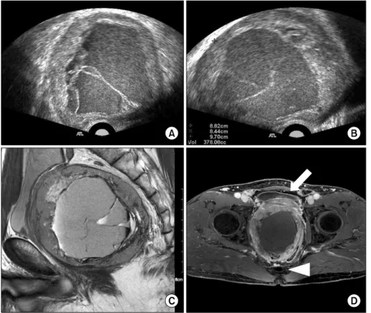
Fig. 1. (A, B) Transrectal ultra- sonography revealed that the huge prostatic mass measuring 97×88×
84 mm was well capsulated with internal hemorrhage. The mass was isolated from the surrounding structures. (C, D) Magnetic re- sonance image of the prostate showed an enlarged prostatic mass with hemorrhagic necrosis. The prostatic mass had a large size (110×85×86 mm) with hetero- geneous enhancement and dis- placed bladder (arrow) anteriorly and rectum (arrowhead) poster- iorly. This implies that the tumor was mainly localized within the prostate and there was no definite evidence of the direct invasion of adjacent organs.
of weak stream, residual urine sensation, and perineal discomfort. Previously, he had been diagnosed with be- nign prostatic hyperplasia and had been treated with med- ication for more than two years at local urologic clinics.
Otherwise, he was generally in good health. For these months, he had been experiencing worsening weak stream and urethral pain without any hematochezia or change in bowel habits. Digital rectal examination re- vealed non-specific findings for the prostate, except that it was apparently enlarged. The serum level of prostate-spe- cific antigen was normal (0.85 ng/mL). Transrectal ultra- sonography revealed that a huge prostatic mass measuring 97×88×84 mm was well capsulated with internal hemor- rhage and that the mass was isolated from the surrounding structures (Fig. 1A, 1B). The radiologist recommended ab- dominopelvic computed tomography (CT) and magnetic resonance (MR) imaging for an evaluation of the prostatic mass. CT scan showed direct invasion to adjacent organs with no metastasis. MR imaging of the prostate showed an enlarged prostatic mass with hemorrhagic necrosis. The prostatic mass was large (110×85×86 mm) with hetero- geneous enhancement, displacing the bladder anteriorly
and rectum posteriorly (Fig. 1C, 1D). This implied that the tumor was mainly localized within the prostate and there was no definite evidence of a direct invasion of adjacent organs. The patient then underwent five-core prostate bi- opsy guided with transrectal ultrasound. Histologically, the tumor was composed of spindle cells with vesicular nuclei and eosinophilic cytoplasm. These cells were ar- ranged in a whirling or fascicular pattern. There was no significant nuclear pleomorphism (Fig. 2A). Mitotic counts were more than five per 50 high-power fields. The tenta- tive diagnosis was prostatic stromal sarcoma. Tumor cells from the biopsy specimen showed strong and diffuse im- munohistochemical reactivity to KIT (CD117) and CD34, while negative immunohistochemical staining results were obtained for desmin, smooth muscle actin, cytoker- atin, and S-100 (Fig. 2B, 2C). These outcomes are con- cordant with the diagnosis of GIST. Therefore, we con- cluded that the final diagnosis was primary prostatic ex- tra-GISTs. Tumor genotyping was not carried out due to the high costs of this examination. To evaluate the tumor stage, the patient underwent gastroscopy and colono- scopy for the primary lesion, and chest CT and bone scan
Fig. 2. (A) The tumor is composed of relatively uniform spindle cells with vesicular nuclei and eosinophilic cytoplasm. These cells are arranged in whorled or fascicular pattern. There is no significant nuclear pleomorphism (H&E, ×200). (B) CD117 (c-kit) staining shows diffuse strong positive immunoreactivity in the cytoplasm of the neoplastic cells (×200). (C) Tumor cells also demonstrate diffuse strong cytoplasmic positive inmmunostaining for CD34 (×200).
for the distant metastasis, but there were no abnormal find- ings in these examinations. We did not start neoadjuvant treatment with imatinib because of its high cost and the pa- tient’s lack of medical expense insurance. Therefore, we planned to perform radical prostatectomy routinely; if the tumor had involved the rectum, we would have addition- ally performed colostomy after complete tumor resection.
The patient changed his mind a day before the operation;
he refused to undergo radical surgery and left the hospital against our advice. Therefore, we have not followed-up on him since then.
DISCUSSION
For a long time, the origin and nomenclature of GIST was a subject of much controversy. The putative cell of ori- gin of GIST is the interstitial cell of Cajal (ICC), which is the pacemaker cell of the gastrointestinal muscles. It is known that ICC expresses the gene product of KIT (CD117), a pro- to-oncogene that encodes the receptor tyrosine kinase, Kit. Because these ICC cells may exist in diverse anatomic sites in addition to the exterior tubular gastrointestinal tract, it is possible to explain unusual cases of extra-GISTs such as those of the uterus, vagina, prostate, and bladder [3].
As the origin of these extra-GISTs is not yet clear, it is dif- ficult to decide whether these tumors are real primary ex- tra-GISTs or secondary sites of GISTs, perhaps apart from the gastrointestinal tract and parasitic growth on another
location [2]. The morphologic features of GISTs are varia- ble, and their biological behavior is also difficult to predict. GISTs of the rectum represent only a small per- centage (5%) of all GISTs and are often found in males old- er than 50 years of age [2]. Large rectal GISTs involving the prostate or extra-GISTs originating from the prostate may be interpreted as primary prostatic tumors by using radio- logical studies and clinical manifestations such as lower urinary tract symptoms, including weak stream, inter- mittency, and residual urine sensation. Prostate enlarge- ment as a result of extra-GISTs is very unusual. We identi- fied 27 published cases of extra-GISTs diagnosed using prostatic tissue specimens from a review of the literature published to date, including papers on pelvic exentera- tion, radical prostatectomy, local excision of tumor, and ultrasound-guided prostatic biopsy (Table 1) [3-8]. We an- alyzed the 20 classified cases reported by Anagnostou et al [2] and found the other seven cases ourselves. The median age of all patients was 56.8 years (age range: 42∼82 years), and the clinical symptoms were mainly lower uri- nary tract symptoms (voiding difficulty, hematuria, and/or dysuria) and/or lower gastrointestinal tract symptoms (perianal discomfort and/or small stool diameter). The tu- mor size of extra-GISTs presenting as prostatic mass varied considerably (range: 1∼15 cm); most of these tumors were very huge, occupying a large volume of the pelvic area. This was observed in our case as well (11 cm).
Tumors from the 20 cases identified by Anagnostou et al [2] involved both the prostate and the rectum; those from
Table 1. Reviewed gastrointestinal stromal tumors diagnosed on prostatic histological examination Case
No. Study (year) Age
(yr) Tumor
size (cm) Tumor location Type of surgery Follow-up
interval (mo) Metastasis 1 Voelzke et al (2002) 62 N/A Anterior rectal wall,
prostate, bladder base Pelvic exenterationa 14 Mesentery 2 Madden et al (2004) 45 15.0 Adherent to rectum CP+portion of rectum 51 Liver 3 Madden et al (2004) 54 5.5 Perirectal soft tissue Pelvic exenterationa 75 Liver 4 Madden et al (2004) 82 7.9 Undetermined rectum,
prostate TURP, pelvic
exenteration 22 None
5 Sandblom et al (2005) 51 13.0 Rectum, prostate RCP+rectum 13 None
6 Van der Aa et al [4]
(2005) 49 14.2 Prostate Nonea 25 Liver
7 Herawi et al [3]
(2006) 64 5.4 Rectum, attached to
prostate APR+RP 48 None
8 Herawi et al [3]
(2006) 50 7.0 Rectum, prostate
effacement APR+CP 42 None
9 Herawi et al [3]
(2006) 61 7.4 Prostate, rectum, bladder,
seminal vesicle RP 12 None
10 Herawi et al [3]
(2006) 51 1.7 Between prostate/rectum Local excision 48 None
11 Herawi et al [3]
(2006) 42 8.5 Undetermined attached
to prostate Nonea 4 N/A
12 Herawi et al [3]
(2006) 48 1.0 Rectum submucosal Nonea 48 N/A
13 Herawi et al [3]
(2006) 65 N/A Unknown Nonea N/A N/A
14 Herawi et al [3]
(2006) 48 N/A Unknown Nonea 4 N/A
15 Herawi et al [3]
(2006) N/A N/A N/A Nonea N/A N/A
16 Herawi et al [3]
(2006) 75 N/A Rectum, prostate TURP, RP N/A N/A
17 Yinghao et al [5]
(2007) 75 6.7 Prostate TURP, RP 6 None
18 Yinghao et al [5]
(2007) 49 8.0 Prostate RPa 14 None
19 Arce-Lara et al [6]
(2007) 61 9.0 Prostate RPa 14 None
20 Arce-Lara et al [6]
(2007) 61 7.7 Prostate RPa N/A None
21 Dickson et al (2008) 48 N/A Rectum, prostate Retropubic
prostatectomy N/A None
22 Yaman et al (2008) 58 N/A Rectum, prostate N/A N/A N/A
23 Park et al [8]
(2008) 58 7.5 Prostate RP, retropubic
prostatectomy 6 None
24 de la Roza et al [7]
(2009) 48 13.0 Rectum, prostate, bladder Pelvic exenterationa 18 None 25 de la Roza et al [7]
(2009) 46 9.0 Rectum, prostate, bladder Nonea N/A Lung
26 Loeb et al (2009) 56 14.0 Rectum, prostate Pelvic exenterationa N/A None
27 Yanovskiy et al (2010) 79 N/A Between rectum/prostate RP N/A N/A
N/A: not available, CP: cystoprostatectomy, TURP: transurethral prostatectomy, RCP: radical cystoprostatectomy, RP: radical prostatectomy, APR: abdominal perineal resection.
aDiagnosis on biopsy.
the two other cases were believed to originate from the space between these two organs. These cases may demon- strate extension into the prostate from a primary rectal origin. A small percentage, five cases (18.5%), of the pub- lished reports reported a primary prostatic origin [4-9].
Further, only three of these cases were confirmed to origi- nate not from rectal GISTs but from primary prostatic GISTs through complete specimens obtained by radical surgery [5,8,9]. In the case reported by Arce-Lara et al [6], the researchers were not confident of their decision re- garding the origin of the tumors and therefore, were un- able to regard the tumors as primary prostatic extra-GISTs due to the proximity of the rectum and the patient’s re- sponse to the neoadjuvant treatment with a tyrosine kin- ase inhibitor such as imatinib.
Further, although the present report may be presumed to be the second case of primary prostatic extra-GIST in Korea, there are limitations to this report. A weakness in the present report is that because the patient did not under- go radical surgery, our case was based only on prostatic bi- opsy and radiological studies, like the case reported by Van der Aa et al [4]. On the basis of the same principle, it is not certain whether this tumor was really a primary pro- static extra-GIST.
On the contrary, because neoadjuvant therapy with a tyrosine kinase inhibitor such as imatinib has a large effect on the patient’s prognosis, it is very important to dis- tinguish between extra-GISTs presenting as prostatic masses and other primary prostatic tumors of spindle cell lesions. Further, immunohistochemical outcomes are the most useful methods for distinguishing between them.
Primary prostatic extra-GISTs presented uniformly strong positive responses for both KIT and CD34 stains, while the other prostatic tumors only presented positive responses for CD34 stain only [5].
Further, Fletcher et al [10] proposed the consensus cri- teria for defining the risk of aggressive behavior in GISTs at the National Institutes of Health. He created a classi- fication of risk groups (low, intermediate, and high) of GISTs using tumor size and mitotic count as predictable values. According to these criteria, the patient fell into the high-risk category and should have been promptly treated with imatinib and radical surgery.
To summarize, we report an unusual case of extra-GIST
originating from the prostate as the primary site. It is im- portant that clinicians take great caution with the diag- nosis of all patients, such that rectal invasion first be ex- cluded in order to confirm the diagnosis of primary pro- static extra-GISTs.
We concluded that extra-GISTs should always be in- cluded in the differential diagnosis of any prostatic spindle cell tumor. Early testing by means of positive KIT (CD117) results will lead to suggestions of immediate imatinib neo- adjuvant therapy or surgical resection.
ACKNOWLEDGEMENTS
This work was supported by a research grant from Jeju National University Hospital.
REFERENCES
1. Hansel DE, Herawi M, Montgomery E, Epstein JI. Spindle cell lesions of the adult prostate. Mod Pathol 2007;20:148- 58.
2. Anagnostou E, Miliaras D, Panagiotakopoulos V. Diagnosis of gastrointestinal stromal tumor (GIST) on transurethral re- section of the prostate: a case report and review of the literature. Int J Surg Pathol 2011;19:632-6.
3. Herawi M, Montgomery EA, Epstein JI. Gastrointestinal stro- mal tumors (GISTs) on prostate needle biopsy: a clin- icopathologic study of 8 cases. Am J Surg Pathol 2006;30:
1389-95.
4. Van der Aa F, Sciot R, Blyweert W, Ost D, Van Poppel H, Van Oosterom A, et al. Gastrointestinal stromal tumor of the prostate. Urology 2005;65:388.
5. Yinghao S, Bo Y, Xiaofeng G. Extragastrointestinal stromal tumor possibly originating from the prostate. Int J Urol 2007;14:869-71.
6. Arce-Lara C, Shah MH, Jimenez RE, Patel VR, Benson DM Jr, Clinton SK, et al. Gastrointestinal stromal tumors involv- ing the prostate: presentation, course, and therapeutic approach. Urology 2007;69:1209.e5-7.
7. de la Roza G, Naqvi A, Clark K. Gastrointestinal stromal tu- mors presenting as a prostatic mass. Can J Urol 2009;16:
4502-6.
8. Park SW, Lee W, Huh GY, Chung MK. Gastrointestinal stro- mal tumor of prostate. Korean J Urol 2008;49:383-5.
9. Lee CH, Lin YH, Lin HY, Lee CM, Chu JS. Gastrointestinal stromal tumor of the prostate: a case report and literature review. Hum Pathol 2006;37:1361-5.
10. Fletcher CD, Berman JJ, Corless C, Gorstein F, Lasota J, Longley BJ, et al. Diagnosis of gastrointestinal stromal tu- mors: a consensus approach. Int J Surg Pathol 2002;10:
81-9.