Printed in the Republic of Korea
http://dx.doi.org/10.5012/jkcs.2013.57.6.721
Structural and Spectral Characterization of a Chromium(III) Picolinate Complex:
Introducing a New Redox Reaction
Mohammad Hakimi*
Chemistry Department, Payame Noor University, 19395-4697 Tehran, I. R. Iran.
*E-mail: [email protected]
(Received August 21, 2013; Accepted September 22, 2013)
ABSTRACT. Reaction between 2-pyridinecarboxylic acid (Hpic) and K3[Cr(O2)4] give complex [Cr(pic)3].H2O (1) which is characterized by elemental analysis and spectroscopic methods (FT-IR, Raman) and X-ray crystallography. In the crystal structure of 1, chromium atom with coordinated by three nitrogen and three oxygen atoms has a distorted octahedral geome- try. Also a water molecule is incorporated in crystal network. Each water molecule acts as hydrogen bond bridging and con- nects two adjacent complexes by two O–H···O hydrogen bonds.
Key words: Chromium(III) complex, Picolinate, Spectral characterization, X-ray crystal structure
INTRODUCTION
Pyridine derivatives are important intermediates widely used in the synthesis of drugs1 and pesticides.2 The struc- tures and chemical properties of metal complexes of some pyridine carboxylates have been widely investigated.3 Some chromium complexes of the pyridine carboxylates have been known as potential bioavailable sources of chromium (III) as a model of glucose tolerance factor (GTF).3a,b
We are interesting in design and synthesis of complexes with pyridine derivatives and also N,O-donor ligands. And in the past few years, we have published a series of paper in this field.4 In this paper preparation of the chromium(III) complex, [Cr(pic)3].H2O (1), with 2-pyridinecarboxylic acid (Hpic, Scheme 1) is presented and spectroscopic charac- teristics (IR, Raman) and crystal structure of this complex are determined.
Study of the CSD structures by ConQuest revealed that the chromium atom has very different coordination numbers including two,5 three,6 four,7 five,8 six,9 seven,10 eight,11 nine,12 ten,13 eleven,14 twelve,15 and thirteen.16 In this paper we endeavored to introduce coordination aspect of the Hpic with chromium atom.
EXPERIMENTAL
All starting chemicals and solvents were reagent or ana- lytical grade and used as received. The infrared spectrum of a KBr pellet in the range 4000–400 cm–1 was recorded with a FT-IR 8400-Shimadzu spectrometer. The carbon, hydrogen and nitrogen contents were determined in a Thermo Finnigan Flash Elemental Analyzer 1112 EA. The Raman spectrum was performed using a Nicolet Model 910 Fourier-transform spectrometer. The melting point was determined with a Barnsted Electrothermal 9200 electri- cally heated apparatus.
Synthesis of [Cr(pic)3].H2O (1)
A solution of 0.37 g (3 mmol) of Hpic, dissolved in H2O (5 mL), was added with stirring to a solution of 0.297 g (1 mmol) of K3[Cr(O2)4] in H2O (15 mL). The reaction mixture was stirred for 1 h. Red crystals suit- able for X-ray diffraction were obtained from the solu- tion after standing for 7 d. Yield: 0.284 g (65%). m.p.
>300oC. Anal. Calcd for C18H14CrN3O7 (%):C, 49.55;
H, 3.23; N, 9.63. Found: C, 48.89; H, 3.23; N, 9.66. IR (cm−1, KBr): 3525 w (νas H2O), 3315 w (νs H2O), 3063 w (ν CH), 1680 s (ν C=O and/or δ H2O), 1605 m (ν C=N), 1470 m (ν C=C), 1327 s, 1288 m, 1157 m (ν CO), 1049 w (ν CN), 856 m, 764 m and 694 w (δ py), 671 w (ρr
H2O), 548 w (ν CrO and/or ρw H2O), 471 m (ν CrN).
Raman: 3025 m (ν CH), 1601 vs (ν C=N), 1485 w (ν C=C), 1205 w (ν CO), 1108 m (ν CN), 980 w, 864 m, 713 w (ρr H2O), 616 m (ν CrO and/or ρw H2O), 471 m (ν CrN), 365 w, 257 w (ρt H2O).
Scheme 1. Chemical structure of the 2-pyridinecarboxylic acid (Hpic).
Crystal Structure Determination and Refinement The data collection for 1 was carried out by Bruker APEX-II CCD diffractometer, using graphite-monochro- mated MoKα (λ = 0.71073Å) radiation at 296 K. The data were integrated with SAINT and corrected for Lorentz polarization and absorption performed using SADABS.17 The structure was solved by Patterson methods, implemented in SHELXS-97.18 Refinement by full-matrix least squares methods based on F2 values against all reflections has been performed by SHELXL-97,18 including anisotropic displacement parameters for all non-H atoms. The position of hydrogen atoms belonging to the carbon atoms Csp2 were geometrically optimized applying the riding model [Csp2–H, 0.93Å; Uiso(H) = 1.2Ueq(C)]. Calculations con- cerning the molecular geometry, the verification of space group, the analysis of hydrogen bonds were performed with PLATON.19
CCDC 880374 for [Cr(pic)3].H2O (1) contain the sup- plementary crystallographic data for this paper. These data can be obtained free of charge from The Cambridge Crystallographic Data Centre via www.ccdc.cam.ac.uk/
data_request/cif.
RESULTS AND DISCUSSION Synthesis and Spectroscopic Characterization
Reaction of Hpic with a water solution of K3[Cr(O2)4] in a molar ratio of 1:3 (M:L) give complex 1 (Scheme 2). The complex was air-stable and soluble in H2O. Similar struc- ture has been reported previously,20 but they used of different precursor, Cr(NO3)3.9H2O, under pH condition. The crys- tallography of 1 has been well done in low temperature than the previously report. Also meanwhile, this paper is presented new redox reaction which is interesting in coor- dination chemistry.
Suggested reaction equation is presented in Scheme 2.
During the reaction, peroxide ligands are replaced by three pic and Cr(V) is reduced to Cr(III) by peroxide anions (Scheme 3). After reduction, buabbles of oxygen molecules are removed from the solution. Other peroxide onions which are not involved in reduction process are neutralized by H+ ions from Hpic and K+ of K3[Cr(O2)4].
In the IR spectrum of Hpic,21 there is a bands at 1722 cm–1 which were assigned to the vibrations of the ν (C=O).
This band is shifted 42 cm–1 to lower energy in 1 indi- cating deprotonation of carboxylic acid group and coor- dination through the oxygen atom.
The presence of a water molecule in 1 affects the IR and Raman spectra in three regions including 3525 and 3315 cm–1 for asymmetric and symmetric OH stretches, 1680 cm–1 for H2O bending and 250–700 cm–1 for “librational modes”. These modes are due to rotational oscillations of the water molecules restricted by interactions with neigh- boring atoms and they are classified into three types (wag- ging (ρw), twisting (ρt) and rocking (ρr)) depending upon the direction of the principal axis of rotation.22 The ring wagging vibrations of the pyridine groups were also observed at 694 and 764 cm–1.
Information about the low frequency of metal-ligand vibrations can be obtained by Raman spectroscopy.23 In the Raman spectra of 1, the bands at 616 and 471 cm–1 were assigned to Cr–O and Cr–N stretching vibrations respectively. Similar results were reported previously.24 The X-ray analysis confirms the higher stretching vibra- tions of Cr–O bond respect to the Cr–N.
Description of the Crystal Structure
The crystal structure of [Cr(pic)3].H2O (1) has been determined by single-crystal X-ray diffraction analysis.
Diagrams of the molecular structure and unit cell were created using Ortep-III25 and Diamond Mercury.26 Crystallographic data and details of the data collection and structure refine- ment are listed in Table 1. Selected bond lengths and angles for complex are listed in Table 2 and hydrogen bond dis- tances, d(H···A), and angles are presented in Table 3.
In the crystal structure of 1 (Fig. 1), the chromium atom is coordinated by three nitrogen and three oxygen atoms of Hpic in distorted octahedral geometry (Fig. 2). Also a water molecule is presence present in this structure and trapped by hydrogen bonds. There are three five-membered chelate rings around the chromium atom which are almost planar (rms deviation 0.080 Å for O1). The Cr–N and Cr–O bond distances in 1 are comparable with the analogous ones reported in the literature for complexes which con- tain dipicolinate coordinated to chromium(III) ion in an O,N,O'-tridentate fashion.27
In the network of 1, each water molecule acts as hydro- Scheme 2. Suggested reaction equation for synthesis of [Cr(pic)3].
H2O (1).
Scheme 3. Reduction of Cr(V) to Cr(III) by peroxide onion.
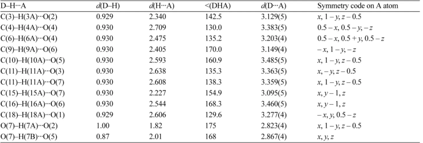
gen bond bridge and connects two adjacent complexes (Fig. 3). The O7 atom of H2O molecule participates in hydrogen bonding as a proton donor and connects O5 atom of one complex to O2 atom in other complex by strong hydrogen bonds (strong hydrogen bonds range in litera- ture is 1.5–2.2 Å28).
Table 1. Crystal data and structure refinement for 1 Empirical formula C18H14CrN3O7
Formula weight (g mol−1) 436.32
Temperature (K) 296
Crystal system Monoclinic
Space group C2/c
Unit cell dimensions (Å, °)
A 30.143(7)
B 8.4769(19)
C 13.870(3)
B 95.479(3)
Volume (Å3), Z 3528.0(14), 8
Calculated density (mg m−3) 1.643 Absorption coefficient (mm−1) 0.70
F(000) 1784
Crystal size (mm−3) 0.20 × 0.10 × 0.10 θ range for data collection (°) 1.4–24.8
h, k, l ranges –30:35, –9:10, –14:16 Reflections collected 8031
Independent reflections 3053
Rint 0.048
Data / restraints / parameters 3053 / 0 / 270 Goodness-of-fit on F2 1.096
Final R indexes [I > 2σ(I)] R1 = 0.0380, wR2 = 0.0848 R indexes (all data) R1 = 0.0635, wR2 = 0.1116 Largest diff. peak and hole (e.Å−3) 0.30 and –0.35
Table 2. Selected bond length (Å) and angles (°) for 1 with esti- mated standard deviations in parentheses
Distances Angles
Cr1–N1 2.063(3) N1–Cr1–O1 81.2(1)
Cr1–N2 2.055(3) N2–Cr1–O3 80.8(1)
Cr1–N3 2.045(3) N3–Cr1–O5 80.5(1)
Cr1–O1 1.945(2) O1–Cr1–N2 170.7(1)
Cr1–O3 1.946(3) O1–Cr1–N3 89.9(1)
Cr1–O5 1.955(2) O1–Cr1–O5 94.4(1)
Table 3. Hydrogen bond geometries (Å, º) for 1
D–H···A d(D–H) d(H···A) <(DHA) d(D···A) Symmetry code on A atom
C(3)–H(3A)···O(2) 0.929 2.340 142.5 3.129(5) x, 1 – y, z – 0.5
C(4)–H(4A)···O(4) 0.930 2.709 130.0 3.383(5) 0.5 – x, 0.5 – y, – z
C(6)–H(6A)···O(4) 0.930 2.475 135.2 3.203(4) 0.5 – x, 0.5 + y, 0.5 – z
C(9)–H(9A)···O(6) 0.930 2.405 170.0 3.149(4) – x, 1 – y, – z
C(10)–H(10A)···O(5) 0.930 2.593 160.9 3.485(5) x, 1 – y, z – 0.5
C(11)–H(11A)···O(3) 0.930 2.638 135.3 3.363(5) x, – y, z – 0.5
C(11)–H(11A)···O(7) 0.930 2.608 138.3 3.359(5) x, 1 – y, z – 0.5
C(15)–H(15A)···O(7) 0.930 2.227 154.9 3.095(5) x, y – 1, z
C(16)–H(16A)···O(6) 0.930 2.544 168.3 3.460(5) x, y – 1, z
C(18)–H(18A)···O(1) 0.929 2.606 129.6 3.277(4) – x, y, 0.5 – z
O(7)–H(7A)···O(2) 1.00 1.82 175 2.823(4) x, 1 – y, z – 0.5
O(7)–H(7B)···O(5) 0.87 2.01 168 2.867(4) x, y, z
Figure 1. The ORTEP-III diagram of the molecular structure of 1. The ellipsoids are drawn at the 40% probability level. The water molecule has been omitted for clarity.
Figure 2. The distorted octahedral geometry for chromium atom in complex 1.
CONCLUSION
In this paper, the preparation of a chromium complex [Cr(pic)3].H2O is presented and its spectral (IR, Raman) and structural properties is described. The crystal structure determination of 1 revealed that this crystal it is assem- bled from a chromium complex and a water molecule. The geometry around the chromium atom is distorted octahedral with CrN3O3 environment. In the crystal of 1, hydrogen bonds give raise to a supramolecular network.
Acknowledgments. We are grateful to Payame Noor University of I. R. Iran for financial support. And the pub- lication cost of this paper was supported by the Korean Chemical Society.
REFERENCES
1. (a) Wachter, G. A.; Davis, M. C.; Martin, A. R.; Franzblau, S. G. J. Med. Chem. 1998, 41, 2436. (b) Jew, S.; et al. Bioorg.
Med. Chem. Lett. 2003, 13, 609.
2. Li, G.; Qian, X.; Cui, J.; Huang, Q.; Zhang, R.; Guan, H.
J. Agric. Food. Chem. 2006, 54, 125.
3. (a) Strearns, D. M.; Armstrong, W. H., Inorg. Chem. 1992, 31, 5178. (b) Gonzales-Vergara, E.; Hegenauer, J.; Saltman, P.; Sabat, M.; Ibers, J. A. Inorg. Chim. Acta 1982, 66, 115.
(c) Grant-Mauk, A.; Coyle, C. L.; Bordignon, E.; Gray, H. B. J. Am. Chem. Soc. 1979, 101, 5054. (d) Libby, E.;
Webb, R. J.; Streib, W. E.; Folting, K.; Huffman, J. C.;
Hendrickson, D. N.; Christou, G., Inorg. Chem. 1989, 28, 4037. (e) Jfns, O.; Johansen, E. S., Inorg. Chim. Acta 1988,
151, 129. (f) Dixit, S. C.; Sharan, R.; Kapoor, R. N. Inorg.
Chim. Acta 1989, 158, 109. (g) Li, W.; Olmstead, M. M.;
Miggins, D.; Fish, R. H. Inorg. Chem. 1996, 35, 51.
4. (a) Hakimi, M.; Mardani, Z.; Moeini, K.; Fernandes, M. A.
J. Coord. Chem. 2012, 65, 2221. (b) Hakimi, M.; Mar- dani, Z.; Moeini, K.; Schuh, E.; Mohr, F. Z. Naturforsch.
2013, 68b, 272. (c) Hakimi, M.; Mardani, Z.; Moeini, K.;
Schuh, E.; Mohr, F. Z. Naturforsch. 2013, 68b, 267. (d) Hakimi, M.; Moeini, K.; Mardani, Z.; Schuh, S.; Mohr, F.
J. Coord. Chem. 2013, 66, 1129. (e) Hakimi, M.; Moeini, K.; Mardani, Z.; Khorrami, F. J. Korean Chem. Soc. 2013, 57, 352. (f) Hakimi, M.; Mardani, Z.; Moeini, K.; Mohr, F.; Fernandes, M. A. Polyhedron 2014, 67, 27.
5. Nguyen, T.; Panda, A.; Olmstead, M. M.; Richards, A. F.;
Stender, M.; Brynda, M.; Power, P. P. J. Am. Chem. Soc.
2005, 127, 8545.
6. Groysman, S.; Villagran, D.; Nocera, D. G. Inorg. Chem.
2010, 49, 10759.
7. Cotton, F. A.; Rice, C. E.; Rice, G. W. Inorg. Chim. Acta 1977, 24, 231.
8. Monillas, W. H.; Yap, G. P. A.; Theopold, K. H. J. Chem.
Cryst. 2011, 41, 415.
9. Hakimi, M.; Kukovec, B.-M.; Minoura, M. J. Chem. Crys- tallogr. 2012, 42, 290.
10. Dingwall, J. G.; Tuck, B. Angew. Chem. Int. Ed. Engl.
1983, 22, 498.
11. Ganesan, M.; Gabbai, F. P. Organometallics 2004, 23, 4608.
12. Koide, H.; Uemura, M. Tetrahedron Lett. 1999, 40, 3443.
13. Barr, R. D.; Green, M.; Marsden, K.; Stone, F. G. A.; Wood- ward, P. J. Chem. Soc., Dalton Trans. 1983, 507.
14. Braunstein, P.; Tiripicchio, A.; Tiripicchio-Camellini, M.;
Sappa, E. Inorg. Chem. 1981, 20, 3586.
15. Braga, D.; Eckert, M.; Fraccastoro, M.; Maini, L.; Grepi- Figure 3. Packing of complex 1, showing the hydrogen bonds in bc plane. Only the hydrogen atoms involved in hydrogen bonding are shown.
oni, F.; Caneschi, A.; Sessoli, R. New J. Chem. 2002, 26, 1280.
16. Aldridge, S.; Hashimoto, H.; Kawamura, K.; Shang, M.;
Fehlner, T. P. Inorg. Chem. 1998, 37, 928.
17. Sheldrick, G. M. SADABS: Program for Empirical Absorption Correction of Area Detector Data, University of Gottin- gen: Gottingen, Germany, 1996.
18. Sheldrick, G. M. SHELXS/L-97: Program for Empirical Absorption Correction of Area Detector Data, University of Gottingen: Gottingen, Germany, 1997.
19. Spek, A. L. PLATON: A Multipurpose Crystallographic Tool, Utrecht University: Utrecht, The Netherlands, 2010.
20. Stearns, D. M.; Armstrong, W. H. Inorg. Chem. 1991, 31, 5178.
21. SAINT+: Bruker Advanced X-ray Solutions, version 7.60A;
Bruker AXS Inc.; Madison, Wisconsin, USA, 2008.
22. (a) Hakimi, M.; Mardani, Z.; Moeini, K.; Mohr, F.; Schuh, E.; Vahedi, H. Z. Naturforsch. 2012, 67b, 452. (b) Hakimi,
M.; Mardani, Z.; Moeini, K. J. Korean Chem. Soc. 2013, 57, 447.
23. Hakimi, M.; Mardani, Z.; Moeini, K.; Minoura, M.; Raissi, H. Z. Naturforsch. 2011, 66b, 1122.
24. Nakamoto, K. In Infrared and Raman Spectra of Inorganic and Coordination Compounds, 6th ed.; John Wiley &
Sons: Hoboken, 2009; p 208.
25. (a) Farrugia, L. J. J. Appl. Crystallogr. 1997, 30, 565. (b) Burnett, M. N.; Johnson, C. K., ORTEP-III, Report ORNL- 6895. Oak Ridge National Laboratory, Oak Ridge, Ten- nessee, U.S.A., 1996.
26. Macrae, C. F.; et al. J. Appl. Crystallogr. 2008, 41, 466.
27. Hakimi, M.; Kukovec, B. M.; Minoura, M. J. Chem. Crys- tallogr. 2012, 42, 290.
28. Desiraju, G. R.; Steiner, T. In The Weak Hydrogen Bond:
IUCr Monographs on Crystallography 9, Oxford University Press: Oxford, 1999; p 12.