The number of patients with end stage renal disease requiring catheter-based hemodialysis has increased in recent decades. Catheter-based hemodialysis is com-
monly used during the time it takes for arteriovenous fistula maturation. Conventional venous access sites such as the subclavian vein (SCV) and internal jugular vein (IJV) had been used for catheter placement.
However, the SCV should be preserved from catheteri- zation, with consideration of the risk of venous stenosis or thrombosis as complications after a procedure in a pa- tient where fistula or graft creation is planned. The rate of complications for an SCV approach is higher than for an IJV approach. The IJV has become the preferred pri-
Placement of a Hemodialysis Catheter using the Dilated Right External Jugular Vein as a Primary Route
1Mi-hyun Park, M.D., Byung Seok Shin, M.D.2
1Department of Radiology, Dankook University Hospital
2Department of Radiology, Chungnam National University Hospital Received March 25, 2010 ; Accepted July 15, 2010
Address reprint requests to : Byung Seok Shin, M.D., Department of Radiology, Chungnam National University Hospital, 640 Daesa-dong, Jung-gu, Daejeon 301- 721, Korea.
Tel. 82-42-280-7333 Fax. 82-42-253-0061 E-mail: [email protected]
Purpose: To evaluate the feasibility that a dilated right external jugular vein (EJV) could be a primary venous access site for large bore hemodialysis catheter placement.
Materials and Methods: Between January 2008 and April 2009, a total of 173 he- modialysis catheters (14.5 F) were placed. Among them, we evaluated the clinical data of 42 patients who underwent placement through a dilated right EJV. We evaluated technical success, duration of catheterization in days, and the presence of complica- tions.
Results: Technical success was achieved for 41 patients (98%). Catheter placement was unsuccessful in one patient due to narrowing of the EJV. The catheter dwell time ranged between 14 and 305 days (mean; 76 days, total catheter days: 3,111 days). A to- tal of 26 hemodialysis catheters were removed due to complications (n=2) and termi- nation of hemodialysis via the hemodialysis catheter (n=24). There was air emboliza- tion (n=1) and catheter kinking (n=3) during procedures and catheter related infec- tions (n=2) during the follow-up period. The incidence of catheter related infection was 0.06 per 100 catheter days. No cases of catheter malfunction or symptomatic ve- nous thrombosis were observed.
Conclusion: We suggest that a dilated right EJV could be considered as a preferred pri- mary route for hemodialysis catheter placement with easy access.
Index words :Catheterization Catheters, Indwelling Jugular Veins
Renal Dialysis
mary venous access site for hemodialysis catheter place- ment with real-time ultrasound guidance, and as a re- sult, can reduce the risk of procedure-related complica- tions (1-4).
Recently, several studies have reported that the exter- nal jugular vein (EJV) could be used as an alternative ve- nous route for catheter placement with an acceptable technical success rate and low complication rate when the use of the IJV was not feasible (5-7). However, no report has evaluated the EJV as a primary venous access site when the use of the IJV is feasible. The purpose of this study was to evaluate the feasibility that a dilated EJV can be a primary venous access site for tunneled he- modialysis catheter placement instead of feasible IJV.
Materials and Methods
We performed the placement of a tunneled hemodial- ysis catheter in 173 patients from January 2008 to April 2009. Among the patients, we attempted to place a large bore tunneled hemodialysis catheter through a dilated right EJV in 42 patients (28 men, 14 women; age range, 22-86 years; mean age, 59 years). Causes of chronic re- nal failure included hypertension in nine patients, dia- betes mellitus in 20 patients, immunoglobulinin A nephropathy in five patients and of unknown origin in eight patients.
Procedure
An interventional radiologist placed all catheters.
Antibiotic prophylaxis were not routinely administered for catheter placement. All patients received an intra- venous injection of pethidine-HCl (Demerol) before a procedure to control for pain. The neck was examined by ultrasound (EnVisor HD; Philips, Bothell, WA USA) using a 5-12 MHz linear phased array transducer prior to the procedure. The course and caliber of the EJV and IJV were also evaluated by ultrasound. The diameter of the IJV was large enough to accommodate the catheter in 34 patients and small (�5 mm) in eight patients. The neck and upper chest were prepared with a standard sterile technique. Local anesthesia was achieved with a 2% lidocaine injection. The EJV approach was tried as a primary venous access route for hemodialysis catheter placement if the dilated EJV diameter was greater than 5 mm. If the dilated EJV diameter was smaller than 5 mm, we used the IJV since it is difficult to access a non-dilat- ed EJV. The right EJV was punctured above the clavicle using a 22G angiocath (BD Angiocath plus, Boin
Medica, Kyungbuk, Korea) (n=33) or a 21 G micropunc- ture needle (Cook, Bloomington, IN, USA) (n=9) under real-time ultrasound guidance in all patients. After the EJV was successfully accessed, a 0.018-inch guidewire (Cook, Bloomington, IN USA) from a micropuncture set was advanced into the superior vena cava, and then a 4 F micropuncture exchange dilator was inserted over the wire. Venograms were obtained for evaluation of the course of the vein and patency of the central venous sys- tem in one case of failure of the peel away sheath inser- tion. The guidewire was exchanged with a stiff 0.038- inch wire from a hemodialysis catheter set and the wire was advanced into the right atrium or inferior vena ca- va. The venotomy site was dilated with a 12 F dilator, and then a 15 F peel-away sheath was inserted carefully over the guidewire under fluoroscopic guidance to en- sure that the guidewire did not kink or cause angulation.
A subcutaneous tunnel that extended from the inci- sion to a point 7 cm to 9 cm above the lateral portion of the chest wall was made and the catheter was inserted through the upper portion of the subcutaneous tunnel with a Dacron cuff placed 1 cm to 2 cm from the lower end of the tunnel. Tunneled hemodialysis catheters (14.5 F Soft-Cell chronic dual-lumen catheters; Bard Access Systems, Salt Lake City, UT, USA) with lengths of 19 cm (n=36) or 24 cm (n=6) were used according to the length that was measured using guidewire from the incision site to the atriocaval junction as determined by fluoroscopy.
We performed catheter insertion through the peel- away sheath using finger-pinch technique during expira- tion. The catheter tips were positioned at the atriocaval junction or in the right atrium. After catheter placement, heparin (100 IU/mL heparin sodium; Green Cross, Seoul, Korea) was injected into each lumen of the catheter for prevention of thrombosis. At the time of in- sertion, we recorded the procedure time, defined as the time that had elapsed from local anesthesia to the final suture, as well as any problems or complications associ- ated with catheter insertion.
Follow-up
Technical success was defined as catheter introduc- tion into the venous system with the tip positioned in the desired location, and with adequate catheter func- tion in the hemodialysis room. A rate of 300 mL/min was considered as an adequate rate of blood flow in adult patients and catheter malfunction was defined as a flow rate of less than 300 mL/min.
Follow-up data included the duration of catheteriza- tion measured in days and complications. Catheteriza- tion days were defined as the number of days from catheter insertion to removal after fistula maturation, re- moval due to complications, or censored observation.
For each patient, the end of follow-up was defined as the date of removal of the catheter, the date after which the patient was unavailable for follow-up, or last day of April 2009 in patients for whom follow-up was avail- able.
The complication rate was calculated per 100 catheter days. Early complications were defined as complica- tions that occurred within the first 30 days of catheter placement and late complications were complications that occurred after 30 days. Early complications includ- ed persistent bleeding at the venous puncture site or catheter exit site, hematoma, cardiac arrthymia, air em- bolus, pneumothorax, or catheter kinking. Late compli- cations included catheter related infection, venous thrombosis, extremity swelling, and catheter occlusion.
Symptomatic venous thrombosis was based on findings of clinical symptoms such as facial edema and arm swelling, or on venogram findings (8-10).
Results
Technical success was achieved in 41 of 42 procedures (98%). All catheters successfully functioned with accept- able blood flow during hemodialysis treatment, and there was no device failure. Catheter insertion times ranged from 10 to 15 minutes, with a mean time of 12 minutes. Catheter placement was unsuccessful in one patient due to tapered narrowing of the EJV and acute angulation at the insertion site to the brachiocephalic vein. We could not pass the large peel-away sheath along the stiff guidewire. As a result, we used a feasible IJV for this case.
The total number of catheter days was 3,111 days.
The catheter dwell time ranged between 14 and 305 days, with a mean dwell time of 76 days (median, 69 days). Five patients were lost to follow-up because of transfer to another hospital. Ten catheters were used for hemodialysis up to the time of investigation. During fol- low-up, 26 catheters were removed of which, 24 were removed after an arteriovenous fistula matured or renal transplantation and 2 were removed due to catheter re- lated infection at 14 and 126 catheter days. One patient had catheter-related sepsis and a positive blood culture caused by staphylococcus aureus. Another patient had
sepsis of unknown origin, probably catheter related.
The incidence of catheter-related infection was 0.06 per 100 catheter days.
There were no major procedure-related complica- tions, and the following minor complications occurred during the procedure: asymptomatic air embolization (n=1) and catheter kinking (n=3). Air embolization was recognized by fluoroscopy and the small amount of air within the pulmonary artery was absorbed in a few minutes without causing any patient symptoms.
Catheter kinking was experienced immediately in three patients (7%). This problem was easily solved by suffi- cient dissection and widening of the subcutaneous space. Gentle pullback of the catheter was also helpful for catheter kinking. No cases of catheter malfunction, symptomatic venous thrombosis, facial or extremity swelling, or death were observed during the follow-up period.
Discussion
The Kidney Disease Outcomes Quality Initiative (K/DOQI) recommends the IJV as the primary site for dialysis catheter placement. In addition, interventional radiologists prefer the IJV as the primary venous access route for tunneled hemodialysis catheter placement be- cause of a higher technical success rate and lower com- plication rate (1). When the use of the IJV is not feasible, other venous routes such as the EJV or femoral vein for catheter placement are usually selected. When conven- tional venous routes are exhausted, unusual venous ac- cess routes such as the brachiocephalic vein, and translumbar and transhepatic routes have been attempt- ed by interventional radiologists with a relatively higher complication rate (5-7, 11-17).
Among the alternative venous routes, the EJV ap- proach has been suggested as a preferred route with an acceptable technical success rate and rate of complica- tions in several studies (5-7, 18-20). Forauer et al. (5) suggested that a dilated EJV could be considered as an acceptable venous access route for hemodialysis catheter placement. Cho et al. (6) attempted to use the right EJV approach when the IJV was not available for placement of the 12 F or 12.5 F catheters. The technical success rate was 96%, and the one case of technical fail- ure was due to previous central venous occlusion, as de- scribed in the study. The EJV is easily detected and ac- cessed in patients with chronic renal failure since it has a bulging contour and superficial location. Because of
easy access and an acceptable complication rate, we used the right EJV as a primary venous access route for hemodialysis catheter placement when the EJV was suf- ficiently dilated for catheter placement.
We suggest that the caliber of the vein be considered an important factor for the decision to take the venous route for large bore hemodialysis catheter placement.
When the diameter of the EJV is small, puncture of the EJV is difficult and requires a prolonged procedure time.
A sufficiently dilated EJV was used in this study. Since the use of the Valsalva maneuver or humming can pro- duce venous distension (21), an EJV with a diameter greater than 5 mm is required for the insertion of a 15 F peel-away sheath for placement of a 14.5 F tunneled he- modialysis catheter.
We experienced one case of technical failure in this study due to tapered narrowing of the EJV and acute an- gulation at the insertion site. For this case, we used a feasible IJV instead of balloon dilatation of the EJV.
Distention of the EJV could indicate the presence of stenosis or occlusion of the insertion site to the central vein or central vein itself. Thus, careful examination of the EJV and brachiocephalic vein by ultrasound before a procedure is required to decide the use of the EJV ap- proach. In addition, Doppler waveform analysis can be helpful in identifying occlusion or stenosis of the bra- chiocephalic vein or superior vena cava (22).
There was a minor complication of transient air em- bolization in one patient and catheter kinking in three patients. An air embolism occurred during insertion of a catheter into the peel-away sheath. However, the pa-
tient had no clinical symptoms and the air in the pul- monary trunk resolved within a few minutes. As a re- sult, special care should be taken during catheter inser- tion when using a large bore peel-away sheath. Vigorous inspiration may cause serious air embolization, especial- ly in a patient with a large arteriovenous shunt in the heart or lungs. We believe the incidence of air emboliza- tion is not associated with the approach route or access site.
We experienced three cases of catheter kinking imme- diately after catheter insertion. In most cases, insuffi- cient space and a fibrous band in the cutaneous and sub- cutaneous layers cause catheter kinking by insufficient dissection. The EJV usually has scanty subcutaneous fat tissue beneath the skin, especially in very thin patients, and can cause catheter kinking. However, kinking was easily solved by sufficient dissection of the subcuta- neous space, breaking of the fibrous band around the entry site of the EJV, and subtle pullback of the catheter.
Two cases of catheter-related infections occurred at catheter days 14 and 126. The incidence of catheter-re- lated infection in this study was 0.06 per 100 catheter days. This value was similar to an incidence of 0.08 per 100 catheter days reported in a large study of right IJV catheterization (23).
Venous stenosis and thrombosis are common compli- cations after catheter insertion. We experienced no cas- es of symptomatic venous thrombosis in this study. Cho et al. (6) also reported no symptomatic venous thrombo- sis in 23 patients who underwent central venous catheter placement through the right EJV. These investi-
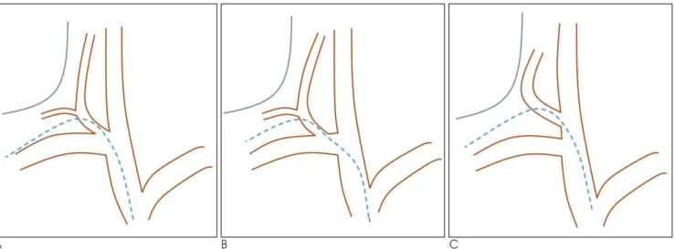
A B C
Fig. 1. Anatomic variations of the external jugular vein.
A. The EJV enters into the jugulo-subclavian venous confluence (type A).
B. Into the SCV at a distance from its junction with the IJV (type B).
C. Into the trunk of the IJV (type C). The dotted line indicates the expected course of the hemodialysis catheter.
gators suggested that thrombosis and occlusion may be more of a concern with a relatively small-caliber of the EJV as compared with the IJV. However, other studies including a large series using the EJV with a surgical ap- proach did not report major complications such as symptomatic thrombosis (18, 19). But Wilkin et al. (24) reported a high prevalence of thrombosis (25.9%) of the right IJV, and 62% of the cases were occluded. Other re- ports have described significant high rates of stenosis or thrombosis in patients with placement of previous catheters (25-27). The initial use of a dilated EJV may provide the benefit of preservation of the usable venous route such as the right IJV for catheterization.
The true incidence of catheter related thrombosis or occlusion is difficult to recognize due to a low incidence of symptoms. Collateral vessels can be responsible for possible pitfalls because the collateral vessels can mask the symptoms of venous thrombosis.
Deslaugiers et al. (28) reported that the EJV usually en- ters into the jugulo-subclavian venous confluence (type A, 60%), into the SCV at a distance from its junction with the IJV (type B, 36%) or into the trunk of the IJV (type C, 4%) (Fig. 1). We speculate that the course of the hemodialysis catheter using the right EJV shows a more obtuse angle than that using IJV.
We could avoid deep neck puncture by use of the EJV instead of the IJV. Although the use of real-time ultra- sound guidance can reduce the risk of complications during IJV puncture, a deep puncture in the neck could lead to procedure-related complications such as arterial puncture or massive hemorrhage, especially in patients with an anatomic variation or an abnormal deep loca- tion of the IJV (29). There were no complications of mas- sive hemorrhage or arterial puncture during EJV catheterization in this study. When bleeding occurred at the insertion site of the vein, we could easily compress the EJV because of its superficial location.
Usually, the SCV approach has a higher complication rate compared to the IJV approach. Even if the EJV is connected to the SCV (type C), we speculate that the EJV approach is different from the SCV approach since the catheter does not pass through the narrow space be- tween the clavicle and first rib, and the right EJV has a straight course to the atrium that is similar to the right IJV.
We also think that the right EJV is a superior access route compared to the left IJV. Several studies have re- ported that the left IJV approach had relatively higher rates of stenosis and thrombosis compared to the right
IJV approach (1, 13, 14, 30, 31). Our thinking is that the catheter passes through the narrow space between the sternum and aortic arch, and can cause injury or may induce stenosis by close approximation of the catheter tip to the wall of the superior vena cava.
This study has limitations for the evaluation of late complications since a hemodialysis catheter was used only until arteriovenous fistula maturation.
Furthermore, the catheter dwell time was relatively short in this study.
The EJV approach could preserve the IJV from catheterization, especially in patients with end stage re- nal disease with a limited number of usable veins for catheterization. We suggest that the dilated right EJV could be considered as the preferred primary for he- modialysis catheter placement based on its ease of ac- cess and an acceptable complication rate. However, fur- ther prospective case-control studies are needed to clari- fy this.
References
1. Vascular Access 2006 Work Group. Clinical practice guidelines for vascular access. Am J Kidney Dis 2006;48 Suppl 1:S176-S247 2. Mondschein JI. Hemodialysis access: catheters and ports. In: Mauro
MA. Image-Guided Interventions. Philadelphia, Pa: Saunder/
Elsevier, 2008:1264-1274
3. Trerotola SO, Kuhn-Fulton J, Johnson MS, Shah H, Ambrosius WT, Kneebone PH. Tunneled infusion catheters: increased inci- dence of symptomatic venous thrombosis after subclavian versus internal jugular venous access. Radiology 2000;217:89-93
4. Ruesch S, Walder B, Trame′r MR. Complications of central venous catheters: internal jugular versus subclavian access - a systematic review. Crit Care Med 2002;30:454-460
5. Forauer AR, Brenner B, Haddad LF, Bocchini TP. Placement of hemodialysis catheters through dilated external jugular and collat- eral veins in patients with internal jugular vein occlusions. AJR Am J Roentgenol 2000;174:361-362
6. Cho SK, Shin SW, Do YS, Park KB, Choo SW, Choo IW. Use of the right external jugular vein as the preferred access site when the right internal jugular vein is not usable. J Vasc Interv Radiol 2006;17:823-829
7. Yevzlin AS, Chan M, Gimelli G. How I do it: preferential use of the right external jugular vein for tunneled catheter placement.
Semin Dial 2008;21:183-185
8. Silberzweig JE, Sacks D, Khorsandi AS, Bakal CW. The members of the society of interventional radiology technology assessment committee. Reporting standards for central venous access. J Vasc Interv Radiol 2003;14:S443-S452
9. Bream PR. Tunneled central venous catheters. In: Mauro MA. Image- Guided Interventions. Philadelphia, Pa: Saunder/Elsevier, 2008:
1237-1244
10. Polderman KH, Girbes AJ. Central venous catheter use. Part 1:
mechanical complications. Intensive Care Med 2002;28:1-17 11. Funaki B, Zaleski GX, Leef JA, Lorenz JN, Van Ha T, Rosenblum
JD. Radiologic placement of tunneled hemodialysis catheters in oc-
cluded neck, chest, or small thyrocervical collateral veins in cen- tral venous occlusion. Radiology 2001;218:471-476
12. Falk A. Use of the brachiocephalic vein for placement of tunneled hemodialysis catheters. AJR Am J Roentgenol 2006;187:773-777 13. Fry AC, Stratton J, Farrington K, Mahna K, Selvakumar S,
Thompson H, et al. Factors affecting long-term survival of tun- nelled haemodialysis catheters--a prospective audit of 812 tun- nelled catheters. Nephrol Dial Transplant 2008;23:275-281 14. Kakkos SK, Haddad GK, Haddad RK, Scully MM. Effectiveness of
a new tunneled catheter in preventing catheter malfunction: a comparative study. J Vasc Interv Radiol 2008;19:1018-1026 15. Zaleski GX, Funaki B, Lorenz JM, Garofalo RS, Moscatel MA,
Rosenblum JD, et al. Experience with tunneled femoral hemodial- ysis catheters. AJR Am J Roentgenol 1999;172:493-496
16. Smith TP, Ryan JM, Reddan DN. Transhepatic catheter access for hemodialysis. Radiology 2004;232:246-251
17. Lund GB, Trerotola SO, Scheel PJ Jr. Percutaneous translumbar in- ferior vena cava cannulation for hemodialysis. Am J Kidney Dis 1995;25:732-737
18. Kuter DJ. Thrombotic complications of central venous catheters in cancer patients. Oncologist 2004;9:207-216
19. Moini M, Rasouli MR, Kenari MM, Mahmoodi HR. Non-cuffed dual lumen catheters in the external jugular veins versus other central veins for hemodialysis patients. Saudi J Kidney Dis Transpl 2009;20:44-48
20. Skandalos I, Amvrosiadis D, Filippidis A, Sioulis A, Tsitsios T.
Mavromatidis K. Insertion of long-term tunneled cuffed hemodial- ysis catheters via the external jugular vein by using a simple, safe and reliable surgical technique. J Vasc Access 2007;8:12-16 21. Lewin MR, Stein J, Wang R, Lee MM, Kernberg M, Boukhman M,
et al. Humming is as effective as Valsalva’s maneuver and Trendelenburg’s position for ultrasonographic visualization of the jugular venous system and common femoral veins. Ann Emerg Med 2007;50:73-77
22. Rose SC, Kinney TB, Bundens WP, Valji K, Roberts AC.
Importance of Doppler analysis of transmitted atrial waveforms prior to placement of central venous access catheters. J Vasc Interv Radiol 1998;9:927-934
23. Trerotola SO, Johnson MS, Harris VJ, Shah H, Ambrosius WT, McKusky MA, et al. Outcome of tunneled hemodialysis catheters placed via the right internal jugular vein by interventional radiolo- gists. Radiology 1997;203:489-495
24. Wilkin TD, Kraus MA, Lane KA, Trerotola SO. Internal jugular vein thrombosis associated with hemodialysis catheters. Radiology 2003;228:697-700
25. Agarwal AK, Patel BM, Haddad NJ. Central vein stenosis: a nephrologist’s perspective. Semin Dial 2007;20:53-62
26. Forauer AR, Glockner JF. Importance of US findings in access planning during jugular vein hemodialysis catheter placements. J Vasc Interv Radiol 2000;11:233-238
27. Jean G, VanelT, Chazot C, Charra B, Terrat JC, Hurot JM.
Prevalence of stenosis and thrombosis of central veins in he- modialysis after a tunneledjugular catheter. Nephrologie 2001;22:
501-504
28. Deslaugiers B, Vaysse P, Combes JM, Guitard J, Moscovici J, Visentin M, et al. Contribution to the study of the tributaries and the termination of the external jugular vein. Surg Radiol Anat 1994;16:173-177
29. Lin BS, Kong CW, Tarng DC, Huang TP, Tang GJ. Anatomical variation of the internal jugular vein and its impact on temporary haemodialysis vascular access: an ultrasonographic survey in uraemic patients. Nephrol Dial Transplant 1998;13:134-138 30. Salgado OJ, Urdaneta B, Colmenares B, Garcia R, Flores C. Right
versus left internal jugular vein catheterization for hemodialysis : complications and impact on ipsilateral access creation. Artif Organs 2004;28:728-733
31. Richard HM 3rd, Hastings GS, Boyd-Kranis RL, Murthy R, Radack DM, Santilli JG, et al. A randomized, prospective evaluation of the Tesio, Ash split, and Opti-flow hemodialysis catheters. J Vasc Interv Radiol 2001;12:431-435
대한영상의학회지 2010;63:351-357
일차적인 경로로 확장된 우측 외경정맥을 이용한 혈액투석도관의 설치술1
1단국대학교병원 영상의학과
2충남대학교병원 영상의학과
박 미 현∙신 병 석2
목적: 확장된 우측 외경정맥이 대구경의 혈액투석도관의 설치술에서 일차적인 접근 경로로서 유용한지 알아보고자 하였다.
대상과 방법: 2008년 1월부터 2009년 4월까지 혈액투석도관(14.5F)을 시행한 173명 중 확장된 우측외경정맥을 이용하여 혈액투석도관 삽입술을 시행한 42명(24%)의 환자의 임상자료를 분석하였다. 기술적 성공률과 도관삽입 기간 그리고 합병증에 대하여 분석하였다.
결과: 총 41명(98%)에서 성공적으로 삽입하였으며 1명에서 외경정맥의 협착으로 삽입에 성공하지 못하였다. 도관 유치기간은 14일-305일(평균 76일, 총 3,111일)이었다. 총 26명에서 합병증(n=2)과 도관을 통한 투석종료 (n=24)로 인하여 도관을 제거하였다. 시술 중 공기색전증(n=1)과 도관의 꺽임(n=3)이 있었으며 관찰 기간에 도 관 관련 감염 (n=2)이 발생하였다. 도관관련 감염의 발생빈도는 100일 도관일 당 0.06이었다. 도관의 기능부전이 나 증상이 있는 정맥혈전증은 관찰되지 않았다.
결론: 확장된 외경정맥은 혈액투석도관 설치술시 쉽게 접근할 수 있는 일차적인 경로로써 고려할 수 있을 것으로 판 단된다.