관상동맥 질환은 서구에서 성인 사망률의 가장 많은 원인이 며, 최근 우리나라에서도 그 발병이 증가되는 추세로 조기 진 단과 치료가 환자의 예후에 매우 큰 영향을 미치고 있다. 관상 동맥의 협착을 관찰하기 위하여 고식적인 관상동맥 혈관조영 술(conventional coronary angiography)이 오랜 기간동안 사 용되어 왔으며, 병변의 위치나 협착 정도를 규명하는데 있어서 많은 비침습적인 검사가 시도되고 있으나 아직은 관상동맥 혈 관조영술을 대체하지 못하고 있다. 기본 검사로 사용 되고 있 는 관상동맥 혈관조영술로도 20%의 환자에서는 관상동맥의 협착을 발견하지 못하고 있다 (1). 또한 관상동맥 혈관조영술 은 침습적인 검사이기 때문에 적기는 하지만 국소적인 혈관 합 병증에서부터 심장이나 대동맥의 관통, 심근 경색 그리고 사망 에 이르기까지 동반된 위험이 있을 수 있고, 조영제와 방사선 조사의 부작용이 있으며 입원이 필요하다 (1). 이에 관상동맥 질환이 의심되거나 관상동맥 질환이 있는 환자에서 질환의 발 견과 추적을 위한 비침습적인 유용한 검사가 필요하다.
자기공명영상(magnetic resonance imaging)은 비침습적인 검사 방법으로서 조영제 사용 없이 훌륭한 조직 대조도를 가
지고 심장을 검사하는 데에 적합하다 (2). 또한 coronary MRA 는 높은 해상도를 가지고 여러 영상축을 검사할 수 있으며, 방 사선 조사가 없고 부작용이 없다는 점에서 같은 비침습적인 검 사인 심초음파 검사나 전자선 단층촬영(Electron beam com- puted tomography)에 비해 우월하다고 할 수 있다. 1980년대 후반 coronary MRA에 대한 초기의 보고들 (3-5) 이후 심장 주위의 큰 혈관 구조물에 의한 간섭, 심장과 호흡운동에 의한 인공음영등 여러 가지 문제점을 극복하고, 관상동맥 분지들의 복잡한 3차원적 구조를 정확하게 표출하려는 다양한 노력이 현 재까지 시도되어 왔다 (6-18). 이에 저자들은 coronary MRA 의 임상적용 가능성을 타진해 보고자 본 연구를 시행하였다.
대상과 방법
대상
2000년 10월부터 2001년 1월까지 11명의 건강한 자원자 를 대상으로 하였다. 자원자의 평균연령은 26.9세(23세-35세) 였고, 모두 남자였다. 문진에 의해 심장이나 폐에 질환이 없는 건강한 자원자를 대상으로 사전에 충분히 연구내용을 설명하 여 동의를 구하였다. 폐쇄공포증이 없고 사전에 심전도를 확인 하여 정상 R 리듬을 확인하였다.
정상 성인의 이차원 호흡정지 관상동맥 자기공명 혈관조영술
1박성빈・최상일・서준범・이인선・이승수・윤진호・임청환・이용철2・임태환
목적: 관상동맥 자기공명 혈관조영술(coronary magnetic resonance angiography; 이하coro- nary MRA로 약함)의 임상적용 가능성을 타진해 보고자 본 연구를 시행하였다.
대상과 방법: 2000년 10월부터 2001년 1월까지 11명의 건강한 자원자를 대상으로 하였다.
Coronary MRA는 1.5 T 심전도 동기 T1 강조 2차원 다중 호흡정지 나선 급속경사에코법 (EKG-gated T1-weighted, 2D multiphase breath-hold spiral fast gradient echo sequence) 을 이용한 훼손경사에코(spoiled gradient echo)를 사용하였다. 관상동맥의 직경을 기시부에서 1 cm 떨어진 부위에서 측정하여 평균을 구하였으며, 모든 영상화된 관상동맥의 영상화 구현 정도와 영상화의 질을 분석하였다.
결과: 관상 동맥 근위부의 영상화 구현 정도는 82%에서 100%의 분포를 보였으며, 중간과 원 위부의 영상화 구현 정도는 36%에서 55%의 분포를 보였다. 관상동맥 근위부의 직경은 대부 분의 경우 고식적 혈관조영술의 참고치와 이전 coronary MRA의 결과와 좋은 연관을 보였다.
좌측 회선 관상동맥의 영상화 정도와 영상의 질은 제한점을 보였다.
결론: 2D Coronary MRA는 대부분의 관상동맥 근위부 분절을 적절하게 영상화 할 수 있다.
1울산대학교 의과대학 서울중앙병원 방사선과
2중앙대학교 의과대학 방사선과
본 연구는 보건복지부 연구비 지원(HMP-98-G-1-028)에 의해 이루어진 것임.
이 논문은 2001년 8월 16일 접수하여 2001년 12월 21일에 채택되었음.
자기공명 영상 촬영
Coronary MRA는 1.5 Tesla Signa CV/i(GE Medical System, Milwaukee, Wis, U.S.A.) 기종으로 심전도 동기 T1 강조 2차원 다중 호흡정지 나선 급속경사에코법(EKG-gated T1-weighted, 2D multiphase breath-hold spiral fast gra- dient echo sequence)를 이용한 훼손경사에코(spoiled gradi- ent echo, SPGR)를 사용하였다. 앙와위에서cardiac phase array coil을 흉곽에 설치한 후 호흡 정지 시상 영상을 얻어 표 면 코일의 위치를 교정 하였다. 이후 반복적인 방법과 해부학 적인 방법을 병용하여 다음과 같은 연속된 스캔을 얻어 관상 동맥을 영상화 하였다.
1) 관상 영상을 얻은 후 관상 정맥동 위치(coronary sinus level)에서 횡축상 영상을 얻어 우측 관상 동맥(right coronary artery)과 좌측 주 관상동맥(left main coronary artery)의 기 시부를 확인 하였다 (Fig. 1). 2) 연속된 중첩 영상으로 우측 폐동맥과 우측 심방 사이에서 우측 관상동맥을 확인 후, 아래 부분으로 우측 방실 고랑(right atrioventricular groove)을 지 나는 것을 확인하여 사선 영상을 얻어 우측 관상동맥을 장축 에 맞게 영상화 하였다. 구불구불한 중간부위 이하 부분을 얻 기 위해서 계속적인 사선영상을 얻었다 (Fig. 2). 3) 횡축상 영 상에서 좌측 주 관상동맥과 좌측 전하행 관상동맥(left ante- rior descending coronary artery) 그리고 좌측 회선 관상동맥 (left circumflex coronary artery)의 기시부를 영상화 한 후
(Fig. 1) , 좌측 폐동맥과 심방을 따라서 좌측 전하행 관상동 맥을 확인 후 사선 관상 영상을 얻어 좌측 전하행 관상동맥과 좌측 회선 관상동맥의 근위부를 영상화 하였다 (Fig. 3). 4) 이 후 연속된 다중 사선 관상 영상으로 좌측 전하행 관상동맥과 좌측 회선 관상동맥의 중간 및 원위부를 확인하였다 (Fig. 4).
5) 우측 관상동맥의 영상화를 위해 부가적으로 해부학적 접근 법을 통해 축상 영상에서부터 비스듬히 장축 영상을 얻었다.
이후 방실 고랑을 지나는 우측 관상동맥을 확인 후 동맥의 주 행을 따라 사선영상을 얻어 혈관의 주행을 영상화 하였다.
관상동맥의 위치를 확인 하는 방법(localization technique) 은 기존의 고식적 방법과 최근 도입된 실시간 interactive con- trol tool인 iDrive pro(GE Medical System, Milwaukee, Wis, U.S.A.)를 병용하였다.
호흡 정지 T1 강조영상을 위한 조건들은 다음과 같이 하였 다;
TR(repetition time): varied according to heart rate, TE(echo time): 4 msec, flip angle: 15°,
slice thickness: 5 mm, interslice gap: 없음, FOV(field of view): 38 × 28.5 cm, matrix No.: 256 × 128
모든 자원자에서 검사는 한시간 이내에 마치는 것을 원칙으 로 하였다.
Fig. 1. 27-year-old healthy man. Transaxial coronary MR an- giography shows the ostium and proximal segment of right coronary artery (arrow) and left main coronary artery (open ar- row).
Fig. 2. 25-year-old healthy man. Oblique sagittal coronary MR angiography shows the right coronary artery. The origin of the right coronary artery (arrow a), proximal (arrow b), middle (ar- row c) and distal (arrow d) segments are shown clearly.
자기공명 영상 분석
자기공명 영상 분석은 관상동맥의 직경을 기시부에서 1 cm 떨어진 부위에서 방사선과 의사 1인이 2번 측정하여 평균을 구하였으며, 모든 영상화된 관상동맥을 미국 심장협회 (American Heart Association)의 기준 (19)에 따라 8분절 (우 측 관상동맥, 좌측 주 관상동맥, 좌측 전하행 관상동맥, 좌측 회선 관상동맥의 근위부와 우측 관상동맥, 좌측 전하행 관상동 맥, 좌측 회선관상동맥의 중간부와 우측 관상동맥의 원위부)로 나누어 영상화 구현 정도(visibility)를 분석하였다.
4개의 주분절(우측 관상동맥, 좌측 주 관상동맥, 좌측 전하 행 관상동맥, 좌측 회선 관상동맥)의 영상화의 질을 아래와 같 은 기준 (11)으로 방사선과 의사 2인이 합의하에 등급화 하였 다.
0: 관상동맥이 영상화 되지 않는 경우
1: 관상동맥이 있을 것으로 예상되는 위치에 선상 구조물이 있는 경우
2: 관상동맥의 분절이 관찰되나, 인공물이 심한 경우 3: 관상동맥의 직경을 관찰할 정도의 작은 분절이 영상화 된
경우
4: 관상동맥이 인공물이나 주위조직과의 겹침이 없이 영상 화 된 경우
5: 근위부와 중간부분까지 인공물 없이 영상화 된 경우
통계적 처리
1등급(grade 1) 이상의 부분적으로 영상화된 분절만 분석하 였으며, 관상동맥의 직경을 Dodge 등(20)에 의한 20명 우측 관상동맥 우월(right dominant coronary anatomy) 자원자에 서의 고식적 관상동맥 혈관조영술의 참고치, Pennell 등(7)의 이전 coronary MRA 결과와 two-sample t-test를 사용하여 통계적 유의성을 알아 보았다.
각 분절에서의 영상화 구현 정도를 알아 보았다. 영상화 구 현정도는 1등급이상의 부분적으로 영상화된 경우와 관상동맥 의 직경을 관찰할 정도의 3등급이상의 경우로 나누어 분석하 여 coronary MRA의 유용성을 알아 보았다. 영상화의 질은 각 분절의 경우를 평균하여 4개의 주분절에서 구하였다.
결 과
각각의 관상동맥 8분절의 영상화 구현정도는 다양한 분포를 보였다 (Table 1). 부분적으로 인식이 가능한 1등급이상을 기 준으로 할 때 관상 동맥 근위부의 영상화 구현 정도는 82%에 서 100%의 분포를 보였으며, 중간과 원위부의 영상화 구현 정 도는 36%에서 55%의 분포를 보였다. 즉 관상 동맥의 근위부 는 좌측 회선 관상동맥에서 11예 중 9예(82%), 우측 관상동 맥, 좌측 주 관상동맥 그리고 좌측 전하행 관상동맥에서 11예 중 11예(100%)로 대부분에서 영상화가 가능하였다 (Figs. 1-
Fig. 3. 28-year-old healthy man. Oblique sagittal coronary MR angiography shows the proximal left anterior descending coro- nary artery (arrow) and the origin of the left circumflex coro- nary artery(open arrow).
Fig. 4. 35-year-old healthy man. Oblique coronary MR angiog- raphy shows the left circumflex coronary artery (arrows).
4).
관상 동맥의 직경의 변화를 알 수 있는 3등급 이상의 경우 를 분석하였을 때, 근위와 중간 우측 관상동맥, 좌측 주 관상 동맥과 근위 좌측 전하행 관상동맥은 각각 11예 중 11예 (100%), 11예 중 9예(82%), 11예 중 10예(91%), 11예 중 8예(73%)로 영상화 구현정도가 우수하였으나, 원위 우측 관 상동맥에서는 11예 중 6예(55%), 중간 좌측 전하행 관상동맥 과 근위와 중간 좌측 회선 관상동맥에서는 각각 11예 중 5예 (45%), 11예 중 4예(36%), 11예 중 2예(18%)로 영상화 구 현정도가 좋지 않았다 (Table 1).
각각의 관상동맥에 대하여 영상의 질은 우측 관상동맥과 좌
측 주 관상동맥에서 평균 3등급이상으로 우수하였다 (Fig. 5).
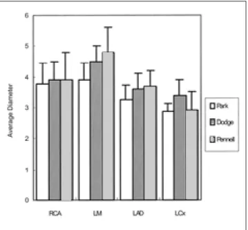
각각의 관상동맥 분절의 근위부 직경은 우측 관상동맥이 3.79
±0.65 mm, 좌측 주 관상동맥 3.91±0.54 mm, 좌측 전하행 관상동맥 3.27±0.45 mm, 좌측 회선 관상동맥 2.85±0.28 mm 의 분포를 보였다.
이전의 Dodge 등(20)의 고식적 혈관조영술상의 관상동맥 직경의 결과치와 Pennell 등(7)의 coronary MRA상의 관상동 맥 직경의 결과치와 비교 하였을 때, 대부분의 분절에서 통계 적인 차이를 보이지 않았다. 좌측 주 관상 동맥과 좌측 회선동 맥에서 Dodge등의 결과와 그리고 좌측 주 관상 동맥과 좌측 전하행 관상동맥에서 Pennell 등(7)의 결과와 통계적으로 유 의한 차이를 보였다 (p < 0.05).
고 찰
최근 MR기법의 발전과 함께 coronary MRA는 많은 영상기 법이 시도되어 왔으며, 2차원 영상과 3차원 영상으로 구별 된 다. 3차원 영상은 좀더 높은 신호 대 잡음비율과 얇은 절편의 획득 그리고 일정한 좋은 질의 영상이 가능하다. 그렇지만 획 득 시간이 길고 영상 자료의 후 처리 과정이 필요하므로 획득 후 바로 영상을 확인 할 수 없는 단점을 가진다.
이에 반해 2차원 영상은 영상 시간이 짧고, 모든 스캔이 한 번의 호흡정지로 가능하므로 호흡이나 심장운동에 의한 인공 영상물을 적게 가져올 수 있고, 또한 관상동맥 내의 예기치 않 은 혈류의 포화도 덜 가져올 수 있다. 그렇지만 2차원 영상은 관상동맥의 구불구불함에 따른 영상상의 한계를 갖는다. 즉, 영 Table 1. Visibility of Different Coronary Arterial Segments with
Coronary MR Angiography
Segment *Visibility (G ≥ 1) (G ≥ 3)
Proximal RCA 11 (100) 11 (100)
LM 11 (100) 10 (091)
Proximal LAD 11 (100) 08 (073)
Proximal LCx 09 (082) 04 (036)
Middle RCA 11 (100) 09 (082)
Middle LAD 07 (064) 05 (045)
Middle LCx 04 (036) 02 (018)
Distal RCA 06 (055) 05 (045)
*Data are numbers of patients in whom the given segment was assessable with MR coronary angiography, based on a total of 11 volunteers. Numbers in parentheses are percentages.
RCA = right coronary artery, LM = left main coronary artery, LAD = left anterior descending coronary artery, LCx = left cir- cumflex coronary artery.
Fig. 6. Graph shows average diameter of visualized portions of the proximal coronary vessels, with use of only segments with a MR image quality grade of 1 or better. Error bars indicate 1 standard deviation. Our data are compared with those of prox- imal diameter measurement by Dodge et al (20) and Pennell et al (7).
Fig. 5. Graph shows average image quality of coronary MR an- giography with a MR image quality of grade 1 or better. Right coronary artery (RCA) and left main coronary artery (LM) are visualized better than left anterior descending artery (LAD) and left circumflex artery (LCx) at coronay MRA.
상면을 벗어난 관상동맥의 치우침에 의하여 국소적 신호 감소 를 가져올 수 있으며, 이를 국소적 협착으로 잘못 판단할 수 있다 (12, 13).
이번 연구에서 2차원 호흡정지 coronary MRA는 대부분의 근위부 혈관을 성공적으로 영상화 할 수 있었으나, 좌측 회선 관상동맥에서는 영상화 구현 정도나 영상의 질에서 많은 제한 점을 보였다. 이는 흉벽에 설치하는 표면 코일과 좌측 회선 관 상동맥간의 거리가 상대적으로 길어 낮은 신호 대 잡음비율을 가지게 되며, 좌측 회선 관상동맥은 관상 정맥동이나 좌심이에 접근해 있으므로 낮은 해상도와 신호 대 잡음비율 그리고 대 조 잡음비에 의해 영상을 방해 받을 수 있는 것에 기인한다.
앞으로 coronary MRA의 임상적용을 위하여서는 좌측 회선 관 상동맥을 보다 명확히 영상화 할 수 있는 새로운 영상기법에 대한 연구가 요구된다.
Manning 등(10)과 Pennell 등(14)의 보고에 의하면 50%이 상의 관상동맥 협착병변의 진단에 있어 coronary MRA는 고 식적 혈관조영술과 비교하여 높은 정확도를 보였다. 또한 Hundley 등(17)의 연구에서는 심근경색후의 상태 평가에 있 어 coronary MRA가 100% 정확하게 심근경색과 연관된 관상 동맥의 협착정도와 측부순환의 유무를 인지할 수 있었다고 보 고하였다. 하지만 coronary MRA의 평가에 있어 다양한 연구 결과가 도출될 수 있는데, 이는 다른 자기공명 영상기법, 환자 들의 호흡정지에서의 협조정도, 부정맥 등의 차이에 의한 것으 로 보인다.
이번 연구 결과에서 Dodge 등(20)의 고식적 혈관조영술상 의 관상동맥 직경의 결과치와 Pennell 등(7)의 coronary MRA 상의 관상동맥 직경의 결과치와 비교 하였을 때, 일부 분절에 서 통계적으로 유의한 차이를 보였다. 이는 직접 고식적 관상 동맥 혈관조영술을 시행하지 않고 이미 전에 측정된 결과와 비 교 하였으므로 여기에 기인한 오차와 측정과정에서의 오차가 있을 수 있으며, 또한 우리나라의 결과의 부재로 외국의 결과 와 비교하였으므로 인종적인 차이도 간과할 수 없다.
이외에도 여러가지 한계점을 가지고 있다. 첫째로, 영상화한 자원자수가 다소 적어서 정확한 반영이 어려울 수 있다. 둘째 로, 환자를 대상으로 한 연구가 아니므로 협착 병변의 영상화 정도를 알 수가 없었다. 셋째로, 호흡정지 영상이므로 자원자 가 아닌 심폐기능의 저하가 심한 환자들에게 적용하기에 무리 가 따를 수 있다.
그렇지만, 이번 연구에서 coronary MRA상 관상동맥의 직경 이 이전 고식적 혈관조영술의 참고치와 coronary MRA의 결 과와 비교할 때 일부 분절에서 통계적으로 유의한 차이를 보 였으나, 대부분의 근위분절 관상동맥의 영상화가 가능하였다.
즉, coronary MRA는 근위 관상동맥을 영상화 할 수 있었으 며, 그 주행과 주위 구조물과의 관계를 충분하게 보여줄 수 있 었다. 또한 정상 관상동맥 직경을 비교적 정확하게 측정할 수 있었으며 이는 관상동맥 질환의 접근에 유용할 수 있음을 시 사한다. 또한 선천성 관상동맥 기형의 영상화 (21-23), 관상 동맥 우회 수술 후 (24, 25)와 관상동맥 스텐트 설치후의 개 방도 (26), 심장이식후의 관상동맥의 해부학적 구조 (27) 그
리고 관상동맥의 혈류의 양적 정량화 (28, 29) 등의 임상적 적 용이 가능할 것으로 생각된다.
결론적으로 coronary MRA는 대부분의 관상동맥 근위부 분 절을 적절하게 영상화 할 수 있었다. 더 많은 수의 정상 자원 자에 대한 연구와 관상동맥 질환을 가진 환자에 있어서도 임 상적인 적용의 시도가 필요할 것으로 사료된다. 향후 coronary MRA는 관상동맥 질환의 진단 및 추적에 있어 비침습적인 진 단방법으로 매우 유용할 것으로 기대된다.
참 고 문 헌
1. Johnson LW, Lozner EC, Johnson S, et al. Coronary arteriography 1984(1987: a report of the Registry of the Society for Cardiac Angiography and Interventions. I. Results and complications.
Cathet Cardiovasc Diagn 1989;17:5-10
2. Duerinckx AJ, Higgins C, Pettigrew R. MRI of the cardiovascular system: the Raven Press MRI Teaching File. New York, NY: Raven, 1993
3. Lieberman JM, Botti RE, Nelson AD. Magnetic resonance imaging of the heart. Radiol Clin North Am 1984;22:847-858
4. Paulin S, von Schulthess GK, Fossel E, Krayenbuehl HP. MR imag- ing of the aortic root and proximal coronary arteries. AJR Am J Roentgenol 1987;148:665-670
5. Alfidi RJ, Masaryk TJ, Haacke EM, et al. MR angiography of pe- ripheral, carotid, and coronary arteries. AJR Am J Roentgenol 1987;149:1097-1109
6. Edelman RR, Manning WJ, Burstein D, Paulin S. Coronary arter- ies: breath-hold MR angiography. Radiology 1991;181:641-643 7. Pennell DJ, Keegan J, Firmin DN, Gatehouse PD, Underwood SR,
Longmore DB. Magnetic resonance imaging of coronary arteries:
technique and preliminary results. Br Heart J 1993;70:315-326 8. Edelman RR, Manning WJ, Gervino E, Li W. Flow velocity quan-
tification in human coronary arteries with fast, breath-hold MR an- giography. J Magn Reson Imaging 1993;3:699-703
9. Edelman RR, Manning WJ, Pearlman J, Li W. Human coronary ar- teries: projection angiograms reconstructed from breath-hold two- dimensional MR images. Radiology 1993;187:719-722
10. Manning WJ, Li W, Edelman RR. A preliminary report comparing magnetic resonance coronary angiography with conventional an- giography. N Engl J Med 1993;328:828-832
11. Duerinckx AJ, Urman MK. Two-dimensional coronary MR angiog- raphy: analysis of initial clinical results. Radiology 1994;193: 731- 738
12. Sakuma H, Caputo GR, Steffens JC, et al. Breath-hold MR cine an- giography of coronary arteries in healthy volunteers: value of mul- tiangle oblique imaging planes. AJR Am J Roentgenol 1994;163:
533-537
13. Hofman MB, Paschal CB, Li D, Haacke EM, van Rossum AC, Sprenger M. MRI of coronary arteries: 2D breath-hold vs 3D respi- ratory-gated acqusition. J Comput Assist Tomogr 1995;19:56-62 14. Pennell DJ, Bogren HG, Keegan J, Firmin DN, Underwood SR.
Assessment of coronary artery stenosis by magnetic resonance imaging. Heart 1996;75:127-133
15. Wielopolski PA, van Geuns RJ, de Feyter PJ, Oudkerk M. Breath- hold coronary MR angiography with volume-targeted imaging.
Radiology 1998;209:209-219
16. Van Genus RJ, de Bruin HG, Rensing BJ, et al. Magnetic resonance imaging of the coronary arteries: clinical results from three dimen- sional evaluation of respiratory gated technique. Heart 1999;82:
515-519
17. Hundley WG, Clarke GD, Landau C, et al. Noninvasive determi- nation of infarct artery patency by cine magnetic resonance an- giography. Circulation 1995;91:1347-1353
18. Post JC, van Rossum AC, Hofman MB, Valk J, Visser CA.
Three(dimensional respiratory(gated MR angiography of coronary arteries: Comparison with conventional coronary angiography.
AJR Am J Roentgenol 1996;166:1399-1404
19. Austen WG, Edwards JE, Frye RL, et al. A reporting system on pa- tients evaluated for coronary artery disease: report of the Ad Hoc Committee for Grading of Coronary Artery Disease, Council on Cardiovascular Surgery, American Heart Association. Circulation 1975;51: 5-40
20. Dodge JT Jr, Brown BG, Bolson EL, Dodge HT. Lumen diameter of normal human coronary arteries: influence of age, sex, anatomic variation, and left ventricular hypertrophy and dilatation.
Circulation 1992;86:232-246
21. Duerinckx AJ, Bogaert J, Jiang H, Lewis BS. Anomalous origin of left coronary artery: Diagnosis by coronary MR angiography. AJR Am J Roentgenol 1995;164:1095-1097
22. Manning WJ, Li W, Cohen SI, Johnson RG, Edelman RR.
Improved definition of anomalous left coronary artery by magnet- ic resonance coronary angiography. Am Heart J 1995;130:615-617 23. McConnell MV, Ganz P, Selwyn AP, Li W, Edelman RR, Manning
WJ. Identification of anomalous coronary arteries and their anato- mic course by magnetic resonance coronary angiography. Circula- tion 1995;92:3158-3162
24. Galjee MA, van Rossum AC, Doesburg T, van Eenige MJ, Visser CA. Value of magnetic resonance imaging in assessing patency and function of coronary artery bypass grafts: an angiographically con- trolled study. Circulation 1996;93:660-666
25. Vrachliotis TG, Bis KG, Aliabadi D, Shetty AN, Safian R, Simonetti O. Contrast-enhanced breath-hold MR angiography for evaluating patency of coronary artery bypass grafts. AJR Am J Roentgenol 1997;168:1073-1080
26. Duerinckx AJ, Atkinson D, Hurwitz R, Mintorovitch J, Whitney W. Coronary MR angiography after coronary stent placement. AJR Am J Roentgenol 1995;165:662-664
27. Davis SF, Kannam JP, Wielopolski P, et al. Magnetic resonance coronary angiography in heart transplant recipients. J Heart Lung Transplant 1996;15:580-586
28. Clarke GD, Eckels R, Chaney C, et al. Measurement of absolute epicardial coronary artery flow and flow reserve with breath-hold cine phase contrast magnetic resonance imaging. Circulation 1995;
91:2627-2634
29. Sakuma H, Blake LM, Amidon TM, et al. Coronary flow reserve : noninvasive measurement in humans with breath-hold velocity- encoded cine MR imaging. Radiology 1996;198:745-750
J Korean Radiol Soc 2002;46:321-327
Address reprint requests to : Tae-Hwan Lim, M.D., Department of Radiology, Asan Medical center, University of Ulsan, College of Medicine, 388-1, Poongnap-dong, Songpa-gu, Seoul 138-736, Korea.
Tel. 82-2-3010-4400 Fax. 82-2-476-4719 E-mail: [email protected]
Two-Dimensional Breath-Hold Coronary MR Angiography in Normal Adults
1Sung Bin Park, M.D., Sang Il Choi, M.D., Joon Beom Seo, M.D., In Sun Lee, M.D., Seoung Soo Lee, M.D., Jin Ho Yoon, R.T., Chung-Hwan Lim, R.T., Yong Chul Lee, M.D2., Tae-Hwan Lim, M.D.
1Department of Radiology, Asan Medical Center, University of Ulsan, College of Medicine
2Department of Radiology, Chung-Ang University, College of Medicine
Purpose: To assess the efficacy of two-dimensional breath-hold coronary magnetic resonance angiography (coronary MRA) in normal rolunteers.
Materials and Methods:During a four-month period, 11 volunteers underwent MRA of the major coronary branches using a 2-D multiphase breath-hold spiral fast-gradient echo sequence. The proximal diameter of each visualized coronary artery was measured, and visibility and image quality were also determined.
Results:Adequate visualization was achieved in 82-100% of proximal coronary arterial branches and in 36- 55% of the middle, distal branches. In general, the diameter of the proximal coronary artery correlated closely with that measured from conventional coronary angiography and using previous coronary MRA data. How- ever, visibility and image quality in the left circumflex coronary artery were limited.
Conclusion:In the majority of subjects, 2-D coronary MRA provides adequate visualization of the proximal segments of the major coronary arterial branches.
Index words :Coronary vessels, MR
Magnetic resonance (MR), vascular studies