Blood Res2017;52:135-50. bloodresearch.or.kr
148 Letters to the Editor
Leebeek FW. Myeloproliferative disease in the pathogenesis and survival of Budd-Chiari syndrome. Haematologica 2006;91:
1712-3.
3. Valla D, Dhumeaux D, Babany G, et al. Hepatic vein thrombosis in paroxysmal nocturnal hemoglobinuria. A spectrum from asymptomatic occlusion of hepatic venules to fatal Budd-Chiari syndrome. Gastroenterology 1987;93:569-75.
4. Hillmen P, Lewis SM, Bessler M, Luzzatto L, Dacie JV. Natural history of paroxysmal nocturnal hemoglobinuria. N Engl J Med 1995;333:1253-8.
5. Hoekstra J, Leebeek FW, Plessier A, et al. Paroxysmal nocturnal hemoglobinuria in Budd-Chiari syndrome: findings from a co- hort study. J Hepatol 2009;51:696-706.
6. Hillmen P, Muus P, Dührsen U, et al. Effect of the complement inhibitor eculizumab on thromboembolism in patients with par- oxysmal nocturnal hemoglobinuria. Blood 2007;110:4123-8.
7. Hillmen P, Hall C, Marsh JC, et al. Effect of eculizumab on hemol- ysis and transfusion requirements in patients with paroxysmal nocturnal hemoglobinuria. N Engl J Med 2004;350:552-9.
8. Singer AL, Locke JE, Stewart ZA, et al. Successful liver trans- plantation for Budd-Chiari syndrome in a patient with parox- ysmal nocturnal hemoglobinuria treated with the anti-comple- ment antibody eculizumab. Liver Transpl 2009;15:540-3.
9. Brodsky A, Mazzocchi O, Sánchez F, Khursigara G, Malhotra S, Volpacchio M. Eculizumab in paroxysmal nocturnal hemoglobi- nuria with Budd-Chiari syndrome progressing despite anticoagulation. Exp Hematol Oncol 2012;1:26.
10. Mandala E, Lafaras C, Goulis I, Tsioni K, Georgopoulou V, Ilonidis G. Treatment of a patient with classical paroxysmal nocturnal he- moglobinuria and Budd-Chiari syndrome, with complement in- hibitor eculizumab: Case Report. Hippokratia 2013;17:81-4.
11. Borowitz MJ, Craig FE, Digiuseppe JA, et al. Guidelines for the diagnosis and monitoring of paroxysmal nocturnal hemoglobi- nuria and related disorders by flow cytometry. Cytometry B Clin Cytom 2010;78:211-30.
12. Lee JW, Jang JH, Kim JS, et al. Clinical signs and symptoms asso- ciated with increased risk for thrombosis in patients with parox- ysmal nocturnal hemoglobinuria from a Korean Registry. Int J Hematol 2013;97:749-57.
13. Ki M, Choi HY, Kim KA, Kim BH, Jang ES, Jeong SH. Incidence, prevalence and complications of Budd-Chiari syndrome in South Korea: a nationwide, population-based study. Liver Int 2016;36:
1067-73.
14. Navenot JM, Muller JY, Blanchard D. Investigation of the survival of paroxysmal nocturnal hemoglobinuria red cells through the im- munophenotyping of reticulocytes. Transfusion 1998;38:337-42.
15. Richards SJ, Hill A, Hillmen P. Recent advances in the diagnosis, monitoring, and management of patients with paroxysmal noc- turnal hemoglobinuria. Cytometry B Clin Cytom 2007;72:291-8.
Ultrasound-guided sternal bone marrow aspiration
TO THE EDITOR:
Bone marrow examination is a common procedure during the diagnosis of hematological disorders such as acute and chronic leukemia, non-Hodgkin lympho- ma, and aplastic anemia. It is usually performed on the posterior iliac crest or the sternum. Although rare, a serious complication may occasionally occur when it is performed on the sternum due to the anatomical localization of the vasculature (ascending aortae) just beneath the sternum [1].
The injury mainly results from the use of an incorrect needle length according to the sternal bone width and is mostly life threatening.
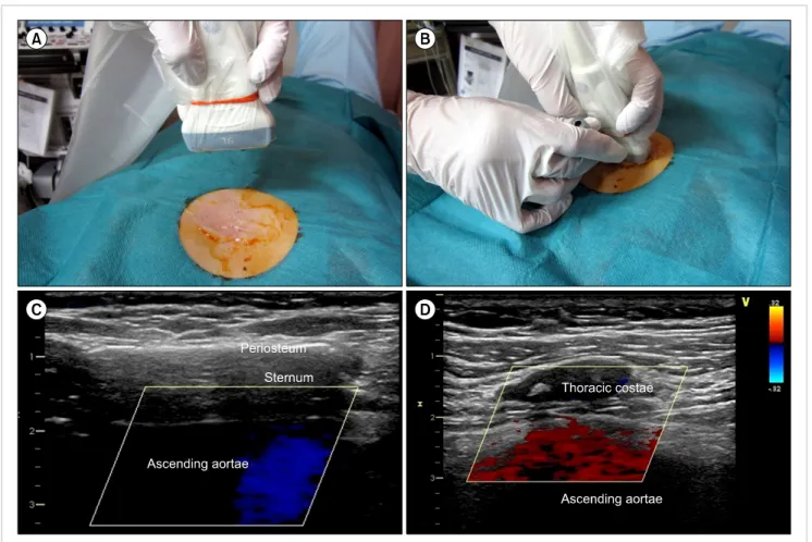
Here we describe a novel sternal bone marrow aspiration technique, named “ultrasound-guided sternal bone marrow aspiration,” which enables the decrease of such risks to nearly zero. The high-resolution ultrasound imaging modal- ity with a linear probe of a frequency higher than 8 MHz and a sterile probe cover (CIV-Flex Transducer Cover, CIVCO, Iowa, USA) were used (Fig. 1A). A linear high-reso- lution ultrasound probe with its frequency at 9 MHz (Vivid-E9, GE, San Jose, CA, USA) was applied perpendicular to the long axis of the sternum to obtain the image (Fig.
1B). The sternum is identified in the center of the figure
as a hypoechoic structure surrounded by the hyperechoic periosteum (Fig. 1C). The image clearly reveals that the depth from the skin to the sternum is approximately 1 cm, and the width of the sternum is 1 cm. Considering the pressure that potentially would decrease the depth from the skin to the sternum during the puncture of the needle, the needle depth was adjusted to 1.3–1.5 cm. Just beneath the sternum, the blood flow of the ascending aortae is clearly visible using a B-mode ultrasonography with a color flow imaging (Fig. 1C). Since bony structures have high ultra- sound absorption coefficients, we suggest that the probe be applied just beside the sternum, longitudinal to the body trunk for cases in which the blood flow and depth of the ascending aortae are not visible. The thoracic costae can be identified as a hypoechoic circle just in the center of the figure (Fig. 1D). The depth of the ascending aortae is measured as 2.5–3 cm from the skin (Fig. 1D). Finally, using an out-of-plane technique [2], an aspiration needle is inserted (Fig. 1B). In brief, the probe is applied perpendicu- lar to the long axis of the sternum and adjusted to show the sternum just in the center of the screen. Next, the needle is positioned in the middle of the transducer and punctures the skin at the same distance from the transducer to the sternum at the depth estimated from the screen.
By rotating the transducer probe, the needle tip is clearly visible during the aspiration procedure.
Recently, ultrasound guidance for central venous cathe-
terization as well as for peripheral nerve block has gained
popularity and been shown to reduce the rate of complica-
bloodresearch.or.kr Blood Res 2017;52:135-50.
Letters to the Editor 149
Fig. 1. (A) A 9 Mhz linear ultrasound transducer probe covered with a sterile probe cover. (B) A sterile linear ultrasound transducer probe was applied perpendicular to the long axis of sternum. The needle is positioned at the middle of the transducer probe and punctures the skin using “out-of-plane”
technique under the total ultrasound guidance. (C) A typical ultrasonogram of sternum. The depth from the skin to the sternum is measured as approximately 1 cm with its width approximately 1 cm. Sternum, a bony structure, has high adsorption coefficient of ultrasound, and is visualized as hypoechoic lesion surrounded by the hyperechoic periosteum. Beneath the sternum, a blood flow of ascending aortae is clearly visualized using B-mode ultrasonogram with color flow imaging. (D) A longitudinal view of the mediastinum just beside the sternum. In the center of the view, a thoracic costae is identified as a hypoechoic circle surrounded by the hyperechoic periosteum. Beneath the thoracic costae, blood flow of the ascending aortae is identified using B-mode sonogram with color flow imaging.
tions [3-6]. We have shown that ultrasound guidance for sternum bone marrow aspiration may potentially eliminate the fatal complications associated with cardiovascular injury.
Yusuke Asakura
1, Maho Kinoshita
2, Yusuke Kasuya
2, Shiori Sakuma
2, Makoto Ozaki
21
Department of Anesthesiology, Nagoya Kyoritsu Hospital, Nagoya,
2Department of Anesthesiology, Tokyo Womens’
Medical University, Tokyo, Japan
Correspondence to: Yusuke Asakura Department of Anesthesiology, Nagoya Kyoritsu Hospital,
1-172, Hokke, Nakagawa-ku, Nagoya, Aichi, 454-0933, Japan E-mail: yasakura@kaikou.or.jp
Received on Jul. 6, 2016; Accepted on Aug. 2, 2016 https://doi.org/10.5045/br.2017.52.2.148
AuthorsÊ Disclosures of Potential Conflicts of Interest No potential conflicts of interest relevant to this article were reported.
REFERENCES
1. Santavy P, Troubil M, Lonsky V. Pericardial tamponade: a rare complication of sternal bone marrow biopsy. Hematol Rep 2013;5:e13.
2. Song IK, Choi JY, Lee JH, et al. Short-axis/out-of-plane or long-axis/in-plane ultrasound-guided arterial cannulation in children: A randomised controlled trial. Eur J Anaesthesiol 2016;33:522-7.
3. Asakura Y, Kandatsu N, Kato N, Sato Y, Fujiwara Y, Komatsu T.
Ultra-sound guided sciatic nerve block combined with lumbar plexus block for infra-inguinal artery bypass graft surgery. Acta Anaesthesiol Scand 2008;52:721-2.
Blood Res2017;52:135-50. bloodresearch.or.kr
150 Letters to the Editor
4. Asakura Y, Mizuno T, Kato N, Kandatsu N, Fujiwara Y, Komatsu T. Respiratory status that facilitates subclavian venous catheterization. Acta Anaesthesiol Scand 2008;52:867-9.
5. Asakura Y, Kandatsu N, Hashimoto A, Kamiya M, Akashi M, Komatsu T. Ultrasound-guided neuroaxial anesthesia: accurate diagnosis of spina bifida occulta by ultrasonography. J Anesth
2009;23:312-3.
6. Asakura Y, Nakamichi Y, Mori K, Ibuki K, Kasuga H, Hori H.
Ultrasound-guided central venous catheterization: efficacy of si- multaneous perioperative ultrasonographic scanning for the presence of carotid plaques in the prevention of the perioperative development of ischemic stroke. J Anesth 2012;26:621-2.