Blood Res2016;51:133-47. bloodresearch.or.kr
142 Letters to the Editor
monary mucosa-associated lymphoid tissue lymphoma success- fully treated with clarithromycin. Chest 2010;138:730-3.
2. Ohe M, Hashino S. A case of follicular B-cell lymphoma treated using clarithromycin. Korean J Hematol 2011;46:203-6.
3. Ohe M, Hashino S, Hattori A. Successful treatment of diffuse large B-cell lymphoma with clarithromycin and prednisolone.
Korean J Hematol 2012;47:293-7.
4. Ohe M, Hashino S. Successful treatment with clarithromycin for Mixed phenotype acute leukemia, T/myeloid, NOS. Rinsho Ketsueki 2010;51:297-9.
5. de Leval L, Gisselbrecht C, Gaulard P. Advances in the under- standing and management of angioimmunoblastic T-cell lymphoma. Br J Haematol 2010;148:673-89.
6. Dogan A, Ngu LS, Ng SH, Cervi PL. Pathology and clinical fea- tures of angioimmunoblastic T-cell lymphoma after successful treatment with thalidomide. Leukemia 2005;19:873-5.
7. Matsumura Y, Kuroda J, Shimura Y, et al. Cyclosporine A and re- duced-intensity conditioning allogeneic stem cell trans- plantation for relapsed angioimmunoblastic T cell lymphoma with hemophagocytic syndrome. Intern Med 2012;51:2785-7.
8. Ohe M, Hashino S. Successful treatment with erythromycin for idiopathic thrombocytopenic purpura. Korean J Hematol 2011;46:139-42.
9. Wu L, Zhang W, Tian L, Bao K, Li P, Lin J. Immunomodulatory effects of erythromycin and its derivatives on human T-lympho- cyte in vitro. Immunopharmacol Immunotoxicol 2007;29:587-96.
10. Ratzinger F, Haslacher H, Poeppl W, et al. Azithromycin sup- presses CD4(+) T-cell activation by direct modulation of mTOR activity. Sci Rep 2014;4:7438.
11. Hoeben A, Landuyt B, Highley MS, Wildiers H, Van Oosterom AT, De Bruijn EA. Vascular endothelial growth factor and angiogenesis. Pharmacol Rev 2004;56:549-80.
12. Zhao WL, Mourah S, Mounier N, et al. Vascular endothelial growth factor-A is expressed both on lymphoma cells and endo- thelial cells in angioimmunoblastic T-cell lymphoma and related to lymphoma progression. Lab Invest 2004;84:1512-9.
13. Aguiar Bujanda D. Complete response of relapsed angioimmuno- blastic T-cell lymphoma following therapy with bevacizumab.
Ann Oncol 2008;19:396-7.
14. Matsune S, Sun D, Ohori J, et al. Inhibition of vascular endothelial growth factor by macrolides in cultured fibroblasts from nasal polyps. Laryngoscope 2005;115:1953-6.
15. Saad AS, Shaheen SM, Elhamamsy MH, Badary OA. An open-la- bel randomized controlled phase II study of clarithromycin (CL) plus CVP in patients (pts) with previously untreated stage III/IV indolent non Hodgkin lymphoma (NHL). J Clin Oncol (ASCO Annual Meeting Abstracts) 2014;32(Suppl):abst e19510.
Bilateral myelomatous pleural effusion: presentation of two cases
TO THE EDITOR: We would like to report 2 cases of multiple
myeloma (MM) complicated by myelomatous pleural effu- sions (MPE). MM is a malignant transformation and pro- liferation of a single clone of plasma cells, which typically infiltrates the bone marrow and produces monoclonal im- munoglobulins (Ig). In the United States, the annual in- cidence of MM is 4-5 cases per 100,000. Myeloma accounts for 1% of all cancers and slightly over 10% of all hematologic malignancies [1]. Recent advancements in treatment have resulted in significantly improved outcomes for both newly diagnosed and relapsed cases [2]. Common complications in MM are renal insufficiency, anemia, infections, skeletal involvement leading to hypercalcemia, pathologic fractures, and neurologic involvement [3]. Pleural effusions in MM are uncommon and MPEs are very rare [4].
CASE 1
An 81-year-old woman presented to her primary care physician with fatigue and muscle soreness in her left thigh.
Routine blood work revealed anemia with a hemoglobin level of 8.9 g/dL and a mean corpuscular volume of 110.7 fL. Serum folate and vitamin B12 levels were within normal ranges. Further workup revealed a total serum protein level of 9.8 g/dL and beta-2 microglobulin level of 3.5 mg/L.
An abnormal protein band was found on immunofixation, which tested positive for IgAkappa.
Bone marrow biopsy examination revealed 60% cellu- larity, with 80% immature plasma cells. Chromosomal anal- ysis revealed a complex karyotype with trisomy 3 and mon- osomy 13. Fluorescence in situ hybridization (FISH) analysis confirmed the presence of a plasma cell clone with the above-mentioned trisomy plus FGFR3/IGH fusion, t(4;14).
This patient was deemed a high-risk patient based on the cytogenetic analyses [5].
She was treated with melphalan and prednisone, but the MM progressed after 1 cycle. She was then switched to bortezomib at a dose of 1.3 mg/m2 with 40 mg of dex- amethasone weekly. She received 4 cycles of the second regimen.
Five months after initial diagnosis, the patient presented to the emergency room with worsening dyspnea. On exami- nation, she was found to be in moderate respiratory distress, with bilateral scattered rales and dullness to percussion over her left lower chest. Chest computed tomography (CT) re- vealed large left-sided and moderate right-sided pleural effu- sions, left upper lobe nodular opacity, and pleural nodularity (Fig. 1).
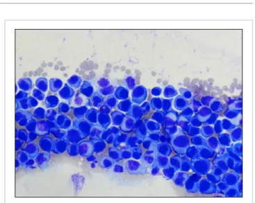
The patient underwent left thoracentesis, which revealed 550 mL of straw-colored fluid. Cytologic analysis of the pleural fluid revealed numerous atypical plasmacytoid cells with multinucleated forms, nucleoli, cytoplasmic vacuoles, and mitotic figures that were consistent with myelomatous cells (Fig. 2). Her condition deteriorated and she died 1 week into her hospitalization.
CASE 2
A 63-year-old woman was admitted because of weakness
bloodresearch.or.kr Blood Res 2016;51:133-47.
Letters to the Editor 143
Fig. 2. Pleural fluid cytology revealing numerous atypical plasmacytoid cells with multinucleated forms, nucleoli, cytoplasmic vacuoles, and mitotic figures.
Fig. 1. Computed tomography image showing extensive bilateral pleural nodularity. A pulmonary mass can be seen on the right anterior chest wall (yellow arrow); bilateral pleural effusions are also present (purple arrows).
she had been experiencing over the previous 2 weeks. She reported having dropped a glass twice from her hand, which prompted her to present to the emergency department. In the initial workup, the patient had a calcium level of 11.6 mg/dL, hemoglobin level of 8.0 g/dL, and creatinine level of 3.6 mg/dL. The patient had a previous diagnosis of mono- clonal gammopathy of undetermined significance based on serum protein electrophoresis.
Serum free light chain analysis revealed an elevated level of lambda light chains (729 mg/L). Serum immunofixation revealed abnormal IgG and lambda light chain bands.
Chromosomal analysis revealed monosomy 13, and FISH analysis revealed an FGFR3/IgH fusion, t(4;14). Both of these genetic abnormalities were also seen in Case 1. On the basis of findings in a review article by Mikhael et al. [5], this patient’s cytogenetic signature placed her in the inter- mediate risk category with a median overall survival of 4–5 years.
The patient was subsequently started on bortezomib at a dose of 1.3 mg/m2 on days 1, 4, 8, and 11; lenalidomide at a dose of 25 mg once daily for 2 weeks on and 1 week off; and dexamethasone at a dose of 40 mg once weekly.
She was also started on monthly infusions of pamidronate at a dose of 60 mg.
Two months later, the patient was readmitted with pro- gressive shortness of breath and fatigue without fevers or chills. Chest plain radiography performed on admission re- vealed new bilateral pleural effusions, and chest CT fol- low-up showed an apical pleural-based mass consistent with widespread myeloma. A therapeutic and diagnostic thor- acentesis was performed. Fluid cytology revealed the pres- ence of malignant atypical plasma cells, which was con- sistent with the involvement of plasma cell myeloma.
The patient continued to have recurrent left pleural effu-
sions, for which a tunneled left pleural drainage catheter was placed. She was discharged and continued on the initial treatment plan. She received 8 cycles of this chemotherapy regimen.
The patient then developed a new paraspinal plasmacyto- ma confirmed by biopsy examination. Because of this pro- gression, we elected to restart the patient on lenalidomide at a dose of 25 mg daily, with dexamethasone at a dose of 40 mg weekly; in addition, she was started on carfilzomib at a dose of 20 mg/m2 on days 1, 2, 8, 9, 15, and 16. To date, the patient has tolerated 2 cycles of carfilzomib without any symptomatic decline.
DISCUSSION
Pleural effusions develop in about 6% of patients with MM. In this subset of patients, less than 1% of effusions are MPEs [6]. Current literature reveals that less than 100 cases of MPE have been reported worldwide. According to these case reports [7, 8], MPE is consistently a poor prognostic indicator, with mean survival of less than 4 months.
MPE has been reported in patients with ages ranging from 22 to 83 years, equally distributed between males and females, and in IgA, IgG, IgD, and light chain subtypes [9, 10]. A case series published by Cho et al. in 2010 [10]
conducted a statistical analysis of 19 patients. In that series, IgA myeloma was most frequently implicated with malig- nant pleural effusions, followed by IgD and IgG; however, these findings are likely not statistically significant because only 19 patients were analyzed.
Kim et al. [9] demonstrated in a case report that despite aggressive treatment with systemic chemotherapy, radia- tion, autologous stem cell transplantation, or direct chemo- therapy injection in the pleural cavity, these effusions often recurred within months and ultimately led to the patient’s death. One case report [11] showed resolution of MPE and
Blood Res2016;51:133-47. bloodresearch.or.kr
144 Letters to the Editor
MM with bortezomib, a proteasome inhibitor known for its remarkable efficacy in treating extramedullary MM.
Although the pathogenesis of MPE is unknown, it is theor- ized that it may be a direct extension of thoracic myeloma- tous involvement. A review of 57 cases [9] demonstrated that half of the patients with MPE had concomitant thoracic skeletal, lung parenchymal, or chest wall plasmacytomas, which would provide a source for MPEs. Similarly, both of our patients had a pulmonary nodule, which likely repre- sented metastatic disease.
Genetic analysis showed that the patient in our first case had a trisomy at chromosome 3 and monosomy at chromo- some 13. In addition to the t(4;14) translocation, this com- plex karyotype is associated with unfavorable prognosis [5].
Given that the median survival time for high-risk patients without malignant pleural effusions is 3 years, it is likely that the progression of the myeloma and development of the pleural effusions contributed significantly to the even- tual death of the first patient. In our second case, the patient had no trisomy, but she did have monosomy of chromosome 13 in addition to the t(4;14) translocation. This chromosome 13 abnormality was also seen in 77.8% of patients in the Cho et al. [10] case series.
Although rare, more cases of MPE are being described in the literature, with evidence indicating its poor prognosis and lack of efficacious treatment [5, 12]. Because of the severity of MPE, we recommend that patients with pleural effusions and suspicion of myeloma undergo protein electro- phoresis, flow cytometry, cytologic examination of the pleu- ral fluid, or pleural biopsy examination to identify MPE and begin treatment promptly [12, 13].
Akshay Amaraneni1, Usman Saeed2, Devin Malik1, Megan Brown3, Sreenivasa R. Chandana4
1Department of Internal Medicine, 2Department of Internal Medicine-Pediatrics, Western Michigan University, Homer Stryker M.D. School of Medicine, Kalamazoo, 3Michigan State University, College of Human Medicine, East Lansing,
4Division of Hematology and Oncology, West Michigan Cancer Center, Kalamazoo, MI, USA Correspondence to: Sreenivasa R. Chandana
Division of Hematology and Oncology, West Michigan Cancer Center, 200 N Park Street, Kalamazoo, MI 49007, USA
E-mail: schandana@wmcc.org
Received on May 9, 2015; Revised on May 27, 2015; Accepted on Jun. 15, 2015 http://dx.doi.org/10.5045/br.2016.51.2.142
AuthorsÊ Disclosures of Potential Conflicts of Interest No potential conflicts of interest relevant to this article were reported.
REFERENCES
1. Siegel R, Naishadham D, Jemal A. Cancer statistics, 2013. CA
Cancer J Clin 2013;63:11-30.
2. Kumar SK, Rajkumar SV, Dispenzieri A, et al. Improved survival in multiple myeloma and the impact of novel therapies. Blood 2008;111:2516-20.
3. Bladé J, Rosiñol L. Complications of multiple myeloma. Hematol Oncol Clin North Am 2007;21:1231-46.
4. Yosunkaya S, Maden E, Toy H, Yazici R, Ozer F, Reisli I. A multi- ple myeloma case presenting with bilateral pleural involvement.
Tuberk Toraks 2007;55:285-9.
5. Mikhael JR, Dingli D, Roy V, et al. Management of newly diag- nosed symptomatic multiple myeloma: updated Mayo strat- ification of myeloma and risk-adapted therapy (mSMART) con- sensus guidelines 2013. Mayo Clin Proc 2013;88:360-76.
6. Kintzer JS Jr, Rosenow EC 3rd, Kyle RA. Thoracic and pulmonary abnormalities in multiple myeloma. A review of 958 cases. Arch Intern Med 1978;138:727-30.
7. Meoli A, Willsie S, Fiorella R. Myelomatous pleural effusion.
South Med J 1997;90:65-8.
8. Kamble R, Wilson CS, Fassas A, et al. Malignant pleural effusion of multiple myeloma: prognostic factors and outcome. Leuk Lymphoma 2005;46:1137-42.
9. Kim YJ, Kim SJ, Min K, et al. Multiple myeloma with myeloma- tous pleural effusion: a case report and review of the literature.
Acta Haematol 2008;120:108-11.
10. Cho YU, Chi HS, Park CJ, Jang S, Seo EJ, Suh C. Myelomatous pleural effusion: a case series in a single institution and literature review. Korean J Lab Med 2011;31:225-30.
11. Mangiacavalli S, Varettoni M, Zappasodi P, Pica G, Lazzarino M, Corso A. A striking response to bortezomib in a patient with pleu- ral localization of multiple myeloma. Leuk Res 2009;33:577-8.
12. Keklik M, Sivgin S, Pala C, et al. Flow cytometry method as a diag- nostic tool for pleural fluid involvement in a patient with multiple myeloma. Mediterr J Hematol Infect Dis 2012;4:e2012063.
13. Oudart JB, Maquart FX, Semouma O, Lauer M, Arthuis- Demoulin P, Ramont L. Pleural effusion in a patient with multiple myeloma. Clin Chem 2012;58:672-4.
A rare case of diffuse large B cell lymphoma-associated
hemophagocytic syndrome initially present in the bone marrow with a favorable clinical course
TO THE EDITOR: Lymphoma-associated hemophagocytic syndrome (LAHS) is a hematological disorder associated with malignant lymphoma. It is characterized by clinical features and laboratory findings associated with hemophago- cytic lymphohistiocytosis (HLH), such as fever, cytopenia, hyperferritinemia, hypofibrinogenemia, and hemophagocy- tosis in the bone marrow (BM) [1]. The development of LAHS can be accounted for by various types of lymphoma,