ISSN: 2233-601X (Print) ISSN: 2093-6516 (Online)
− 243 −
Received: November 19, 2018, Revised: March 16, 2019, Accepted: March 20, 2019, Published online: August 5, 2019
Corresponding author: Chang Hyun Kang, Department of Thoracic and Cardiovascular Surgery, Seoul National University Hospital, Seoul National University College of Medicine, 101 Daehak-ro, Jongno-gu, Seoul 03080, Korea
(Tel) 82-2-2072-3010 (Fax) 82-2-764-3664 (E-mail) [email protected]
© The Korean Society for Thoracic and Cardiovascular Surgery. 2019. All right reserved.
This is an open access article distributed under the terms of the Creative Commons Attribution Non-Commercial License (http://creativecommons.org/
licenses/by-nc/4.0) which permits unrestricted non-commercial use, distribution, and reproduction in any medium, provided the original work is properly cited.
Primary Extraskeletal Osteosarcoma in the Anterior Mediastinum: A Case Report and Review
Seohee Joo, M.D., Jae Won Song, M.D., Kwon Joong Na, M.D., Samina Park, M.D., In Kyu Park, M.D., Young Tae Kim, M.D., Chang Hyun Kang, M.D.
Department of Thoracic and Cardiovascular Surgery, Seoul National University Hospital, Seoul National University College of Medicine, Seoul, Korea
Extraskeletal osteosarcoma (ESOS) is a malignant soft tissue neoplasm producing osteoid, without any con- tinuity with the bone or periosteum. Primary ESOS presenting in the mediastinum is an extremely rare, yet aggressive malignant tumor associated with a poor prognosis. We report a case of primary ESOS arising from the thymus in a 63-year-old male patient.
Key words: 1. Osteosarcoma 2. Mediastinum
Case report
In this study informed consent was obtained form the patient.
A 63-year-old man was referred to Seoul National University Hospital for further evaluation of a grow- ing chest mass found during an abdominal aortic aneurysm work-up. The chest mass was first identi- fied as an incidental finding 5 years previously and surgical resection was recommended. However, the patient was lost to follow-up. The patient had a his- tory of hypertension and hyperlipidemia. His physical examination was unremarkable and a routine labo- ratory test showed no abnormal results. The patient’s initial contrast-enhanced chest computed tomography (CT) taken 5 years previously showed a 2.6-cm, het- erogeneously enhancing mass with no invasion of the adjacent vessels. However, the chest CT scan taken upon presentation to our department revealed a well-defined, 5.5-cm, heterogeneously enhancing mass in the anterior mediastinum that contained calcifica-
tions and encased the left brachiocephalic vein (Fig.
1).
The tumor was preoperatively diagnosed as a T3 advanced thymoma or thymic cancer, and robot-as- sisted total thymectomy via a subxiphoid approach was performed. Intraoperative findings revealed a tu- mor encasing the left brachiocephalic vein, internal thoracic vein and artery, and left phrenic nerve. The mass was completely excised with adequate resection margins by en bloc resection, and the vessels and nerve were sacrificed. Histology showed a grossly en- capsulated tumor measuring 5.6 cm×5.0 cm×4.8 cm, with direct invasion of the brachiocephalic vein.
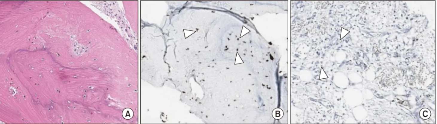
There was no invasion of the phrenic nerve, internal thoracic vessels, or lymphatic system. No lymph node metastasis was identified. Hemorrhage and necrosis was detected from 20% of the tumor’s cut surface, and calcification was present (Fig. 2). Histopathologic findings showed osteoid formation within the tumor and immunohistochemical studies showed a positive stain for vimentin, a focal positive stain for cytoker-
Korean J Thorac Cardiovasc Surg 2019;52:243-246 □ CASE REPORT □
https://doi.org/10.5090/kjtcs.2019.52.4.243
Seohee Joo, et al
− 244 −
Fig. 1. Chest radiographs. (A) A chest X-ray showing a mass-like opacity at the anteroposterior window and (B) a chest CT axial view showing a 5.6-cm irregular mass with calcification encasing the left brachiocephalic vein, without any attachment to the bony thorax at the time of admission. (C) A CT axial view, taken 5 years previously, showing a 2.6-cm mass with no involvement of nearby vessels. CT, computed tomography.
Fig. 2. Operative findings and gross findings of the mass. (A) Dissection of the left brachiocephalic vein (ar- rowhead). (B) Dissection of the in- ternal thoracic vein (arrowhead).
(C) Complete removal of the tumor (arrowhead). (D) A grossly encapsu- lated tumor, measuring 5.6 cm×5.0 cm×4.8 cm and exhibiting calcifica- tions, hemorrhage, and necrosis on its cut surface.
atin, and negative stains for CD5, CD117, MUC1, and desmin (Fig. 3).
The pathological findings were consistent with osteosarcoma. The tumor showed no continuity with the thoracic bones, and no other extra-thoracic pri-
mary tumors were present. The tumor was diag- nosed as a primary osteosarcoma of the thymus. The patient started radiotherapy 1 month after the oper- ation and is scheduled to receive chemotherapy.
Extraskeletal Osteosarcoma in the Mediastinum
− 245 −
Fig. 3. Microscopic findings of the mass. (A) Osteoid formation, a definitive characteristic of osteosarcoma (H&E staining, ×20 magnifica- tion). (B) Positive staining for vimentin (×20 magnification; arrowheads). (C) Focal positive staining for cytokeratin (×20 magnification;
arrowheads).
Discussion
Extraskeletal osteosarcoma (ESOS) is a malignant soft tissue neoplasm that produces osteoid, bone, or chondroid materials in places without direct attach- ment to bone or periosteum. ESOS is a rare malig- nant tumor, accounting for 1%–2% of all soft tissue sarcomas and 2%–4% of all osteosarcomas [1].
Unlike conventional osteosarcoma, which mainly af- fects teenagers and young adults, ESOS most com- monly affects patients older than 30.
ESOS is difficult to diagnose due to its asympto- matic nature, and is usually found due to its mass effect. ESOS is diagnosed by the following criteria:
(1) the presence of a uniform morphological pattern of sarcomatous tissue without the possibility of ma- lignant mesenchymoma, (2) production of malignant osteoid or bone by the sarcoma, and (3) exclusion of a primary osseous tumor [2]. Because calcification or osteoid formation occurs in approximately 50% of patients with this tumor, the tumor may be best de- tected by chest CT. However, calcification is a com- mon feature found in tumors with other etiologies, including thymomas [3]. Therefore, ESOS cannot be diagnosed based on a single chest CT scan showing malignant features accompanied by calcification. A definite diagnosis of ESOS requires histopathologic confirmation. ESOS shares the same histopathologic features as osteosarcoma originating from the skel- etal system. It usually has a uniform sarcomatous pattern and produces osteoid. The immunophenotypes of ESOS, which are also similar to those of osteo- sarcoma, include expression of CD99, alkaline phos-
phatase, osteocalcin, and vimentin. In many cases, ac- tin, desmin, S-100 protein, epithelial membrane anti- gen, and keratin are expressed as well [4].
Furthermore, a frozen biopsy during the operation may aid in diagnosing and obtaining an adequate re- section margin. Obtaining an accurate and quick sec- tion of an osteosarcoma, however, is considered to be a challenge due to the calcified characteristics of the tissue.
The prognosis of ESOS remains poor, with a re- ported 5-year survival rate varying from 25% to 66%. Although little is known about the factors af- fecting survival in patients with ESOS, complete sur- gical resection is considered to be the optimal treat- ment to improve survival, since ESOS is not partic- ularly sensitive to chemotherapy or radiotherapy [3,5]. The effectiveness of chemotherapy and radio- therapy is currently being debated, as a study has shown a favorable prognosis for ESOS [6].
Our case of a primary ESOS arising in the media- stinum is extremely rare. Only 10 cases have been reported in the international literature. Of the 10 pa- tients, 6 were male and 4 were female. The median age was 47 years (range, 19–77 years) and the me- dian size of the tumors was 9.25 cm (range, 5.5–16 cm). The outcome of ESOS arising from the media- stinum remains poor, with overall 1-, 2-, and 5-year survival rates of merely 44%, 22%, and 11%, respectively. The median overall survival was 4.0 months.
In conclusion, primary ESOS in the mediastinum is extremely rare, difficult to diagnose, and associated with a poor prognosis. In this case, the mass was ini-
Seohee Joo, et al
− 246 − tially considered to be a thymoma. However, physi-
cians should be aware of the possibility of ESOS, and surgical resection should be considered as a primary choice in cases where ESOS is suspected.
Conflict of interest
No potential conflict of interest relevant to this ar- ticle was reported.
ORCID
Seohee Joo: https://orcid.org/0000-0002-1066-2011 Jae Won Song: https://orcid.org/0000-0002-3530-0623 Kwon Joong Na: https://orcid.org/0000-0003-4158-9790 Samina Park: https://orcid.org/0000-0001-9625-2672 In Kyu Park: https://orcid.org/0000-0003-3550-5554 Young Tae Kim: https://orcid.org/0000-0001-9006-4881 Chang Hyun Kang: https://orcid.org/0000-0002-1612-1937
References
1. Fletcher CD, Unni KK, Mertens F. Extraskeletal osteosarcoma.
In: Fletcher CD, Unni KK, Mertens F, editors. World Health Organization classification of tumours: pathology and ge- netics of tumours of soft tissue and bone. Lyon: IARC Press; 2002. p. 182-3.
2. Allan CJ, Soule EH. Osteogenic sarcoma of the somatic soft tissues: clinicopathologic study of 26 cases and review of literature. Cancer 1971;27:1121-33.
3. Lee CH, Park CR, Kim JW, Suh JH, Lee YJ, Jung JP.
Extraskeletal osteosarcoma arising from the pleura.
Korean J Thorac Cardiovasc Surg 2014;47:320-4.
4. Mavrogenis AF, Papagelopoulos PJ. Soft tissue tumors: ex- traskeletal osteosarcoma. Atlas Genet Cytogenet Oncol Haematol 2014;18:443-6.
5. Burt M, Ihde JK, Hajdu SI, et al. Primary sarcomas of the mediastinum: results of therapy. J Thorac Cardiovasc Surg 1998;115:671-80.
6. Qian J, Zhang XY, Gu P, Shao JC, Han BH, Wang HM.
Primary thoracic extraskeletal osteosarcoma: a case re- port and literature review. J Thorac Dis 2017;9:E1088-95.