pISSN 2288-9272 eISSN 2383-8493 J Oral Med Pain 2021;46(3):93-97 https://doi.org/10.14476/jomp.2021.46.3.93
Myositis Associated with Infratemporal Space Abscess in Patient with Myxofibrosarcoma of Nasal Cavity: Case Report
Jiyeon Kim, Min Chang, YounJung Park, Hyung-Joon Ahn, Seong-Taek Kim, Jong-Hoon Choi, Jeong-Seung Kwon
Department of Orofacial Pain and Oral Medicine, Dental Hospital, Yonsei University College of Dentistry, Seoul, Korea
Received September 6, 2021 Revised September 19, 2021 Accepted September 19, 2021
The limited mouth opening, also known as trismus, can result from temporomandibular joint disorders, infection, neoplasm, trauma, and abnormal anatomic structure like coro- noid hyperplasia. Head and neck cancer patients often complain of limited mouth opening, which is usually induced by myofibrotic contracture of masticatory muscle. But clinicians should consider any reasons such as infection or cancer growth and metastasis if trismus gets worse or pain develops. We report the case of the patient, who was diagnosed with myxofibrosarcoma on nasal cavity. He had suffered from trismus after concurrent chemo- radiotherapy. However, pain had developed and trismus had worsened. He was diagnosed with infratemporal space abscess and myositis of masticatory muscles.
Key Words:
Key Words: Head and neck neoplasms; Myositis; Trismus
Correspondence to:
Jeong-Seung Kwon
Department of Orofacial Pain and Oral Medicine, Dental Hospital, Yonsei University College of Dentistry, 50-1 Yonsei-ro, Seodaemun-gu, Seoul 03722, Korea Tel: +82-2-2228-3111
Fax: +82-2-393-5673 E-mail: [email protected]
https://orcid.org/0000-0003-4584-7355
JOMP
Journal of Oral Medicine and PainCopyright Ⓒ 2021 Korean Academy of Orofacial Pain and Oral Medicine.
CC This is an open-access article distributed under the terms of the Creative Commons Attribution Non-Commercial License (http://creativecommons.org/licenses/by-nc/4.0/), which permits unrestricted non-commercial use, distribution, and reproduction in any medium, provided the original work is properly cited.
INTRODUCTION
The limited mouth opening (LMO) is a condition in which the mouth opening is limited owing to various causes, in- cluding trauma, infection, neurologic disorders, neoplasms, anatomic structures, and temporomandibular joint disor- ders [1-3]. Head and neck cancer patients often suffer from trismus, especially after radiation therapy. It is known that the main reason to cause trismus is myofibrotic masticatory muscle contracture, which occurs as a result of radiation- induced fibrosis of muscles [4-8].
In these patients, clinicians should consider not only muscle contracture but also myositis due to infection as a cause of LMO because they are immunocompromised.
Myositis is an inflammatory muscle disease, which has clinical characteristics of inflammation or infection such as edema, erythema, and increased temperature.
In this paper, we present the case of a patient, who al- ready had the LMO after concurrent chemoradiotherapy
(CCRT) for myxofibrosarcoma (MFS) of nasal cavity.
However, pain had developed and the LMO had worsened.
He was diagnosed with infratemporal space abscess and myositis of masticatory muscles.
The protocol of this study was approved by the Insti- tutional Review Board of Yonsei University Dental Hospital (IRB no. 2-2021-0090).
CASE REPORT
A 41-year-old man visited the Department of Orofacial Pain and Oral Medicine of Yonsei University Dental Hospital (Seoul, Korea) with a complaint of pain in his right masse- ter area and LMO.
He underwent partial removal of extensive MFS arising
in right nasal cavity by the otolaryngologist 4 years and 6
months before the visit. He had also received CCRT for 5
weeks after surgery and kept taking an anti-cancer medi-
cation Pazopanib (Votrient; Novartis, Ljubljana, Slovenia).
Although he had experienced some discomfort and limita- tion during opening mouth after CCRT, he started to feel pain in right masseter area when opening mouth and chew- ing and LMO worsened 2 months before the visit without any reason. Even though he had already taken strong anal- gesics (Targin CR [oxycodone hydrochloride and naloxone hydrochloride], IRcodon [oxycodone hydrochloride], Mypol [acetaminophen/codeine phosphate hydrate/ibuprofen]), his pain had aggravated. He did not show any inflammatory signs in orofacial area and blood tests including complete blood count, routine chemistry were normal except low red blood cell count and hemoglobin.
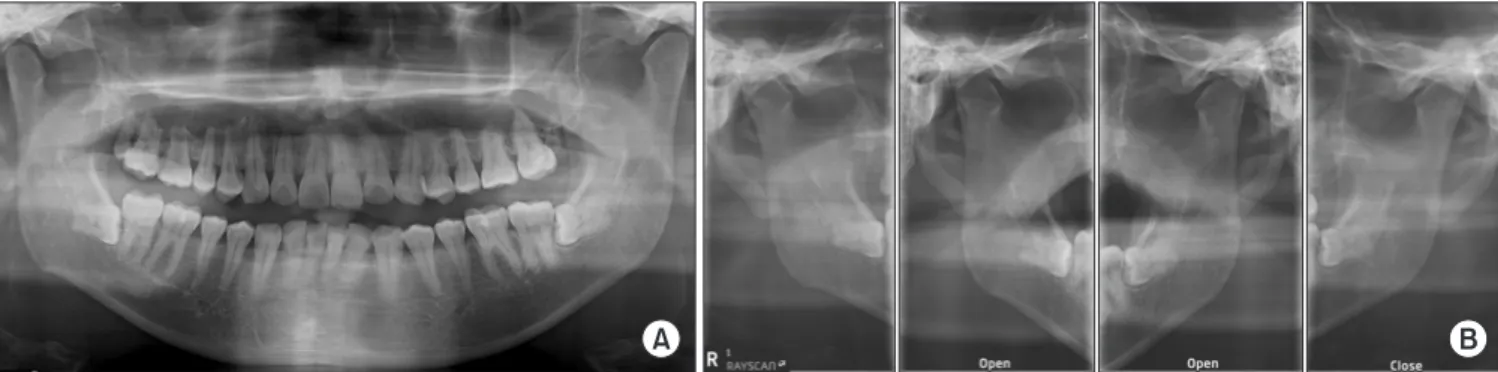
On clinical examination, the unassisted maximal interin- cisal distance was about 24 mm with pain in right masseter area. The assisted maximal interincisal distance was 26 mm with hard end feel. The maximum range of protrusive, right lateral, and left lateral movement were 4 mm, 2 mm, and 3 mm respectively. The protrusive and right lateral movement induced pain in both and right temporomandibular joint pain respectively. The palpation revealed familiar pain in the origin of the right masseter. The occlusal contacts were stable in maximum intercuspation. The panoramic radio- graph on the first visit revealed that right maxillary sinus walls were extensively disappeared and haziness in right maxillary sinus was noted (Fig. 1).
Based on history and clinical examinations, the patient was diagnosed with a myofascial pain on right masseter and myofibrotic contracture due to radiation therapy. He underwent physical therapy, and practiced mouth opening exercise with wooden tongue depressors. However, symp- toms did not improve. Cone beam computed tomography
revealed that right maxillary sinus was occupied with radi- opaque lesion infiltrated to surrounding structures without pathologic findings in mandible condyle (Fig. 2). We looked at again the post-operative magnetic resonance (MR) imag- es taken at the department of oncology for follow-up pur- pose and they revealed high signal intensity with diffuse in- flammatory change on infratemporal space and right tem- poralis, and lateral pterygoid muscle (Fig. 3). Therefore, he was diagnosed with myositis caused by infratemporal space abscess, and then he was referred to oncologist for evalua- tion and further treatment.
A B
Fig. 1.
Fig. 1. Panoramic radiograph on the first visit. (A) Destruction of right maxillary sinus wall and haziness in right sinus were noted. (B) Panoramic temporomandibular joint radiograph revealed limitation of condylar translation in both temporomandibular joints.
Fig. 2.
Fig. 2. Axial view of cone beam computed tomography on the first
visit. Destruction of right maxillary sinus posterior wall was shown,
but no definite pathologic bony change on both condyle was
found.
DISCUSSION
Myositis, or muscle inflammation is characterized by the symptoms such as swelling, redness and local heat on mus- culoskeletal area [9]. Most common etiologies are infectious agents including picornaviruses (coxsackievirus), echovi- rus, retroviruses (human immunodeficiency virus, human T-cell lymphotropic virus type 1), borrelia, toxoplasma, and adenoviruses. Nevertheless, non-infectious etiologies such as foods, drugs (D-penicillamine, cimetidine), occupational exposure, and medical devices should also be considered [9]. In addition, oral surgery or an intramuscular injection sometimes lead to infection of masticatory muscles as a post-operative complication [10].
Myositis of the masticatory muscles causes the limitation of the mandibular movement mainly due to pain. In head and neck cancer patient, it can be assumed that cancer and cancer treatments such as radiotherapy and chemotherapy induce immunosuppressed condition, which causes an in- fectious myositis. The myositis can exacerbate trismus and pain.
In this case, we took a closer look at MR images before the first visit because pain had developed and the LMO had worsened. Upon T2 images 2 years and 8 months before the first visit, fatty layer of right lateral pterygoid muscle showed significant atrophic change compared to the op- posite side (Fig. 4). The fatty layer showed further atro- phic change in the MR image taken 1 year and 10 months before the visit (Fig. 5), suggesting the possibility of the post-operative inflammation [11,12]. Whole-body positron
emission tomography/computed tomography did not re- veal other lesions. Furthermore, it showed more hyperin- tense T2-weighted signals compared to the previous imag- ing. Therefore, it was presumed that recent occurrence of the pain and aggravated LMO were caused by myositis as- sociated with infratemporal space abscess related to known malignancy.
Since there are a number of various causes of the LMO (Fig. 6), the clinicians should consider all possible causes in patient with the LMO. In addition, the clinicians should also be aware that the LMO can be caused by more than one factor. In patient who have head and neck cancer and
A B
Fig. 3.
Fig. 3. Axial T2-weighted magnetic resonance images taken at the de- partment of oncology. (A) Two months before the first visit. It revealed high signal intensity on right infratemporal space, temporalis (arrowhead), and lateral pterygoid muscle (arrows). (B) 3 days after first visit. It showed more increased high signal intensity with diffuse inflammatory change compared to 2 months before. A, anterior view;
P, posterior view; R, right view; L, left view.
Fig. 4.
Fig. 4. Axial T2-weighted magnetic resonance images 2 years and 8
months before the first visit. It revealed loss of the fat plane signal
(arrows) adjacent to the right lateral pterygoid muscle, indicating
more inflammatory changes compared to the left side.
undergo radiotherapy, the LMO is most commonly caused by myofibrotic contracture [4–8,13], but the possibility of infectious myositis also has to be ruled out considering im- munocompromised state of the patients. The infectious myositis should also be suspected, especially if the LMO be- comes worse, or the pain occurs or changes into a different pattern than before. In such cases, MR or contrast computed tomography images should be taken for accurate diagnosis.
CONFLICT OF INTEREST
No potential conflict of interest relevant to this article was reported.
ORCID
Jiyeon Kim
https://orcid.org/0000-0002-2150-6882
Limited mouth opening
Temporomandibular
joint disorder Others
Temporomandibular joint Muscle Infection Neoplasm Anatomic
structure Trauma
Disc displacement
without reduction
Ankylosis
Adhesion Adherence
Muscle contracture
Fibromyalgia
Myofibrotic
Myostatic
Myositis
Cause
Cellulitis Condylar hyperplasia
Coronoid impedance
Eagle s syndrome Trauma
Autoimmune Bacteria Virus