ABSTRACT
Background: We aimed to assess the performance of the 2015 American College of
Rheumatology (ACR)/European League Against Rheumatism (EULAR) classification criteria for gout in Korean patients with acute arthritis and to compare the performance of the ACR/
EULAR criteria to that of other sets of criteria for gout classification.
Methods: Patients with acute arthritis who underwent diagnostic arthrocentesis at one of the four participating rheumatology clinics were consecutively enrolled between February and December 2017. Crystal-proven gout was diagnosed upon confirming the presence of monosodium urate (MSU) crystals in patients with a clinical impression of gout as judged by the rheumatologist. The performance of the ACR/EULAR and other gout classification criteria, including the Rome, New York, American Rheumatism Association (ARA), Mexico, and Netherlands criteria, was analyzed regardless of the presence/absence of MSU crystals.
Results: The study enrolled 118 gout patients (all crystal-proven) and 95 non-gout patients.
According to the area under the curve, the diagnostic performance was the highest for the ACR/EULAR classification criteria (sensitivity, 80.5%; specificity, 95.8%; area under the curve, 0.966), followed by the Netherlands, Rome, ARA, New York, and Mexico criteria. All six sets of criteria demonstrated lower sensitivity in patients exhibiting the first episode of acute arthritis.
Conclusion: In Korean patients with acute arthritis, the ACR/EULAR classification criteria outperformed other sets of gout classification criteria even in the absence of information regarding the presence of MSU crystals. However, to enhance diagnostic sensitivity, synovial fluid analysis should be considered in patients with the first episode of acute arthritis.
Keywords: Gout; Classification Criteria; Sensitivity; Specificity; Monosodium Urate
Original Article
In Ah Choi ,1 Ji Hyoun Kim ,2 Yun Jong Lee ,2 Eun Ha Kang ,2 You-Jung Ha ,2 Kichul Shin ,3 Jeong Seok Lee ,4 Eunyoung Emily Lee ,4 Jin Kyun Park ,4 Eun Young Lee ,4 Eun Bong Lee ,4 and Yeong Wook Song 4
1 Division of Rheumatology, Department of Internal Medicine, Chungbuk National University Hospital, Cheongju, Korea
2 Division of Rheumatology, Department of Internal Medicine, Seoul National University Bundang Hospital, Seongnam, Korea
3 Division of Rheumatology, Department of Internal Medicine, Seoul Metropolitan Government-Seoul National University Boramae Medical Center, Seoul, Korea
4Division of Rheumatology, Department of Internal Medicine, Seoul National University Hospital, Seoul, Korea
Performance of the 2015 American College of Rheumatology/European League against Rheumatism
Classification Criteria for Gout in Korean Patients with Acute Arthritis
Received: Mar 24, 2019 Accepted: May 14, 2019 Address for Correspondence:
Yeong Wook Song, MD, PhD
Division of Rheumatology, Department of Internal Medicine, Seoul National University, College of Medicine, 101 Daehak-ro, Jongno-gu, Seoul 03087, Korea.
E-mail: [email protected]
© 2019 The Korean Academy of Medical Sciences.
This is an Open Access article distributed under the terms of the Creative Commons Attribution Non-Commercial License (https://
creativecommons.org/licenses/by-nc/4.0/) which permits unrestricted non-commercial use, distribution, and reproduction in any medium, provided the original work is properly cited.
ORCID iDs In Ah Choi
https://orcid.org/0000-0003-4662-4065 Ji Hyoun Kim
https://orcid.org/0000-0003-0765-347X Yun Jong Lee
https://orcid.org/0000-0001-7615-8611 Eun Ha Kang
https://orcid.org/0000-0001-9697-1159 You-Jung Ha
https://orcid.org/0000-0001-6107-9523 Kichul Shin
https://orcid.org/0000-0002-6749-7598
Immunology, Allergic
Disorders & Rheumatology
Jeong Seok Lee
https://orcid.org/0000-0001-8261-7044 Eunyoung Emily Lee
https://orcid.org/0000-0002-1575-9007 Jin Kyun Park
https://orcid.org/0000-0003-2167-9393 Eun Young Lee
https://orcid.org/0000-0001-6975-8627 Eun Bong Lee
https://orcid.org/0000-0003-0703-1208 Yeong Wook Song
https://orcid.org/0000-0002-5384-3437 Funding
This research was supported by a grant of the Korea Health Technology R&D Project through the Korea Health Industry Development Institute (grant No. HI14C1277), funded by the Ministry of Health & Welfare, Republic of Korea.
Disclosure
The authors have no potential conflicts of interest to disclose.
Author Contributions
Conceptualization: Choi IA, Song YW. Data curation: Choi IA, Kim JH, Lee YJ, Shin K, Lee JS, Lee EE. Formal analysis: Choi IA.
Investigation: Choi IA, Kim JH, Lee YJ, Kang EH, Ha YJ, Shin K, Lee JS, Lee EE, Park JK, Lee EY, Lee EB, Song YW. Methodology: Choi IA, Lee YJ, Shin K, Lee JS, Park JK, Lee EY, Lee EB, Song YW. Writing - original draft: Choi IA.
Writing - review & editing: Choi IA, Kim JH, Lee YJ, Kang EH, Ha YJ, Shin K, Lee JS, Lee EE, Park JK, Lee EY, Lee EB, Song YW.
INTRODUCTION
Gout is the most common type of inflammatory arthritis among men while its incidence is gradually increasing in many countries, including Korea.1,2 The global burden of gout has been increasing in parallel with its prevalence.3 Without appropriate treatment, patients may experience relapsing arthritis and the disease progresses to chronic tophaceous gout, with joint destruction and renal insufficiency. However, the presenting symptoms of several diseases such as pseudogout, septic arthritis, and rheumatoid arthritis with foot involvement can mimic those of gout, which delays the diagnosis and timely intervention with adequate treatment for gout. Thus, it is important to accurately differentiate between gout and other diseases.
A definite diagnosis of gouty arthritis can be established based on the presence of
monosodium urate (MSU) crystals in joint fluid extracted from the inflamed area, which is evaluated under a polarizing microscope. However, the aspiration and visualization of MSU crystals required specially trained personnel. In addition, the process of extracting joint fluid is painful to the patient. Furthermore, the first metatarsophalangeal joint, which is the most commonly involved site, is a small joint with a very small amount of synovial fluid, which makes it difficult to obtain sufficient joint fluid for analysis. Finally, the test may appear negative if the uric acid crystals have already dissolved the delayed visit to the clinic. To overcome these limitations of synovial fluid analysis, substantial effort has been done for the development of diagnostic criteria based mainly on clinical symptoms of gouty arthritis.
Since the publication of the Rome criteria in 1963,4 several sets of gout classification criteria have been developed, including the New York,5 1977 American Rheumatology Association (ARA),6 Mexico,7 and Netherlands8 criteria, although none of these scoring systems have become widely adopted in clinical practice. Meanwhile, in 2015, the American College of Rheumatology (ACR) and European League Against Rheumatism (EULAR) published a new set of classification criteria for gout,9 based on laboratory and imaging tests in addition to clinical symptoms and, if available, polarizing microscopy evaluation, to enable early diagnosis and treatment. The 2015 ACR/EULAR classification criteria were internally validated on a sample of 983 patients with arthritis (MSU positive, 509; MSU negative, 474) with available polarizing microscopy data; of these, 623 patients were White/European/
Caucasian and 180 were East Asian (Taiwanese).10 To date, the clinical efficacy of the 2015 ACR/EULAR classification criteria has not been verified in Koreans.
In the present study, we aimed to determine whether the 2015 ACR/EULAR gout
classification criteria can effectively distinguish gout from non-gout arthritis among Korean patients with acute arthritis in the absence of synovial fluid analysis. Additionally, we aimed to compare the performance of six currently available sets of gout classification criteria in the same population.
METHODS
Patients and study design
Patients with acute arthritis who underwent diagnostic and/or therapeutic arthrocentesis at any of the four participating rheumatology clinics were consecutively enrolled between February and December 2017. Those who were previously diagnosed and have been treated
for arthritis were also allowed to participate if they present with acute exacerbation and need diagnostic arthrocentesis for differential diagnosis or therapeutic arthrocentesis.
Polarizing microscopy examination of synovial fluid extracted from the affected joint or bursa was conducted by experienced rheumatologists and/or pathologists. Additional SF examinations, including white blood cell counts, Gram staining and cultures were performed for differential diagnosis.
The patients were stratified into the gout group (diagnosis established based on the
confirmed presence of MSU crystals in the synovial fluid) and the non-gout group (diagnosis based on the clinical impression of certain non-gout disease and confirmed non-presence of MSU crystals in the synovial fluid). Patients having gout combined with non-gout arthritis were excluded from the study because their mixed clinical characteristics would have biased the clinical classification. Subgroup analyses were conducted in patients with the first episode of acute arthritis and in those with non-tophaceous disease.
Data collection
Data were collected using the predetermined questionnaire containing 12 items regarding the pattern of joint involvement (first metatarsophalangeal joint, ankle/midfoot, or other joints/polyarticular involvement), characteristics of the symptomatic episode (whether they have erythema overlying the affected joint, cannot tolerate touch or pressure to the affected joint or have great difficulty with walking or inability to use the affected joint), time course of the arthritis episodes (once or recurrent typical episodes), and clinical evidence of tophus (Supplementary Data 1, written in Korean), and the clinical record form (CRF) containing 12 items to review age, gender, clinical diagnosis, colchicine response, presence of tophus and synovial fluid culture/crystal exam, serum uric acid, radiograph, ultrasonography, and DECT data (Supplementary Data 2). The questionnaire and the CRF were compiled based on the six existing sets of gout classification criteria. The review of the medical records was done by the investigators themselves or by the fellow in the clinic (if there are any), who was also involved in the enrollment process. To avoid missing data for the analysis, the data abstraction tool (Supplementary Data 3, with instructions written in Korean) were also used.
Overviews of the ACR/EULAR gout classification criteria and other sets of gout classification criteria published to date are available in Supplementary Data 4 and 5, respectively. Since the presence of MSU crystals alone is sufficient to diagnose gout according to the ACR/EULAR, ARA, New York, and Mexico classification criteria, we considered that the results of the positive polarizing microscopy investigation were not available for the primary performance analysis. Therefore, the performance of “ACR/EULAR classification” was tested using clinical domain, serum uric acid in laboratory domain, and imaging domain (radiographic, ultrasound, or DECT imaging), while MSU results in laboratory domain scored as 0 (not done). The performance of the “ACR/EULAR—clinical only—classification” used clinical domain and serum uric acid, but not included MSU crystal results or imaging domain. In the Rome criteria, patients were considered to have gout only if at least two of three clinical criteria were satisfied (excluding the presence of MSU crystals).
Statistical analyses
Sample size was calculated using the formula n = Z2 × P(1−P)∕d2, with a 95% confidence level (Z = 1.96) and a margin of error of 5% (d = 0.05). Based on the ACR/EULAR classification criteria, the sensitivity of the clinical-only criteria was 0.85 (P = 0.85)10; therefore, a sample size of at least 49 patients was required for this study.
Categorical variables are described in terms of frequency (percentage), while data regarding continuous variables are presented as mean ± standard deviation. Comparisons between the gout and non-gout groups were performed using χ2 or Fisher's exact test for categorical variables and Student's t-test for continuous variables. The performance of each set of classification criteria was assessed in terms of sensitivity, specificity, positive predictive value, negative predictive value, and area under the receiver operating characteristic curve.
Statistical analyses were performed using statistical software (MedCalc, version 18.6;
MedCalc Software bvba, Ostend, Belgium). Graphs were visualized using GraphPad Prism, version 7.00, for Windows (GraphPad Software, La Jolla, CA, USA).
Ethics statement
The present study protocol was reviewed and approved by the Institutional Review Board (IRB) of the relevant hospitals (CBNUH IRB approval No., 2016-09-001; SNUH IRB approval No., 1611-055-807; SNUBH IRB approval No., B-1705/395-402; SMG-SNU Boramae Medical Center IRB approval No., 20170414/16-2017-62/051). Informed consent was submitted by all subjects when they were enrolled.
RESULTS
Baseline and clinical characteristics
Among the 215 patients who underwent arthrocentesis, one patient having clinical rheumatoid arthritis combined with gout and another patient having spondyloarthropathy combined with gout were excluded. Finally, the study enrolled 118 patients with the clinical impression of gout and confirmed presence of MSU crystals in the synovial fluid (gout group), as well as 95 patients with a clinical impression of gout or non-gout disease but with absence of MSU crystals from the synovial fluid (non-gout group). The baseline demographics and clinical characteristics of the enrolled patients are summarized in Table 1.
Conventional radiography data were available for all patients. However, ultrasonography was performed only in 35.2% of patients and DECT was not available in participating hospitals.
Only 11.8% of patients had double-contour sign in the US study.
Overall performance of gout classification criteria: the 2015 ACR/EULAR criteria outperformed other gout classification criteria
The performance of the six sets of gout classification criteria was analyzed in the entire study sample (Fig. 1A and Table 2). In the primary analysis of performance, synovial fluid microscopy was considered not performed, to prevent circular reasoning. Among the six sets evaluated, the 2015 ACR/EULAR criteria showed the best performance, regardless of imaging results (Table 2). When synovial fluid microscopy results were added, the sensitivities of the ACR/EULAR, ARA, New York, and Mexico criteria improved to 100%, that of the Rome criteria improved to 97.5%, and that of the Netherlands criteria improved to 99.2%;
additionally, the specificity of the ACR/EULAR criteria improved to 100% (Fig. 1A).
Performance of gout classification criteria in subgroups: all gout classification criteria have lower sensitivity in patients with first-time arthritis
The performance of the six sets of gout classification criteria was analyzed in the subgroup of patients with first-time arthritis (Fig. 1B and Table 3) and in the subgroup of patients with non-tophaceous disease (Fig. 1C and Table 4). Once again, synovial fluid microscopy analysis was considered not performed for the primary performance analysis.
Table 1. Baseline demographics and clinical characteristics of acute arthritis patients stratified according to gout incidence
Characteristics Gout (n = 118) Non-gout (n = 95) P value
Age, yr 57.9 ± 15.3 63.1 ± 13.8 0.624
Gender, Men, % 94.1 21.3 < 0.001
Clinical diagnosis, % Gout (100) Rheumatoid arthritis (57.9) -
Acute CPPD arthritis (21.1) Spondyloarthropathy (6.3) Osteoarthritis (5.3) Sjogren's disease (2.1) Behcet's disease (2.1) Septic arthritis (1.1) Dermatomyositis (1.1) Lupus arthritis (1.1) Adult-onset Still's disease (1.1) Palindromic rheumatism (1.1)
First arthritis episode 25 (21.2) 21 (22.1) 0.870
Joint involved -
First metatarsophalangeal 68 (57.6) 15 (15.8)
Ankle/midfoot 70 (59.3) 47 (49.5)
Other joint involvement only 13 (11.0) 44 (46.3)
Pattern of joint involvement 0.040
Monoarthritis 32 (27.1) 17 (17.9)
Oligoarthritis 68 (57.6) 51 (53.7)
Polyarthritis 18 (15.3) 27 (28.4)
Serum uric acid, mg/dL < 0.001
< 4 1 (0.8) 30 (31.6)
≥ 4 and < 6 3 (2.5) 32 (33.7)
≥ 6 and < 8 18 (15.3) 27 (28.4)
≥ 8 and < 10 44 (37.3) 3 (3.2)
≥ 10 52 (44.1) 3 (3.2)
Serum creatinine, mg/dL 1.18 ± 0.60 1.04 ± 1.20 0.256
Comorbidities
Cardiovascular disease (including hypertension) 57 (48.3) 37 (38.9) 0.161
Diabetes mellitus 24 (20.3) 17 (17.9) 0.660
Chronic kidney disease 27 (22.9) 12 (12.6) 0.056
History of urinary tract stones 5 (4.2) 0 -
Medications
Urate-lowering agents 60 (55.5) 0 -
Diuretics 7 (5.9) 9 (9.5) 0.173
Aspirin 7 (5.9) 7 (7.4) 0.184
Presence of clinical tophus 35 (29.7) 0 -
MSU crystals 118 (100) 0 -
Bone erosion on X-ray 30 (25.4) 0 -
Changes on ultrasound or DECT 14 (11.8) 0 -
Data shown as incidence (yes/no or percentage) or mean ± standard deviation or number (%). A threshold score of ≥ 8 classifies an individual as having gout.
P values are calculated by Student's t-test or χ2 test, as appropriate.
CPPD = calcium pyrophosphate dehydrate, MSU = monosodium urate, DECT = dual-energy computed tomography.
Table 2. Performance of existing sets of gout classification criteria
Criteria Sensitivity, % Specificity, % PPV, % NPV, % AUC
ACR/EULARa 80.5b 98.9c 99.0 80.3 0.968
ACR/EULAR, clinical-onlyd 75.4 98.9 98.9 76.4 0.964
ARAa 77.1b 89.5 90.1 75.9 0.905
New Yorka 61.0b 94.7 93.5 66.2 0.836
Romea 70.3e 97.9 97.7 72.7 0.910
Mexicoa 73.7b 71.6 76.3 68.7 0.808
Netherlandsa 89.8f 83.2 86.9 86.8 0.941
PPV = positive predictive value, NPV = negative predictive value, AUC = area under the receiver operating characteristic curve, ACR/EULAR = American College of Rheumatology/European League Against Rheumatism, ARA = American Rheumatism Association,
aWithout synovial fluid microscopy; dWithout synovial fluid microscopy or imaging; When taking into account the presence of monosodium urate crystals, the sensitivities of these classification criteria were b100%, e97.5%, or f99.2%, while the specificity was f100%.
0.8 1.0
0
0 0.1 0.2 0.3 0.4 0.5
True positive rate (sensitivity)
A
0.4 0.6
0.2
False positive rate (1-specificity)
0.8 1.0
0
0 0.1 0.2 0.3 0.4 0.5
True positive rate (sensitivity)
B
0.4 0.6
0.2
False positive rate (1-specificity)
0.8 1.0
0
0 0.1 0.2 0.3 0.4 0.5
True positive rate (sensitivity)
C
0.4 0.6
0.2
False positive rate (1-specificity) With synovial fluid microscopy With synovial fluid microscopy With synovial fluid microscopy
Mexico Rome ARA Netherlands New York
ACR/EULAR ACR/EULAR -clinical
Fig. 1. Performance of different sets of gout classification criteria in Korean patients with acute arthritis. Receiver operating characteristic plots are shown.
Box shows the results obtained when taking into consideration the positive findings on polarizing microscopy (sensitivity: 100% for the ACR/EULAR, ARA, New York, Rome, and Mexico criteria; 96.0% for the New York criteria). (A) Whole sample of patients (n = 213). The 2015 ACR/EULAR criteria showed high sensitivity and the best specificity among currently available sets of gout classification criteria. (B) Subgroup of patients with first-time arthritis (n = 46). The sensitivity of the 2015 ACR/EULAR scoring system was substantially lower in this subgroup than that noted in the overall sample, while specificity was preserved; a similar decrease in sensitivity was noted for the Netherlands criteria, whereas a more substantial decrease was noted for the ARA, New York, Rome, and Mexico criteria. (C) Subgroup of patients with non-tophaceous disease (n = 178). The diagnostic performance was preserved for all sets of existing criteria in this subgroup, except for the New York criteria.
ARA = American Rheumatism Association, ACR/EULAR = American College of Rheumatology/European League Against Rheumatism.
Table 3. Performance of gout classification criteria in 46 Korean patients with first-time arthritis
Criteria Sensitivity, % Specificity, % PPV, % NPV, % AUC
ACR/EULARa 52.0b 100 100 63.6 0.888
ACR/EULAR, clinical-onlyc 48.0 100 100 61.8 0.882
ARAa 32.0b 76.2 61.5 48.5 0.628
New Yorka 16.0b 71.4 40.0 41.7 0.546
Romea 24.0d 100 100 52.5 0.788
Mexicoa 28.0b 85.7 70.0 50.0 0.697
Netherlandsa 60.0e 100 100 67.7 0.901
This analysis included 25 gout and 21 non-gout patients.
PPV = positive predictive value, NPV = negative predictive value, AUC = area under the receiver operating characteristic curve, ACR/EULAR = American College of Rheumatology/European League Against Rheumatism, ARA = American Rheumatism Association.
aWithout synovial fluid microscopy; cWithout synovial fluid microscopy or imaging; When taking into account the presence of monosodium urate crystals, the sensitivities of these classification criteria were b100%, d100%, or
e96.0%.
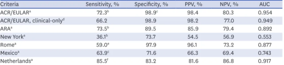
Table 4. Performance of gout classification criteria in 178 Korean patients with non-tophaceous disease
Criteria Sensitivity, % Specificity, % PPV, % NPV, % AUC
ACR/EULARa 72.3b 98.9c 98.4 80.3 0.954
ACR/EULAR, clinical-onlyd 66.2 98.9 98.2 77.0 0.949
ARAa 73.5b 89.5 85.9 79.4 0.892
New Yorka 36.1b 73.7 54.5 56.9 0.553
Romea 59.0e 97.9 96.1 73.2 0.877
Mexicoa 63.9c 71.6 66.3 69.4 0.743
Netherlandsa 85.5f 83.2 81.6 86.8 0.917
This analysis included 83 patients with non-tophaceous gout and 95 non-gout patients.
PPV = positive predictive value, NPV = negative predictive value, AUC = area under the receiver operating characteristic curve, ACR/EULAR = American College of Rheumatology/European League Against Rheumatism, ARA = American Rheumatism Association.
aWithout synovial fluid microscopy; dWithout synovial fluid microscopy or imaging; When taking into account the presence of monosodium urate crystals, the sensitivities of these classification criteria were b100%, e96.4%, or
f98.7%, while the specificity was c100%.
In patients with first-time arthritis (n = 46; gout prevalence, 54.3%), the sensitivity of the 2015 ACR/EULAR scoring system was substantially lower than that noted in the overall sample (52% vs. 80.5% for the full criteria without MSU results; 48% vs. 75.4% for the clinical-only criteria), while specificity was preserved; a similar decrease in sensitivity was noted for the Netherlands criteria, whereas a more substantial decrease was noted for the ARA, New York, Rome, and Mexico criteria (Table 3). When the synovial fluid microscopy results were added, the sensitivities of ACR/EULAR, ARA, New York, Rome, and Mexico criteria improved to 100%, while that of the Netherlands criteria improved to 96.0%
(Fig. 1B). In patients with non-tophaceous disease (n = 178; gout prevalence, 46.6%), the diagnostic performance was preserved for all sets of existing criteria, except for the New York criteria (Fig. 1C and Table 4).
DISCUSSION
To our knowledge, the present investigation represents the first external validation of the 2015 ACR/EULAR gout classification criteria in Korean patients with acute arthritis.
Furthermore, while validation studies for the ACR/EULAR criteria in other populations have been published,11,12 our study was the first to conduct extensive analysis involving a direct comparison of the performance of the currently available gout classification criteria including the 2015 ACR/EULAR classification.
In this multicenter study enrolling over 200 Korean patients with acute arthritis, the performance of the 2015 ACR/EULAR classification criteria for gout was superior to that of the ARA, New York, Rome, Mexico, and Netherlands criteria, showing high specificity and fair sensitivity (98.5% and 80.5%, respectively) even when the synovial fluid microscopy results were not taken into consideration. These findings suggest that the 2015 ACR/EULAR classification criteria are useful for diagnosing gout even in the absence of information regarding the presence of MSU in the synovial fluid. However, we found decreased sensitivity for all six sets of gout classification criteria in the sub-group with first-time arthritis because the presence of recurrent arthritis is an important criterion in clinical diagnosis of gout, indicating that synovial fluid microscopy is recommended in such patients.
Although this may be due to the characteristics of the population participating in this study, all six existing criteria for gout showed a tendency to have lower sensitivity and higher specificity in this study, compared to previous validation studies in different population.11,12 Since most of the criteria had a sufficient specificity in our study, the differences in the performance were mainly based on sensitivity. Considering the time sequence in which the diagnostic criteria were developed, it is interesting to see that the sensitivity of the diagnostic criteria gradually improved over time, from the 1963 Rome and 1966 New York criteria, with the improvement of 1977 ARA and 2010 Mexico criteria, and further improvement of the 2010 Netherlands and 2015 ACR/EULAR classification criteria. As the performance improves, the number of items of the diagnostic criteria has increased and the method of calculating scores has become increasingly complicated. However, with the exception of men gender and cardiovascular disease status in the Netherlands criteria, most items are duplicated in each clinical criterion.
Because ultrasonography or computed tomography (CT) was performed limitedly in this study, this study cannot reveal their role in the classification criteria, and this is the major
limitation of this study. Second, this study was conducted at four rheumatology clinics affiliated with university hospitals (two tertiary and two secondary centers). Therefore, our study population likely included patients with more severe patterns of gout than usually seen in primary care, which represents another limitation of this study. Furthermore, the disease distribution in the non-gout group included in our study may differ from that normally encountered in the primary care setting (e.g., more than half of the non-gout patients had rheumatoid arthritis). On the contrary, all patients were clinically diagnosed (gout vs. non- gout arthritis) by experienced rheumatologists, which represents a major strength of the present study. Even when the arthrocentesis procedure is completed without complications13 and the microscopy examination is conducted by a rheumatologist and/or pathologist, there may be sampling errors, which can lead to the misclassification of MSU-negative gout patients as non-gout patients (i.e., false-negative result) if the gout diagnosis is established solely based on the presence of MSU crystals in the synovial fluid evaluated using polarizing microscopy.14 The likelihood of such an error is expected to be higher among patients with undifferentiated arthritis. The conclusions of the present study are unlikely to be biased by false-negative results associated with the inclusion of MSU-negative gout patients into the non-gout group because there were no cases of undifferentiated arthritis and because the diagnosis of non-gout arthritis in every patient allocated to the non-gout group was established by a rheumatologist.
In conclusion, our present findings suggest that the 2015 ACR/EULAR classification criteria serve as a useful diagnostic tool for gout among Korean patients with acute arthritis in the setting of specialized rheumatology clinics. This represents the first external validation of the 2015 ACR/EULAR classification criteria in this population, which was achieved using a prospective multicenter cohort covering various types of gout with mono-, oligo-, and polyarticular involvement, as well as various non-gout rheumatic diseases. Future research involving primary care clinics is warranted.
ACKNOWLEDGMENTS
The results of the interim analysis were presented as poster in the 2018 Annual European Congress of Rheumatology, which can be accessed from https://ard.bmj.com/content/77/
Suppl_2/1046.1 and Annals of the Rheumatic Diseases 2018;77:1046.
SUPPLEMENTARY MATERIALS
Supplementary Data 1 Patient questionnaire Click here to view
Supplementary Data 2 Clinical record form Click here to view
Supplementary Data 3 Data abstraction tool Click here to view
Supplementary Data 4
Overview of the ACR/EULAR gout classification criteria Click here to view
Supplementary Data 5
Overview of other sets of gout classification criteria published to date Click here to view
REFERENCES
1. Elfishawi MM, Zleik N, Kvrgic Z, Michet CJ Jr, Crowson CS, Matteson EL, et al. The rising incidence of gout and the increasing burden of comorbidities: a population-based study over 20 years. J Rheumatol 2018;45(4):574-9.
PUBMED | CROSSREF
2. Kim JW, Kwak SG, Lee H, Kim SK, Choe JY, Park SH. Prevalence and incidence of gout in Korea: data from the national health claims database 2007–2015. Rheumatol Int 2017;37(9):1499-506.
PUBMED | CROSSREF
3. Smith E, Hoy D, Cross M, Merriman TR, Vos T, Buchbinder R, et al. The global burden of gout: estimates from the Global Burden of Disease 2010 study. Ann Rheum Dis 2014;73(8):1470-6.
PUBMED | CROSSREF
4. Kellgren JH, Jeffery MR, Ball J. The Epidemiology of Chronic Rheumatism. Oxford, United Kingdom: Blackwell Scientific Publications; 1963.
5. Decker JL. Report from the subcommittee on diagnostic criteria for gout. In: Bennett PH, Wood PHN, editors. Population Studies of the Rheumatic Diseases. Proceedings of the Third International Symposium; 1966 Jun 5–10; New York, NY. Amsterdam, The Netherlands: Excerpta Medica Foundation; 1968, 385-7.
6. Wallace SL, Robinson H, Masi AT, Decker JL, McCarty DJ, Yü TF. Preliminary criteria for the classification of the acute arthritis of primary gout. Arthritis Rheum 1977;20(3):895-900.
PUBMED | CROSSREF
7. Peláez-Ballestas I, Hernández Cuevas C, Burgos-Vargas R, Hernández Roque L, Terán L, Espinoza J, et al.
Diagnosis of chronic gout: evaluating the American College of Rheumatology proposal, European League Against Rheumatism recommendations, and clinical judgment. J Rheumatol 2010;37(8):1743-8.
PUBMED | CROSSREF
8. Janssens HJ, Fransen J, van de Lisdonk EH, van Riel PL, van Weel C, Janssen M. A diagnostic rule for acute gouty arthritis in primary care without joint fluid analysis. Arch Intern Med 2010;170(13):1120-6.
PUBMED | CROSSREF
9. Neogi T, Jansen TL, Dalbeth N, Fransen J, Schumacher HR, Berendsen D, et al. 2015 Gout Classification Criteria: an American College of Rheumatology/European League Against Rheumatism collaborative initiative. Ann Rheum Dis 2015;74(10):1789-98.
PUBMED | CROSSREF
10. Taylor WJ, Fransen J, Jansen TL, Dalbeth N, Schumacher HR, Brown M, et al. Study for Updated Gout Classification Criteria (SUGAR): identification of features to classify gout. Arthritis Care Res (Hoboken) 2015;67(9):1304-15.
PUBMED | CROSSREF
11. Janssens HJ, Fransen J, Janssen M, Neogi T, Schumacher HR, Jansen TL, et al. Performance of the 2015 ACR-EULAR classification criteria for gout in a primary care population presenting with monoarthritis.
Rheumatology (Oxford) 2017;56(8):1335-41.
PUBMED | CROSSREF
12. Louthrenoo W, Jatuworapruk K, Lhakum P, Pattamapaspong N. Performance of the 2015 American College of Rheumatology/European League Against Rheumatism gout classification criteria in Thai patients. Rheumatol Int 2017;37(5):705-11.
PUBMED | CROSSREF
13. Taylor WJ, Fransen J, Dalbeth N, Neogi T, Ralph Schumacher H, Brown M, et al. Diagnostic arthrocentesis for suspicion of gout is safe and well tolerated. J Rheumatol 2016;43(1):150-3.
PUBMED | CROSSREF
14. Berendsen D, Neogi T, Taylor WJ, Dalbeth N, Jansen TL. Crystal identification of synovial fluid aspiration by polarized light microscopy. An online test suggesting that our traditional rheumatologic competence needs renewed attention and training. Clin Rheumatol 2017;36(3):641-7.
PUBMED | CROSSREF