Comparison of Changes in Physical Characteristics of Daily Disposable and 3-day Usable Contact Lenses over 3 Days of in Vitro Use
Sun-jung Song
1,a, Ki-hong Kim
2,3,b, and Byoung-sun Chu
2,3,c,*1
Dept. of Optometry & Vision Science, Daegu Catholic University, Lecturer, Gyeongsan 38430, Korea
2
Dept. of Optometry & Vision Science, Daegu Catholic University, Professor, Gyeongsan 38430, Korea
3
Optical Convergence Technology Center, Daegu Catholic University, Professor, Gyeongsan 38430, Korea (Received February 23, 2020: Revised March 19, 2020: Accepted June 17, 2020)
···
Purpose: The study compared changes in physical properties of daily disposable and 3-day-lenses (3-day wearing lenses) over 3 days in vitro. Methods: Artificial Tear Solution (ATS) was prepared to simulate the actual composition of tears.
Three types of single-use disposable contact lenses (senofilcon A, delefilcon A, filcon II 3) and 3-day wearing lenses (silicone tripolymer) were soaked in the artificial tear solution, inside an incubator at 37oC with 150 rpm for 12, 24, and 48 hours. Oxygen transmissibility and visible light transmittance were measured, and the lens surface was observed over time.
Results: The initial oxygen permeability (Dk/t) of the 3-day wearing lenses was 13.23, which was significantly lower than that of the three types of disposable lenses at all times. All four lenses showed more than 97% visible light transmission, but after 24 hours, the transmittance of delefilcon A was markedly reduced to less than 80%. Surface observations showed an increase in deposits over time which was greatest for Filcon II 3. Conclusions: Three-day wearing lenses seem likely to cause problems due to hypoxia as a result of the low oxygen permeability. Delefilcon A is expected to blur vision after 24 hours, and filcon II 3 in the high water content group should be avoided in patients with high protein levels in tears. In addition, the safety of the 3-day wearing lenses could not be determined in this study.
Key words: Daily disposable lenses, 3-days lenses, Oxygen transmissibility, Visible light transmittance, SEM (scanning elec- tron microscope)
···
서 론
실리콘 하이드로겔 렌즈는 기존의 하이드로겔 단량체에 실리콘을 첨가시켜 만듦으로써, 높은 산소투과도를 얻을 수 있어 기존 하이드로겔 렌즈에 비해 오랜 시간 안전한 착용을 가능하게 하였다.[1-3] 그리고 종래의 정기 교체용 렌즈의 경우 정기적인 세척에도 불구하고, 콘택트렌즈 착 용 시 발생되는 침착물에 의해 거대 유두 결막염,[4] 각막
염[5,6] 및 각막 궤양[7]등의 부작용이 발생되었다.[8] 이러한
문제는 교체주기가 짧은 정기 교체용 콘택트렌즈 및 일회 용 콘택트렌즈의 사용함에 따라 줄어들었다.[9-11]
세계적으로 렌즈의 사용 편리성과 안전성 측면에서 일 회용 렌즈의 판매는 꾸준히 증가하고 있으며, 2018년 글 로벌 기준 판매렌즈의 32%가 일회용으로 보고되었다.[12]
그러나 실리콘 하이드로겔 재질의 일회용 렌즈의 사용에 도 불구하고, 권장 착용시간(8시간)을 초과하여 착용하거
나 밤샘 착용렌즈가 아님에도 불구하고 밤샘 착용 하는 경우, 일회용 렌즈를 재사용하는 경우 등 준수 사항을 지 키지 않을 시 안질환 발생에 크게 노출됨을 보고하고 있 다.[13]
최근 콘택트렌즈 관련 실험 시 in vitro 방법에 있어 실 제 눈물과 유사한 인공 눈물의 사용의 중요성을 언급하고 있다.[14] In vitro 연구에 사용되는 인공누액은 생리학적으 로 적절한 성분을 함유하여 용액의 특성을 유지함으로써 콘택트렌즈의 팽창 및 수축, 렌즈의 두께 변화를 발생시킬 수 있다. 또, 렌즈의 재질에 따라 눈물 막 구성요소와의 증 착 패턴 및 렌즈 상호작용이 다르게 나타난다. 최근 연구 에 따르면 지질, 단백질, 뮤신 및 완충액의 혼합물로 구성 된 인공누액을 이용한 실험과 단독 지질 및 단백질을 사 용한 실험의 결과가 크게 다름을 보고하고 있다.[14,15]
이에 본 연구에서는 in vitro 조건 하에서 눈물의 구성 비 율대로 조성된 인공누액을 이용하여 시간 경과에 따른 렌
<초청논문>
*Corresponding author: Byoung-sun Chu, TEL: +82-53-850-2553, E-mail: [email protected]
Authors ORCID: ahttps://orcid.org/0000-0002-9965-9803, bhttps://orcid.org/0000-0003-0876-6169, chttps://orcid.org/0000-0001-9419-2194
즈의 물리적 특성변화(산소투과도, 가시광선 투과율의 변 화 및 표면 관찰)를 관찰하였다. 시간 경과에 따른 물리적 특성을 알아봄으로써 3종의 일회용 렌즈 간의 비교 및 권 장시간 초과착용 따른 위험성을 알아보고자 하였다. 그리 고 이미 시장에 출시 된 3일 착용렌즈에 있어 3일간 착용 에 관한 안전성에 관한 검증은 없는바 일회용 렌즈와의 비교를 통해 3일 착용의 안전성에 관한 검증을 하고자 하 였다.
연구방법
1. 실험재료
1) 실험용 콘택트렌즈
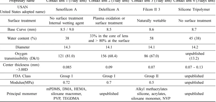
본 연구에서는 시중에 판매되고 있는 일회용(1일 착용) 렌즈 3종과 3일 착용렌즈 1종을 대상으로 하였다. 실험에 이용 된 렌즈는 모두 실리콘 하이드로겔 소재였으며, 일회 용 렌즈 3종은 미국연방식품의약국(FDA) 분류상 그룹 I과 II군에 분류되어 있으며, 3일 착용렌즈는 기준에 분류되어 있지 않았다. contact lens 1과 contact lens 2는 그룹 I(저
함수, 비이온성)로써 senofilcon A, delefilcon A 재질이며, contact lens 3은 그룹 II(고함수, 비이온성)로 filcon II 3 재 질의 렌즈이며, 각 렌즈의 시료명과 일반적인 특성을 Table 1에 나타내었다.
2) 인공누액(Artificial Tear Solution; ATS)
본 연구에서 사용된 인공누액은 총 3단계로 조제되었다.
첫 번째 단계로 농축지질(lipid stock solution; LSS)을 만 드는 것이다. 본 연구에 사용된 LSS는 500X농축하였으며, 인공누액에 사용된 지방성분은 Table 2와 같다.
다음 단계로 지질 인공누액(lipid artificial tear solution;
LTS)을 만드는 것이다. LSS에 완충 용액(phosphate buffered saline; PBS, PH 7.4)을 첨가하고, LSS가 PBS에 완전히 혼 입될 때까지 37oC에서 초음파 처리하였다.
마지막 단계로 단백질과 점액을 첨가하는 것이다. 사용 된 특정 단백질과 점액 및 최종 ATS에서의 그 농도는 Table 2. 3에 개략적으로 기술되어 있으며, 인공누액에 사 용된 모든 재료는 시그마 알드리치에서 구입하였다.
Table 1. Characteristics of silicone hydrogel lenses
Proprietary name Contact lens 1 (1-day lens) Contact lens 2 (1-day lens) Contact lens 3 (1-day lens) Contact lens 4 (3-days lens) USAN
(United States adopted name) Senofilcon A Delefilcon A Filcon II 3 Silicone Tripolymer Surface treatment No surface treatment
Internal wetting agent
Plasma oxidation or
surface treatment Naturally wettable No surface treatment
Base Curve (mm) 8.5 / 9.0 8.5 8.6 8.7
Water content (%) 38 33% in the core of lens
and > 80% at the surface 58 45 (38)
Diameter 14.3 14.1 14.1 14.2
Oxygen
transmissibility (Dk/t) 121 (81.0) 156 (68.4) 86 (67.0) unpublished
(13.2) Center thickness (mm)
−3.00D 0.085 0.09 0.07 0.07 - 0.13
FDA Class Group I Group I Group II unpublished
Modulus(MPa) 0.72 0.7 0.5 unpublished
Principal monomer
mPDMS, DMA, HEMA, siloxane macromer,
PVP, TEGDMA
unpublished
Alkyl methacrylates silicone, acrylates, siloxane monomer, NVP
unpublished
( ): this experimental measurement DMA (N,N-dimethylacrylamide) EGDMA (ethyleneglycol dimethacrylate) HEMA (poly-2-hydroxyethyl methacrylate) mPDMS (monofunctional polydimethylsiloxane) NVP (N-vinyl pyrrolidone)
TEGDMA (tetraethyleneglycol dimethacrylate)
TPVC (tris-(trimethylsiloxysilyl) propylvinyl carbamate) TRIS (trimethylsiloxy silane); NCVE (N-carboxyvinyl ester) PBVC (poly[dimethysiloxy] di [silylbutanol] bis[vinyl carbamate]) PVP (polyvinyl pyrrolidone)
2. 실험방법
4종의 실험용 콘택트렌즈를 2 mL의 ATS에 각 1개씩 넣 어 진탕 배양기를 이용하여 37oC, 150 rpm의 속도로 흔들 어주면서 침지시켰다.
시간 경과에 따른 물리적 특성변화를 알아보기 위해 12h, 24h, 48h 오염 후 산소투과도(oxygen transmissibility), 가시광선 투과율(visible light transmittance), 주사 전자현 미경(scanning electron microscope; SEM)을 이용한 표면 관찰을 각 3회씩 실시하였다.
3. 특성분석
1) 산소투과도(oxygen transmissibility)
산소투과도는 polarographic 방법으로 측정하였다. PBS 에 담긴 콘택트렌즈와 polarographic cell, cell mounting fixture를 항온 항습기(WL1000S, with lab 습도 95%, 온도 35oC)에 넣고 2시간 이상 평형 상태를 유지시킨 후, polaro- graphic cell위에 렌즈를 올려놓았다. 렌즈는 nylon mesh를 씌운 cell mount ting fixture로 고정하였고, permeo-meter (Model 201T O2permeometer, CHREATECH, USA)를 이용 하여 전류 값을 측정했다. 렌즈의 중심두께는 압력 다이얼 게이지(Litematic: Model VL-50, Mitutoyo)를 이용하여 측정 하였다. 동일한 재질을 동일 조건에서 렌즈 전류 값을 3번씩 반복 측정하였고, 모든 방법은 한국 식품의약품안전처 (ministry of food and drug safety; MFDS)의 기준에 따랐다.[16]
2) 가시광선 투과율(visible light transmittance)
오염 된 렌즈는 PBS에 수화된 상태에서 실시하였다. 가
시광선 투과율 측정 시 UV-Visible Spectrophotometer(Evolution 201, USA)를 이용하였고, 시험에 이용 된 4개의 렌즈(seno- filcon A, delefilcon A, filcon II 3, silicone tripolymer)를 370~790 nm까지의 파장 범위에서 각각의 렌즈에 대해 3번 씩 측정한 후 평균하여 사용하였다.
3) 주사 전자현미경(scanning electron microscope; SEM) 을 이용한 표면 관찰
SEM은 전자 현미경의 한 종류로, 전자 빔을 주사(走査)하 여 표본의 상(象)을 얻게 된다. 전자들과 표본의 원자들과 의 상호 반응에 따른 다양한 표면정보를 제공한다.
각 렌즈(senofilcon A, delefilcon A, filcon II 3, silicone tripolymer)는 자연 건조시킨 후, SEM(Scanning Electron Microscope: Mira III, Czech)을 이용하여 시간별 표면변화 를 500X 확대 관찰하였다.
결 과
1. 산소투과도(oxygen transmissibility)
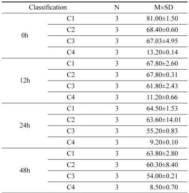
산소투과도(Dk/t = 10−9(cm/sec)(mlO2/ml × mmHg)는 착 용 전 senofilcon A(Dk/t = 81.0)의 산소투과도가 가장 높았 으며 다음으로 delefilcon A(Dk/t = 68.40)와 filcon II 3(Dk/t
= 67.03), silicone tripolymer(Dk/t = 13.30)가 가장 낮은 산소 투과도를 나타내었다. 12h 경과 후 senofilcon A, delefilcon A(Dk/t = 67.80)의 산소투과도가 동일하게 가장 높았으며, 다음으로 filcon II 3(Dk/t = 61.80), silicone tripolymer(Dk/
t = 11.20)순으로 나타났다. 24 h, 48h 경과 후senofilcon A(Dk/
Table 2. Molecular and experimental details of the specific lipids used for all lipid doping solutions[15]
Triolein Cholesterol Oleic acid Oleic acid methyl
ester Cholesteryl oleate Phosphatidyl choline Lipid type Triglyceride Sterol Fatty acid Fatty ester Cholestyl ester Phospholipid
Formular C57H104O6 C27H46O C18H34O2 C19H36O2 C45H78O2 C42H82NO8P Molecular
Weight (g/mol) 885.5 386.7 282.5 296.5 651 760.1
Lipid Stock
Concentration (mg/mL) 8.0 0.9 0.9 6.0 12.0 0.25
Final ATS
Concentration (mg/mL) 0.016 0.0018 0.0018 0.012 0.024 0.0005
Sigma Product Number c7140 c8667 112-80-1 112-62-9 c9253 p2772
Table 3. Protein and mucin concentration in ATS[15]
Bovine Albumin Hen Egg Lysozyme Bovine Mucin Bovin Colostrum Lactoferrin
Bovine Immunoglobulin G
Molecular Weight (kDa) 66.4 14.3 3×10⁵ to 4×10⁷ 83.1 161
Concentration (mg/mL) 0.20 1.90 0.15 1.80 0.02
Sigma Product Number A7888 L6876 M3895 L4040 I5506
t=64.50, 63.80), delefilcon A(Dk/t=63.60, 60.30), filcon II 3(Dk/t=55.20, 54.00) 대비 silicone tripolymer(Dk/t=9.20, 8.50)의 산소투과도가 현저히 낮게 나타났다(Fig. 1)(Table 4).
2. 가시광선 투과율(visible light transmittance)
착용 전 각 렌즈(senofilcon A, delefilcon A, filcon II 3, silicone tripolymer)의 가시광선 투과율은 senofilcon A(97.21%), delefilcon A(97.66%), filcon II 3(98.45%), silicone tripolymer (97.65%) 모두 97% 이상의 투과율을 보였다. 12h 경과 후 filcon II 3(96.88%)의 가시광선 투과율이 가장 높았으며 delefilcon A(90.61%)의 가시광선 투과율이 가장 낮게 나 타났다. 24h 경과 후 filcon II 3(97.16%), silicone tripolymer (94.75%), senofilcon A(90.91%), delefilcon A(77.27%)순
이었다. 48h 경과 시 delefilcon A(77.03%)의 가시광선 투 과율이 현저히 낮게 나타났다(Fig. 2)(Table 5).
3. 주사 전자현미경(scanning electron microscope;
SEM)을 이용한 표면 관찰
시간 경과됨에 따라 모든 렌즈에서 침착물의 증가를 보 였으나 일관된 형태를 보이지는 않았다. senofilcon A, delefilcon A, filcon II 3은 시간이 경과됨에 따라 표면의 침착물이 뭉쳐져 덩어리를 형성하는 모습을 보였으며, C4 는 뭉쳐짐 없이 흰색의 침착물이 퍼진 형태로 증가하는 모습을 보였다. Fig. 3에서 보이는 흰색 침착물은 지방과 단백질이 결합된 형태로 보여 진다.[17-19]
Fig. 1. Oxygen transmissibility of each contact lens over time.
C1: contact lens 1, C2: contact lens 2, C3: contact lens 3, C4: contact lens 4.
Table 4. Oxygen transmissibility of each contact lens over time
Classification N M±SD
0h
C1 3 81.00±1.50
C2 3 68.40±0.60
C3 3 67.03±4.95
C4 3 13.20±0.14
12h
C1 3 67.80±2.60
C2 3 67.80±0.31
C3 3 61.80±2.43
C4 3 11.20±0.66
24h
C1 3 64.50±1.53
C2 3 63.60±14.01
C3 3 55.20±0.83
C4 3 09.20±0.10
48h
C1 3 63.80±2.80
C2 3 60.30±8.40
C3 3 54.00±0.21
C4 3 08.50±0.70
C1: contact lens 1, C2: contact lens 2, C3: contact lens 3, C4:
contact lens 4
Fig. 2. Visible light transmittance of each contact lens over time. C1: contact lens 1, C2: contact lens 2, C3:
contact lens 3, C4: contact lens 4.
Table 5. Visible light transmittance of each contact lens over time
Classification N M±SD
0h
C1 3 97.21±0.19
C2 3 97.66±0.84
C3 3 98.45±0.23
C4 3 97.65±0.23
12h
C1 3 93.06±1.32
C2 3 90.61±0.63
C3 3 96.88±1.39
C4 3 95.00±3.26
24h
C1 3 90.91±1.47
C2 3 77.26±1.90
C3 3 97.16±0.46
C4 3 94.75±1.00
48h
C1 3 88.00±1.53
C2 3 77.03±1.53
C3 3 86.56±7.14
C4 3 90.87±0.28
결과 및 고찰
눈에 직접 접촉하는 콘택트렌즈 표면은 눈물에 포함되
어 있는 단백질, 지방, 뮤신, 칼슘 등이 침착되며, 특히 소 프트렌즈의 경우 눈물성분의 침착이 많다. 소프트렌즈의 재질 특성 및 표면 특성에 따라 눈물성분의 침착 정도가 Fig. 3. Random deposit spots on the surface of fouled lens over 3 days. Scanning electron micrographs (SEM; magnification
×500). C1: contact lens 1, C2: contact lens 2, C3: contact lens 3, C4: contact lens 4.
크게 달라지는데 일반적으로 고함수. 이온성 렌즈의 경우 침착량이 가장 많다고 보고하고 있다.[20-24]
현재 시장에서 가장 많이 이용되고 있는 실리콘 하이드 로겔 렌즈는 렌즈 내 실록산(siloxane)그룹을 함유하는 재
질로[25-30] 소수성의 표면을 형성하여 렌즈 표면에 단백질
의 침착은 적지만 지방 침착량이 많다.[31-35] 지방의 침착 은 건조감, 이물감의 원인이 되며 침착량의 증가에 따라 시야 흐림의 증상이 나타날 수 있어 지방이 많이 부착되 는 실리콘 하이드로겔 렌즈는 착용 후 시간이 지남에 따 라 시력의 질이 처음보다 저하된다고 보고되었다.[36-39]그 리고 단백질의 침착은 콘택트렌즈 표면 및 pore 특성을 변 화시켜 함수율에도 영향을 미칠 수 있다. 역으로 함수율이 단백질의 침착량에 영향을 미치는 것으로 알려졌다. 따라 서 소프트렌즈의 함수율에 의해 결정이 되는 산소투과도에 연차적으로 영향을 미치게 되어 단백질의 침착량이 증가할 수록 산소투과도가 저하된다고 보고하였다.[40]
본 실험에서 측정한 초기 산소투과도의 결과는 제조사 에서 제시한 결과 값과 차이가 있었다. 이는 본 실험 측정 시 한국 식품의약품안전처(MFDS; ministry of food and drug safety)의 기준에 따라 측정을 하였기 때문이다. 한국 식품의약품안전처 기준은 콘텍트렌즈의 렌즈의 두께에 따 른 절대치 값을 제시하므로, 렌즈 두께에 따른 표준화 곡 선을 이용한 국제표준화규격(ISO 18369)에 비해 산소투과 도 값이 낮게 측정된다. ATS (artificial tear solution) 노출 전 산소투과도는 senofilcon A(Dk/t=81.0)가 가장 높았으 며, silicone tripolymer(Dk/t=13.2)가 가장 낮았다. 일반적 으로 실리콘 하이드로겔 렌즈의 경우 렌즈 내 실록산의 첨가에 따라 높은 산소투과도가 가장 큰 장점이며 일반적 인 실리콘 하이드로겔 렌즈의 산소투과도는 Dk/t=60.0이 상을 나타낸다.[13] 그러나 silicone tripolymer(Dk/t=13.2)의 경우 현저히 낮은 산소투과도를 가졌다.[17] 시간 경과에 따라 4종의 렌즈 모두에서 산소투과도의 감소가 나타났 고, 이는 시간 경과에 따른 침착물에 따른 것으로 여겨진 다. 특히, 눈물 내의 단백질의 침착은 콘택트렌즈의 산소 투과도에 영향을 미치며, 침착되는 눈물 단백질 침착량이 증가할수록 산소투과도가 감소한다고 하였다.[16,40] 그러나 이러한 산소투과도의 변화는 렌즈의 재질 특성에 따라 정 도의 차이가 있다.[16,40] Allansmith 등[41]은 함수율이 증가 할수록 단백질 침착량과 침착되는 단백질 종류가 달라져 재질에 따라 산소투과도 변화의 차이를 나타낼 수 있다고 하였다. 본 연구 결과 filcon II 3만이 고함수율 군으로 함 수율에 따른 단백질 침착이 비교적 많았을 것으로 예상되 며 이에 따라 산소투과도의 감소폭이 비교적 크게 나타난 것으로 생각된다. senofilcon A, delefilcon A, filcon II 3는 시간 경과에 따라(12h, 24h, 48h) 산소투과도의 감소를 보
이긴 하였으나 48h 이후에도 Dk/t가 50 이상으로 산소투과 도에 있어 안전성이 유지되었다. 그러나 silicone tripolymer 의 초기의 Dk/t=13.2, 48h 경과 후 Dk/t=8.5로 매일 착용렌 즈에 있어 각막부종을 막기 위한 최소 산소투과도(Dk/t=24)
에[42] 못 미치는바 저산소증에 따른 질환의 발생으로 이어
질 수 있다.
ATS 노출 전 최초 가시광선 투과율은 4종 모두 97%이 상이었다. 12h 경과 후 filcon II 3(96.88%)의 가시광선 투 과율이 가장 높았으며 delefilcon A(90.61%)의 가시광선 투과율이 가장 낮았다. 24h 경과 후 delefilcon A(77.27%) 를 제외한 3종 모두는 90% 이상의 가시광선 투과율을 가 졌으며, 48h 경과 후 delefilcon A(77.03%)을 제외한 3종 모두는 85% 이상의 가시광선 투과율을 나타내었다. 그러 나 12h 경과 후 delefilcon A의 가시광선 투과율이 비교적 크게 감소하였으며, 24 h 경과 후 77.26%로 현저한 감소 폭을 나타냄으로써 24 h 경과 후 시야 흐림 현상을 보일 것으로 예상된다. 가시광선 투과율 측정 시 delefilcon A의 경우 각 파장대에 대하여 불안정한 투과율을 나타내었는 데 이는 렌즈의 표면처리에 따른 것으로 생각된다.
SEM을 이용한 표면 관찰 시 모든 렌즈에서 시간 경과 에 따른 침착물의 증가를 확인할 수 있었다. 표면에서 보 이는 침착의 양상은 조금 다르게 나타났는데 senofilcon A, delefilcon A, filcon II 3의 경우 시간이 지남에 따라 침 착물이 증가하면서 둥근형태의 덩어리 진 모습을 나타내 었다. 그러나 silicone tripolymer는 뭉쳐짐 없이 흰색의 침 착물이 점진적으로 증가하는 모습을 보였다. 표면 침착물 은 누액 내 지방과 단백질, 점액의 다양한 성분의 혼합된 형태로 나타나는데, 표면 침착 시 단백질과 지질 사이에 경쟁적인 침착도 발생하지만 협력적인 부분도 있다고 하
였다.[43] 지방의 분자량은 단백질과 점액에 비해 극히 작은
크기이므로 지방만의 침착을 확인하기는 어렵다.[18,44] 그 러나 지방은 여러 단백질을 서로 뭉쳐주는 결합제의 역할 을 함으로 지방이 증가함에 따라 침착된 단백질간의 결합 을 도와주어[42]시간 경과에 따라 증가된 침착물간의 결합 에 의해 덩어리를 형성하게 된다. 표면에서 보여지는 흰색 덩어리는 단백질과 지방이 결합된 형태이며 senofilcon A 과 delefilcon A의 경우 표면에서 흰색의 침착물이 적은 것으로 보아 비교적 단백질의 침착이 적었을 것으로 여겨 지고, 침착물이 덩어리진 것으로 보아 지방 침착량 증가에 따른 단백질과의 결합의 결과로 보인다. 그러나 filcon II 3 와 silicone tripolymer의 표면의 흰 색 침착물이 비교적 많 으나 덩어리짐은 비교적 적었다. 이로 단백질의 침착이 많 고 상대적으로 지방의 침착이 적어 단백질과 지방의 결합 이 적었을 것으로 예상된다. 표면 관찰 시 filcon II 3에서 단백질의 침착이 가장 많고, 이는 유일하게 고함수군의 렌
즈로 함수율이 영향을 미쳤을 것으로 생각된다. 다음으로 산소투과도의 결과로써 비교적 실리콘의 함유율이 적을 것으로 예상되는 silicone tripolymer가 단백질 침착이 많 았을 것으로 생각된다.
결 론
시간 경과에 따른 4종 렌즈의 변화 관찰 시, 일회용 렌 즈 3종은 48h 경과 시까지도 양호한 산소투과도를 유지하 였다. 그러나 3일 착용렌즈(silicone tripolymer)의 경우 모 든 시간에서 일회용 3종에 비하여 현저히 낮은 산소투과 도를 가졌고 이는 매일 착용렌즈의 산소투과도 안전치 기 준(Dk/t=24)에 미달 된 바 저산소증에 의한 질환 발생으로 이어질 수 있다.
가시광선 투과율의 결과로 48h 경과 후부터 일회용 3종 에서 시야 흐림 증상이 나타날 것으로 예상되며, 3일 착용 렌즈는 48h 경과 시 까지도 90% 이상의 가시광선 투과율 을 유지한 바 3일 착용 시에도 시야 흐림에 대한 불편증 상은 없을 것으로 생각된다.
표면 관찰 시 filcon II 3와 silicone tripolymer에 단백질 침착이 많았을 것으로 보인다.
이에 일회용 3종의 경우 48h 이후 시야 흐림에 대한 불 편 증상이 예상되고, 3일 착용렌즈(silicone tripolymer)는 낮은 산소투과도의 결과로 저산소증에 의한 질환이 우려 된다. 일회용 3종간의 비교에서 delefilcon A에서 가장 먼 저 시야 흐림 증상이 발생될 것으로 예상되며, filcon II 3 의 경우 시간경과에 따른 침착물의 증가가 큼으로 권장시 간 초과 착용 시 미생물에 의한 질환 발생이 우려된다.
일회용 렌즈와 3일 착용렌즈의 시간 경과에 따른 물리 적 특성 변화 비교 결과 본 실험에 이용 된 3일 착용에 대 한 안전성은 찾아 볼 수 없었다.
감사의 글
이 논문은 산업통상자원부 창의산업거점기관지원사업 지원으로 수행한 [안광학렌즈 소재기술 및 신뢰성기반구 축사업] 연구결과입니다.(No. R0004080")
REFERENCES
[1] Fonn D, Sweeney D. The benefits of silicone hydrogel daily disposable Lenses, 2015. https://www.clspectrum.com/
issues/2015/december-2015/the-benefits-of-silicone-hydro- gel-daily-disposable(December 1, 2015)
[2] Holden BA, Stretton S, Jara P, et al. The future of contact lenses: Dk really matters, 2006. https://www.clspectrum.com/
supplements/2006/february-2006/protecting-your-patient- s-eye-health/the-future-of-contact-lenses-dk-really-matters (February 1, 2006)
[3] Pozuelo J, Compañ V, González-Méijome JM, et al. Oxy- gen and ionic transport in hydrogel and silicone-hydrogel contact lens materials: An experimental and theoretical study. J Membrane Sci. 2014;452:62-72. DOI: https://
doi.org/10.1016/j.memsci.2013.10.010
[4] Papas E. On the relationship between soft contact lens oxygen transmissibility and induced limbal hyperaemia.
Exp Eye Res. 1998;67(2):125-131. DOI: https://doi.org/
10.1006/exer.1998.0504
[5] Papas EB. The significance of oxygen during contact lens wear. Cont Lens Anterior Eye. 2014;37(6):394-404. DOI:
https://doi.org/10.1016/j.clae.2014.07.012
[6] Mishima S. Clinical investigations on the corneal endo- thelium. Ophthalmology. 1982;89(6):525-530. DOI: https://
doi.org/10.1016/S0161-6420(82)34755-7
[7] Iskeleli G, Karakoc Y, Ozkok A, et al. Comparison of the effects of first and second generation silicone hydrogel contact lens wear on tear film osmolarity. Int J Ophthal- mol. 2013;6(5):666-670. DOI: https://doi.org/10.3980/
j.issn.2222-3959.2013.05.22
[8] Sheng H, Bullimore MA. Factors affecting corneal endo- thelial morphology. Cornea. 2007;26(5):520–525. DOI:
https://doi.org/10.1097/ICO.0b013e318033a6da
[9] Amann J, Holley GP, Lee SB, et al. Increased endothelial cell density in the paracentral and peripheral regions of the human cornea. Am J Ophthalmol. 2003;135(5):584- 590. DOI: https://doi.org/10.1016/S0002-9394(02)02237-7 [10] Wiffen SJ, Hodge DO, Bourne WM. The effect of contact
lens wear on the central and peripheral corneal endothe- lium. Cornea. 2000;19(1):47-51. DOI: https://doi.org/10.
1097/00003226-200001000-00010
[11] Dillehay SM, Miller MB. Performance of Lotrafilcon B silicone hydrogel contact lenses in experienced low-Dk/t daily lens wearers. Eye Cont Lens. 2007;33(6):272-277.
DOI: https://doi.org/10.1097/ICL.0b013e31802f78c2 [12] Morgan PB, Woods CA, Tranoudis IG, et al. International
contact lens prescribing in 2018, 2019. https://www.clspec- trum.com/issues/2019/january-2019/international-contact- lens-prescribing-in-2018(18 June 2020).
[13] Efron N, Morgan PB. Rethinking contact lens aftercare.
Clin Exp Optom. 2017;100(5):411-431. DOI: https://doi.
org/10.1111/cxo.12588
[14] Mirejovsky D, Patel AS, Rodriguez DD, et al. Lipid adsorption onto hydrogel contact lens materials-advan- tages of nile red over oil red O in visualization of lipids.
Optom Vis Sci. 1991;68(11):858-864. DOI: https://doi.org/
10.1097/00006324-199111000-00005
[15] Lorentz HI. Modeling in vitro lipid deposition on silicone hydrogel and conventional hydrogel contact lens materi- als. PhD Thesis. University of Waterloo, Waterloo. 2012;
p81-p86.
[16] Lee SE, Kim SR, Park M. Influence of tear protein depo-
sition on the oxygen permeability of soft contact lenses. J Ophthalmol. 2017;5131764:1-6. DOI: https://doi.org/10.1155/
2017/5131764
[17] Rebeix V, Sommer F, Marchin B, et al. Artificial tear adsorption on soft contact lenses: methods to test surfac- tant efficacy. Biomaterials. 2000;21(12):1197-1205. DOI:
https://doi.org/10.1016/s0142-9612(99)00221-5
[18] Peng CC, Fajardo NP, Razunguzwa T, et al. In vitro spoilation of silicone-hydrogel soft contact lenses in a model-blink cell. Optom Vis Sci. 2015;92(7):768-780.
DOI: https://doi.org/10.1097/OPX.0000000000000625 [19] Teichroeb JH, Forrest JA, Ngai V, et al. Imaging protein
deposits on contact lens materials. Optom Vis Sci. 2008;
85(12):1151-1164. DOI: https://doi.org/10.1097/OPX.0b013 e31818e8ad6
[20] Bontempo AR, Rapp J. Protein and lipid deposition onto hydrophilic contact lenses in vivo. CLAO J. 2001;27(2):
75-80.
[21] Jones L, Evans K, Sariri R, et al. Lipid and protein depo- sition of N-vinyl pyrrolidone-containing group II and group IV frequent replacement contact lenses. CLAO J.
1997;23(2):122-126.
[22] Minno GE, Eckel L, Groemminger S, et al. Quantitative analysis of protein deposits on hydrophilic soft contact lenses: ?. comparison to visual methods of analysis. ?.
deposit variation among FDA lens material groups. Optom Vis Sci. 1991;68(11):865-872. DOI: https://doi.org/10.1097/
00006324-199111000-00006
[23] Myers RI, Larsen DW, Tsao M, et al. Quantity of protein deposited on hydrogel contact lenses and its relation to visible protein deposits. Optom Vis Sci. 1991;68(10):776- 782. DOI: https://doi.org/10.1097/00006324-199110000- 00003
[24] Minarik L, Rapp J. Protein deposits on individual hydro- philic contact lenses: effects of water and ionicity. CLAO J. 1989;15(3):185-188.
[25] Jones LW, Dumbleton KA. Silicone hydrogel contact lenses: Part 1 evolution and current status. Optometry Today. 2002;20:26-31.
[26] Fonn D, Dumbleton K, Jones L, et al. Silicone hydrogel material and surface properties, 2002. https://www.clspec- trum.com/issues/2002/march-2002/silicone-hydrogel-material -and-surface-properties(15 November 2019).
[27] Edwards K, Atkins N. Silicone hydrogel contact lenses:
Part 2 therapeutic applications, 2002. http://www.sjsup- port.org/pdf/softlens/bandage004.pdf(18 June 2020).
[28] Sweeney D, Fonn D, Evans K. Silicone hydrogels: the evolution of a revolution, 2006. https://www.clspectrum.
com/supplements/2006/february-2006/protecting-your-patient- s-eye-health/silicone-hydrogels-the-evolution-of-a-revolu- tion(15 November 2019).
[29] Kunzler JF. Silicone hydrogels for contact lens applica- tion. Trends in Polymer Science. 1996;4(2):52-59.
[30] Jones L. Modern contact lens materials: A clinical perfor- mance update, 2002. https://www.clspectrum.com/issues/
2002/september-2002/modern-contact-lens-materials-a-clinical
-performa(15 November 2019)
[31] Maziarz EP, Stachowski MJ, Liu XM, et al. Lipid deposi- tion on silicone hydrogel lenses, part I: quantification of oleic acid, oleic acid methyl ester, and cholesterol. Eye Contact Lens. 2006;32(6):300-307. DOI: https://doi.org/
10.1097/01.icl.0000224365.51872.6c
[32] Cheung SW, Cho P, Chan B, et al. A comparative study of biweekly disposable contact lenses: silicone hydrogel ver- sus hydrogel. Clin Exp Optom. 2007;90(2):124-131. DOI:
https://doi.org/10.1111/j.1444-0938.2006.00107.x
[33] Carney FP, Nash WL, Sentell KB. The adsorption of major tear film lipids in vitro to various silicone hydro- gels over time. Cornea. 2008;49(1):120-124. DOI: https://
doi.org/10.1167/iovs.07-0376
[34] Iwata M, Ohno S, Kawai T, et al. In vitro evaluation of lipids adsorbed on silicone hydrogel contact lenses using a new gas chromatography/mass spectrometry analytical method. Eye Contact Lens. 2008;34(5):272-280. DOI: https:
//doi.org/10.1097/ICL.0b013e318182f357
[35] Zhao Z, Carnt NA, Aliwarga Y, et al. Care regimen and lens material influence on silicone hydrogel contact lens deposition. Optom Vis Sci. 2009;86(3):251-259. DOI: https:
//doi.org/10.1097/OPX.0b013e318196a74b
[36] Jones L, Franklin V, Evans K, et al. Spoilation and clinical performance of monthly vs. three monthly Group II dispos- able contact lenses. Optom Vis Sci. 1996;73(1):16-21.
DOI: https://doi.org/10.1097/00006324-199601000-00003 [37] Pritchard N, Fonn D, Weed K. Ocular and subjective
responses to frequent replacement of daily wear soft con- tact lenses. CLAO J. 1996;22(1):53-59.
[38] Nilsson SEG, Andersson L. Contact lens wear in dry envi- ronments. Acta Ophthalmol. 1986;64(2):221-225. DOI:
https://doi.org/10.1111/j.1755-3768.1986.tb06904.x
[39] Gellatly KW, Brennan NA, Efron N. Visual decrement with deposit accumulation of HEMA contact lenses. Am J Optom Physiol Opt. 1988; 65(12):937-941. DOI: https://
doi.org/10.1097/00006324-198812000-00003
[40] Lee JY, Lee JI, Kim SR, et al. Correlation between tear proteins deposition and oxygen transmissibility of soft con- tact lenses. J Korean Ophthalmic Opt Soc. 2017;22(2):97- 103. DOI: https://doi.org/10.14479/jkoos.2017.22.2.97
[41] Allansmith MR, Korb DR, Greiner JV, et al. Giant papil- lary conjunctivitis in contact lens wearers. Am J Ophthal- mol. 1977;83(5):697-708. DOI: https://doi.org/10.1016/
0002-9394(77)90137-4
[42] Holden BA, Mertz GW. Critical oxygen levels to avoid corneal edema for daily and extended wear contact lenses.
Invest Ophthalmol Vis Sci. 1984;25(10):1161-1167.
[43] Franklin VJ, Bright AM, Tighe B. Hydrogel polymers and ocular spoilation processes. Trend in Polymer Science.
1993;1:9-16.
[44] Panthi S, Nichols JJ. An imaging-based analysis of lipid deposits on contact lens surfaces. Cont Lens Anterior Eye.
2018;41(4):342-350. DOI: https://doi.org/10.1016/j.clae.
2017.12.014
3-Days lens와 daily disposable lens(1-day)의 시간 경과 별 물리적 특성 변화의 비교
송선정1, 김기홍2,3, 추병선2,3,*
1
대구 가톨릭대학교 안경광학과, 강사, 경산 38430
2
대구 가톨릭대학교 안경광학과, 교수, 경산 38430
3
대구 가톨릭대학교 안광학기술사업단, 교수, 경산 38430
투고일(2020년 2월 23일), 수정일(2020년 3월 19일), 게재확정일(2020년 6월 17일)
목적: 시중에 판매되고 있는 실리콘 하이드로겔 소재의 일회용 렌즈 3종과 3일 착용렌즈 1종에 있어 시간 경과에 따른 물리적 특성 변화를 비교하였다. 방법: 인공누액은 실제의 눈물의 조성비를 기준으로 준비하였으며, 일회용 렌 즈(senofilcon A, delefilcon A, filcon II 3)와 3일 착용렌즈(silicone tripolymer)를 준비된 인공누액에 담구어 배양기에 서 37oC, 150 rpm의 속도로 흔들어 주면서 12h, 24h, 48h 동안 침지시킨 후 시간 경과에 따른 산소투과도, 가시광선 투과율, 표면 변화를 관찰하였다. 결과: 3일 착용렌즈의 최초 산소투과도(Dk/t)는 13.23이었으며, 모든 시간에 있어 일 회용 렌즈와 비교 시 현저히 낮은 산소투과도를 나타내었다. 가시광선 투과율의 경우 오염 전 4종 모두 97% 이상의 투과율을 가졌으나 24h 경과 후 delefilcon A의 투과율이 80%이하로 현저한 감소를 나타내었다. 표면 관찰 시 시간 경과에 따라 침착물의 증가가 관찰되었으며, filcon II 3에서 가장 많은 침착물이 관찰 되었다. 결론: 본 연구에서 사 용된 3일 착용렌즈는 낮은 산소투과도의 결과로 저산소증에 의한 문제가 우려된다. 일회용 3종간의 비교에서 24h 경 과 후 delefilcon A에서 가장 먼저 시야 흐림 증상이 발생될 것으로 예상된다. 표면침착은 고함수율군의 filcon II 3에 서 큰 폭의 증가를 보였다. 일회용 렌즈와 3일 착용렌즈의 시간 경과에 따른 물리적 특성 변화 비교 결과, 3일 착용 렌즈의 3일 착용여부에 관한 안전성은 검증되지 않았다.
주제어: 일회용 렌즈, 3일 착용렌즈, 산소투과도, 가시광선 투과율, 주사전자현미경
![Table 2. Molecular and experimental details of the specific lipids used for all lipid doping solutions [15]](https://thumb-ap.123doks.com/thumbv2/123dokinfo/4746773.270385/3.892.66.827.145.370/table-molecular-experimental-details-specific-lipids-doping-solutions.webp)