45(1) : 55∼ 61 (2014)
55
가열 추출법과 마이크로웨이브 추출법을 이용한 황금 물 추출물의 뇌세포 보호 및 항산화 효과 비교 연구
이동성1#·고원민2#·김경수2·김동철2·윤치수2·조광호2·최 향2,3·오현철1,2,4·김윤철1,2,4*
1원광대학교 한방체액조절연구센터, 2원광대학교 약학대학, 3연변대학교 약학원, 4원광대학교 약학대학 약품연구소
The Comparison between Hot-Water Extracts and Microwave Extracts of Scutellaria radix for Antioxidant and Neuroprotective Effects
Dong-Sung Lee1#, Wonmin Ko2#, Kyoung-Su Kim2, Dong-Cheol Kim2, Chi–Su Yoon2, Kwangho Cho2, Xiang Cui2,3, Hyuncheol Oh1,2,4 and Youn-Chul Kim1,2,4
*
1Hanbang Body-Fluid Research Center, Wonkwang University,
2College of Pharmacy, Wonkwang University,
3College of Pharmacy, Yanbian University,
4Institute of Pharmaceutical Research and Development, College of Pharmacy, Wonkwang University
Abstract − Microwaves are non-ionizing electromagnetic waves of frequency between 300MHz to 300GHz and positioned between the X-ray and infrared rays in the electromagnetic spectrum. In recent years, the use of microwave for extraction of ingredient from plant material has shown remarkable research interest and potential. Scutellaria radix has been used as a tra- ditional medicine for a variety of diseases. It has been reported to exert beneficial health effects, such as anti-bacterial, anti- viral, anti-inflammatory, and free-radical scavenging. Oxidative stress or the accumulation of reactive oxygen species (ROS) leads neuronal cellular death and dysfunction, and it contributes to neuronal degenerative disease such as Alzheimer’s disease, Parkinson’s disease and stroke. In this study, we aimed to compare the neuroprotective and antioxidant effect of Scutellaria radix extracted by different methods using hot-water extraction (SBE-DW) or microwave extraction (SBE-DW-MW). As a result, we first examined HPLC analysis of hot-water and microwave extracts of Scutellaria radix. The hot-water and microwave extracts of Scutellaria radix showed the discernible difference patterns of HPLC analysis. Microwave-water extracts of Scutel- laria radix increased DPPH radical scavenging activity more than hot-water extraction. Microwave-water extracts of Scutellaria radix also showed neuroprotective effects and ROS inhibition against glutamate-induced oxidative stress in mouse hippocampal HT22 cells, but hot-water extraction not showed. In addition, the phosphorylation of MAPKs induced by glutamate insult was prevented by microwave-water extracts of Scutellaria radix. Thus, these results suggested that microwave extraction can be uti- lized for improving the extraction efficiency and biological activity of Scutellaria radix.
Key words − Scutellaria radix, Hot-water extraction, Microwave extraction, Antioxidant, Neuroprotection, HT22
황금(Scutellaria radix)은 여러해살이 초본식물 중 꿀풀과 에 속하는 황금(黃芩, Scutellaria baicalensis)의 주피를 벗 긴 뿌리를 건조한 것으로서 중국이 원산지로 알려져 있으 며 한국, 중국, 몽골, 및 시베리아 동부 등지에 분포한다.1) 예로부터 동아시아 지역에서 해열, 이뇨, 지사, 이담 및 소 염제의 효능으로 민간처방 및 한약재로써 사용해왔으며, 주
성분은 flavonoid계 화합물로서 baicalein, baicalin, wogonin, wogonoside, oroxylin-A 및 chrysin 등의 30여 종이 분리된 것으로 알려져 있다.2) 최근에 밝혀진 연구에서는 황금의 활 성성분으로 알려진 baicalein, baicalin, wogonin 등에 의해 서 항염증, 항산화, 항고혈압, 항알레르기, 항균, 항바이러스 및 항당뇨 등의 효과를 가지는 것으로 보고된 바 있다.3-9) 이와 같이 황금의 함유성분과 효능에 관한 연구는 많이 보 고 되었으나 추출 방법에 따른 성분 및 효능에 관한 비교 연구는 거의 없었다.
#
These authors contributed equally to this work.
*교신저자(E-mail):[email protected]
(Tel):+82-63-850-6823
한약재나 식물체의 활용에 있어서 가장 기본이 되는 처리 방법은 원재료로부터 유용 성분을 분리하기 위한 추출 공 정이며, 주로 열수에 의한 추출방법을 사용하고 있다. 그러 나 열수 추출법에 경우 추출에 장시간이 소요되고 추출 고 형분의 수율이 낮다는 단점이 있다. 이러한 단점들을 보완 하기 위해 최근에는 마이크로웨이브를 이용한 추출 방법에 대한 연구가 많이 진행되고 있다.10-11) 마이크로파는 주파수 가 300 MHz~300 GHz인 전자기파를 말하며 물체에 조사되 면 성분의 쌍극자 모멘트를 유도하여 급속한 유전가열을 발 생시키는 특성을 가져 식품의 조리, 해동, 건조, 배소, 데치 기, 살균 등의 여러 분야에 사용된다.12)마이크로웨이브를 이용한 추출(microwave-assisted extraction, MAE)은 물질 내의 목적 성분만을 선택적으로 가열하여 추출할 수 있다 는 점에서 매우 효과적인 방법이다.13) 본 연구에서는 마이 크로웨이브 추출과 기존의 일반 가열 추출과의 차이를 비 교하고자 하였다.
미토콘드리아 내의 산화환원 효소계, 외부 항원에 노출된 면역세포에 의해 그리고 외부적으로는 방사선 또는 여러 화 합물 등에 의해 생체 내에는 활성 산소종(reactive oxygen species, ROS)이 생성된다. 활성 산소종이 너무 많이 생성 되거나 항산화 시스템의 기능이 저하되어 활성 산소종의 생 성과 제거 사이의 균형이 파괴되면 생체는 활성 산소종에 의해 산화적 스트레스(oxidative stress)를 받게 된다.14) 산화 적 스트레스는 심장병, 암, 당뇨 등 다양한 질병의 발생과 진행을 촉진한다. 특히, 지금까지의 많은 연구들에 따르면 산화적 스트레스 및 ROS는 신경 세포의 파괴와 기능이상 을 초래하여 알츠하이머, 파킨슨병, 뇌졸중 등의 신경 퇴행 성 질환을 유발하는 것으로 알려져 있다.15-17) 중추 신경계 (central nervous system, CNS)의 대표적인 흥분성 신경전달 물질인 글루타메이트는 시넵스에서 신경전달, 뉴런의 형성 과 생장, 행동이나 학습 및 기억력 등에 매우 중요한 역할 을 한다.18)본 실험에서는 글루타메이트를 처리했을 때 산 화적 스트레스로 인한 세포의 손상을 확인할 수 있는 쥐의 해마유래인 HT22 세포주를 사용하였다.19)
따라서, 본 연구에서는 황금을 이용하여, 가열 추출방법 과 마이크로웨이브 추출 방법의 두 가지 방식으로 추출하 고 그에 따른 성분과 활성의 차이가 나타나는지를 알아보 고자 하였다. HPLC 패턴 분석을 통해 성분의 차이가 나타 나는지 알아보았고, DPPH 소거능 실험과 HT22 세포주에 서 글루타메이트로 유도한 산화적 스트레스에 대한 뇌 세 포 보호 효과를 실험하여 추출 방법에 따른 활성의 차이를 탐색하였다.
재료 및 방법
실험재료 − 본 실험에 사용한 황금은 2012년 9월 익산시
소재 대학한약국에서 옴니허브(경북 영천) 제품으로 구입 하여 사용하였다.
시약 및 기기 − DMEM 배지와 trypsin-ethylenediaminete- traacetic acid(EDTA)는 Gibco Laboratories사에서 구입하였 으며, fetal bovine serum(FBS)는 Hyclone Laboratories사에 서 구입하였다. L-glutamate, Trolox와 3'-(4,5-dimethylthiazol- 2-yl)-2,5-diphenyltetrazolium bromide(MTT)는 Sigma사에서 구입하였다. 96-Well tissue culture plates와 기타 tissue culture dishes는 Nunc사 제품을 이용하였다. 흡광도는 BioRad사의 Microplate Reader를 이용하여 측정하였다.
황금 가열 추출물과 마이크로웨이브 추출물 제조 − 황금 10 g을 물 500 ml로 80oC에서 15분 동안 가열추출(가열 온 도가 80oC 도달 후 15분)하고 여액을 감압 농축하여 황금 물 가열 추출물(SBE-DW) 2.533 g(25.33%)을 얻었다. 또 한, 황금 10 g을 물 500 ml와 함께 비커에 넣고 비커 윗부 분을 랩으로 막은 후 구멍을 뚫는다. 이 때, 물 500 ml만을 담은 비커도 함께 두고, 마이크로웨이브(2,450 MHz)를 이 용하여 15분 추출하고 여액을 감압 농축하여 황금 물 마이 크로웨이브 추출물(SBE-DW-MW) 3.801 g(38.01%)을 얻 었다.
HPLC 분석 − 본 연구에서 사용한 황금 물 가열 추출물 (SBE-DW)과 마이크로웨이브 추출물(SBE-DW-MW)은 HPLC를 사용하여 함유 성분의 패턴을 분석하였다. 사용한 HPLC 장치는 YOUNGLIN-YL9100(Younglin, Anyang, Korea) 이었으며, 컬럼은 Shiseido Capcell Pak C18 column (4.6×150 mm, 5µm)을 사용하였다. 이동상은 MeOH(JT Baker, 영국)과 Water를 사용하였으며, 온도는 실온이었으 며, 유속은 분당 0.7 ml이었다. 크로마토그램은 254 nm 파 장에서 검출하였다. 이동상 용매로는 10% MeOH[MeOH:
water(1:9)]에서 100% MeOH의 조성으로 50분간 변화시켰 다.
DPPH Radical 소거활성 측정 − 2,2-diphenyl-1-picryl- hydrazyl(DPPH) radical 소거활성은 Malterud의 방법에 따 라 측정하였다. DPPH 용액(45 µg/mL methanol)을 각각의 추출물과 혼합한 다음, ELISA microplate reader(Bio Rad Laboratories Inc., California, USA)를 사용하여 515 nm에서 흡광도의 감소를 5분 간격으로 30분간 측정하였다. Free
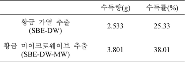
Table I. Comparison between the extract amount or yield of SBE-DW and SBE-DW-MW
수득량(g) 수득률(%) 황금 가열 추출
(SBE-DW) 2.533 25.33
황금 마이크로웨이브 추출
(SBE-DW-MW) 3.801 38.01
radical scavenging activity(%)는(1 − 시료 첨가 흡광도/시료 무첨가 흡광도)×100 식으로 구하였다.
HT22 세포배양 및 뇌 세포 보호활성 측정 − 생쥐 해마 유래 HT22 세포주는 Dr. Inhee-Mook(Seoul National University, Seoul, Korea)로부터 분양하여 사용하였으며, 글 루타메이트로 독성을 유발한 세포주에 대한 보호활성 측정 은 정 등의 방법20)에 따라 실시하였다. 간략하게 설명하면, HT22 세포(2×10⁵cells/well)를 10% heat-inactivated FBS, penicillin G(100 IU/ml)와 streptomycin(100 µg/ml)을 함유한 DMEM 배지에 분주하고 5% CO2 배양기 내에서 37oC에서 24시간 배양한 다음, 각각의 시료 용액(5, 10, 20, 40 µg/ml) 과 5mM Glutamate를 처리한 후 12시간 동안 5% CO2 배 양기 내에서 배양하였으며, 세포생존율은 MTT법을 활용하 여 측정하였으며, 양성대조약물로는 Trolox 50 µM를 사용 하였다. 또한, 모든 실험치는 대조군에 대한 세포보호율을 mean±S.D.로 표시하였으며, 각각 3회 반복 실험치를 이용 하여 계산하였다.
ROS 측정 − 배양된 세포를 PBS로 세척한 후, 10 µM 2',7'-dichlofluorescein diacetate(DCFDA, 35845)를 포함하는 Hank’s balanced salt 용액에 30분 동안 암실에서 반응한 후 세포의 형광광도(SpectramaxGemini XS, Molecular Devices, Sunnyvale, CA, U.S.A.)를 측정한다(excitation wavelength:
490nm; emission wavelength: 525 nm).
Western Blot Analysis − HT22 세포를 60 mm dish에 3×10⁵cells/well 농도로 24시간 배양한후 각각의 시료를 농 도별로 처리하였다. HT22 세포에 RIPA buffer를 첨가한 다 음, 4oC, 14,000×g에서 25분간 원심분리하고 상등액을 튜브 에 옮겼다. 단백질 정량은 BSA 단백질 실험 키트를 이용하 였고 각각의 시료를 12% SDS-polyacrylamide gel에서 영동 하고 Nitrocellulose membrane(NC membrane)으로 전사하 였다. 전사된 NC membrane을 5% 무지방유가 포함된 신선 한 blocking buffer(0.1% Tween 20 in Tris-buggered saline) 에서 blocking한후 p-JNK, p-p38, p-ERK, JNK, p38, ERK antibody를 1:1000으로 희석하여 넣고 1시간 동안 반응시켰 다. 다시 2차 안티바디(Anti-mouse, Anti-rabbit IgG)를 1:1000으로 희석하여 넣고 1시간 동안 반응한 다음, ECL 용액을 1:1로 잘 섞어서 NC membrane위에 부어서 발광시 키고 암실에서 X선 필름에 감광한 후 현상하였다. 같은 방 법으로 Actin antibody를 이용하여 Actin을 측정한다.
통계처리 − 본 실험의 통계처리는 GraphPad Prism, version 3.03(GraphPad Software Inc., San Diego, CA)을 사용하였다. 각 실험군의 결과는 평균치와 표준오차로 나타 내었으며, 각 실험군 간의 결과는 ANOVA test를 사용하여 분석하고 유의적인 차이가 있는 항목에 대해서만 검정하였 다. 실험군 간의 차이는 95% 수준(p<0.05)에서 유의성 있 는 것으로 하였다.
결과 및 고찰
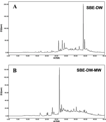
예로부터 생약재를 비롯한 많은 식품의 추출방법으로 사 용되어온 방법은 환류 냉각 추출(reflux extraction) 방법이 었으나 1회 추출하는데 2시간 이상의 긴 시간을 요하는 단 점이 있었다. 이에 비하여 마이크로웨이브 추출 방법은 적 은 용매를 사용하여 단시간에 원하는 물질을 추출할 수 있 는 이점이 알려져 있으며 마이크로웨이브를 사용함으로써 선택적인 가열이 가능하고 물질 내에 깊이 침투한 마이크 로웨이브가 분자간의 결합 고리를 끊어줌으로써 쉽게 추출 이 가능하도록 도와준다. 이와 같은 방법은 환류냉각 추출 방법을 비롯한 기존의 추출방법보다 추출효율이 높은 것으 로 보고 되고 있다.21-23) 본 연구에서의 결과에서도 황금 10 g 을 물 500 ml를 이용하여 가열 추출과 마이크로웨이브 추 출을 하였을 때, 가열 추출물(SBE-DW) 2.533 g(25.33%)에 비해 마이크로웨이브 추출물(SBE-DW-MW) 3.801 g(38.01%) 이 더 높은 추출효율을 보였다. 또한, 황금의 일반 가열 추 출과 마이크로웨이브를 활용한 추출물 간의 함유 구성 성 분의 차이를 알아보고자 HPLC chromatogram 분석으로 통 해 패턴 분석을 비교 하였다(Fig. 1). 그 결과 황금 물 가열 추출물(SBE-DW) 주요성분의 머무름 시간(retention time)은 40분인 반면, 마이크로웨이브 추출물(SBE-DW-MW) 주요 성분의 머무름 시간(retention time)은 27분으로 나타났다.
두 추출물은 같은 분석 조건에서 서로 다른 peak 패턴을 보
Fig. 1. HPLC chromatograms of the SBE-DW (A) and SBE- DW-MW (B).
였으며 이를 통해 추출 방법에 따른 함유 성분이 차이가 있 음을 확인하였다. 추후 연구를 통해 주요 성분의 확인이 필 요할 것으로 사료 된다.
다음으로 황금 물 가열 추출물(SBE-DW)과 마이크로웨이 브 추출물(SBE-DW-MW)의 함유성분 차이에 따른 활성 비 교 연구를 진행하고자 하였다. 호기성 호흡을 하는 생물체 들은 끊임없이 산소 분자(O2)를 세포 내로 받아들여 미토콘 드리아 내의 산화환원 효소계 또는 외부 항원에 노출된 면 역세포에 의해, 그리고 외부적으로는 방사선 또는 여러 화 합물 등에 의해 활성 산소종(reactive oxygen species, ROS) 을 생성한다. 체내 ROS가 많이 생성되거나 항산화 시스템 의 기능이 저하되면서 체내의 산화계와 항산화계의 불균형 으로 인해 산화적 스트레스(oxidative stress)가 일어나게 된 다.14) 체내에 과도하게 축적되는 활성산소는 세포의 비정상 적인 파괴를 유발하고 만성, 급성 질병의 주요한 원인으로 보고 되어 있다.7) 따라서, 황금 물 가열 추출물(SBE-DW) 과 마이크로웨이브 추출물(SBE-DW-MW)의 DPPH radical 소거능을 비교 하고자 하였다(Fig. 2). 황금 물 가열 추출물 (SBE-DW)을 10, 50, 200 µg/ml 농도로 처리하였을 때, 10.3, 15.2, 52.3% DPPH radical 소거능을 보였으나, 황금 물 마이크로웨이브 추출물(SBE-DW-MW)을 10, 50, 200 µg/ml 농도로 처리 하였을 때 15.9, 51.6, 90.6% DPPH radical 소거능을 보여서 대조군으로 사용한 vitamin C와 유 사한 소거능을 보였다. DPPH radical 소거능을 비교한 결과 황금 물 마이크로웨이브 추출물(SBE-DW-MW)이 가열 추 출물(SBE-DW)에 비해 더 우수한 DPPH radical 소거능을 보인다는 것을 확인 하였다.
체내에 발생한 활성 산소종은 산화적 스트레스를 유발하 여 세포 손상을 야기 시킨다. 이러한 산화적 스트레스에 의 한 세포 손상은 알츠하이머 증후군, 파킨슨 증후군, 헌팅턴 증후군과 같은 중추 신경계의 퇴행성 뇌질환의 중요한 요 인으로 알려져 있으며, 그 중 글루타메이트에 의한 산화적
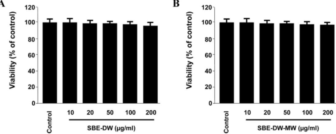
스트레스로 유발되는 뇌세포 손상은 퇴행성 뇌질환의 주요 한 요인으로 보고되고 있다.25,26) 산화적 스트레스에 의한 뇌 손상의 기전으로 신경흥분 독성(Excitotoxicity)이 주요한 기 전 중 하나로 알려져 있는데 이는 과다 분비된 glutamate 등 흥분성 아미노산에 의한 수용체들의 과도한 자극으로 인한 신경세포 사멸(neuronal cell death)이 일어나게 한다.27,28) 글 루타메이트는 central nervous system(CNS)의 대표적인 흥 분성 신경전달 물질로 시넵스에서의 신경전달, 뉴런의 형성 과 생장 및 생존, 행동이나 학습 및 기억력 등에 중요한 역 할을 한다. 이러한 생리학적인 기능에도 불구하고 글루타메 이트의 독성이 신경세포에 손상을 주거나 급·만성 퇴행성 신경질환을 유발한다고 알려져 있다.29) 글루타메이트가 유 도하는 신경 세포 손상은 크게 두 가지 요인이 있는데, 글 루타메이트 수용체의 과다 흥분에 의한 독성과 수용체의 매 개 없이 산화적 스트레스를 유발해 손상을 주는 것이다.30) 본 실험에 사용한 생쥐의 해마유래 세포주인 HT22 세포주 는 글루타메이트 수용체가 없는 세포주로서 글루타메이트 와 함께 처리할 때 신경흥분독성이 아닌 산화적 스트레스 로 인하여 세포가 손상을 받으며, 산화적 스트레스로 인한 퇴행성 뇌질환 연구에 있어서 유용한 실험모델중의 하나이 다. 이러한 산화적 스트레스로부터 뇌 보호 물질을 찾아 그 기전을 밝히려는 많은 연구가 진행 되고 있다. 따라서, 본 연구에서는 황금 물 가열 추출물(SBE-DW)과 마이크로웨 이브 추출물(SBE-DW-MW)의 생쥐의 해마유래 세포주인 HT22세포에서 산화적 스트레스에 대한 보호 효과 비교를 해보고자 하였다. 먼저 HT22 세포에 황금 물 가열 추출물 (SBE-DW)과 마이크로웨이브 추출물(SBE-DW-MW)만을 각 각 처리 하였을 때 세포 독성이 나타나는지 살펴 보았다 (Fig. 3). 그 결과 두 추출물 모두 200 µg/ml 농도까지 세포 독성을 보이지 않았다(Fig. 3). 따라서, 모든 실험은 200 µg/
ml를 최고 농도로 진행 하였다. 다음으로 글루타메이트로 유발하는 HT22 세포 독성에 황금 물 가열 추출물(SBE-DW) 과 마이크로웨이브 추출물(SBE-DW-MW)의 보호 효과를 비교 해보고자 하였다(Fig. 4). 그 결과 황금 물 마이크로웨 이브 추출물(SBE-DW-MW)을 10, 50, 100, 200 µg/ml 농도 로 전처리 하였을 때 보호 효과가 증가(Fig. 4B)하는 반면, 황금 물 가열 추출물(SBE-DW)을 10, 50, 100, 200 µg/ml 농도로 전처리 하였을 때는 보호효과를 보이지 않았다(Fig.
4A). 또한, 글루타메이트로 유발하는 HT22 세포에서의 ROS 생성 억제 효과에서도 황금 물 가열 추출물(SBE-DW)은 억 제 효과가 없었지만(Fig. 5A), 황금 물 마이크로웨이브 추 출물(SBE-DW-MW)은 농도 의존적으로 ROS생성을 억제 하였다(Fig. 5B).
산화적 스트레스를 포함한 다른 여러 스트레스 요인들은 mitogen-activated protein kinase(MAPK) 전달계를 자극하는 데, MAPK 전달계는 세포 외부로부터의 자극을 세포 내로 Fig. 2. DPPH radical scavenging effects of the SBE-DW,
SBE-DW-MW, and vitamin C (positive control). Each value represents the mean ± S.D. of three experiments. *P<0.05 vs.
control.
전달하여 세포 내 반응을 유도하는데 중요한 역할을 하며 세포 성장, 증식, 분화 및 세포 사멸 등 다양한 경로 조절에 관여하는 경로로 알려져 있다. MAPK 전달계는 extracellular
signal-regulated kinase(ERK), p38, c-Jun N-terminal kinase (JNK)의 세가지 family로 구성된다.31-33) 생쥐의 해마유래 세 포주인 HT22 세포주에서 글루타메이트로 유발한 세포 손 Fig. 3. Cytoprotective effect of SBE-DW (A) and SBE-DW-MW (B) in HT22 cells. Cytotoxicity was assessed after 24 h incubation period with indicated-concentrations of the SBE-DW or SBE-DW-MW. Each value represents the mean ± S.D. of three experiments.
Fig. 4. Effects of SBE-DW (A) and SBE-DW-MW (B) on glutamate-induced oxidative neurotoxicity in HT22 cells. Cells were pre- treated for 3h with SBE-DW or SBE-DW-MW, and then incubated for 12 h with glutamate (5 mM). Each value represents the mean ± S.D. of three experiments. *P<0.05 vs. control.
Fig. 5. Effects of SBE-DW (A) and SBE-DW-MW (B) on glutamate-induced reactive oxygen species generation in HT22 cells.
Cells were pre-treated for 3h with SBE-DW or SBE-DW-MW, and then incubated for 12 h with glutamate (5 mM). Each value rep- resents the mean ± S.D. of three experiments. *P<0.05 vs. control. Trolox (50
µM) was used as the positive control.
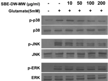
상 기전에 MAPK 전달계가 주요한 경로로 알려져 있다.34) 따라서, 세포 보호 효과와 ROS생성 억제 효과가 우수한 황 금 물 마이크로웨이브 추출물(SBE-DW-MW)이 MAPK에 미치는 영향에 대해 알아 보았다. 그 결과 글루타메이트에 의해 증가하던 p38, JNK, ERK의 인산화가 황금 물 마이크 로웨이브 추출물(SBE-DW-MW)을 농도 별로 전처리 하였 을 때, p38과 JNK의 인산화를 억제 하였으며 ERK의 인산 화 억제에는 영향을 주지 않았다(Fig. 6). 따라서, 황금 물 마이크로웨이브 추출물(SBE-DW-MW)의 글루타메이트에 대한 HT22세포보호 효과는 MAPK 경로 가운데 p38과 JNK 의 인산화를 억제함을 통해 나타났을 것으로 사료 된다. 따 라서, 본 연구 결과 황금 물 마이크로웨이브 추출물(SBE- DW-MW)은 황금 물 가열 추출물(SBE-DW)에 비해 더 높 은 수득률을 보였으며, 더 우수한 항산화 및 뇌세포 보호 활 성을 나타냈다.
결 론
본 연구에서는 황금의 물 가열 추출물(SBE-DW)과 마이 크로웨이브 추출물(SBE-DW-MW)을 제조하여 추출방법에 따른 구성 성분의 차이, 그에 따른 유효 활성 차이를 비교 해보고자 하였다. 황금의 물 가열 추출물(SBE-DW)과 마이 크로웨이브 추출물(SBE-DW-MW)은 함유성분 비교 분석을 위한 HPLC chromatogram분석에서 서로 다른 패턴을 보였 으며, 황금 물 가열 추출물(SBE-DW)에 비해 마이크로웨이 브 추출물(SBE-DW-MW)의 수득률이 더 높았다. 또한, DPPH radical 소거능, 글루타메이트에 의한 독성으로부터
HT22 세포보호 및 ROS 생성억제 효과도 마이크로웨이브 추출물(SBE-DW-MW)이 우수하였다. 이와 같은 HT22 세 포 보호 및 ROS생성억제는 MAPK 경로 가운데 p38과 JNK 의 인산화를 억제에 의한 것으로 판단 된다. 이와 같은 연 구결과는 황금 물 추출물을 제조하는 경우 기존 가열 추출 에 비해 마이크로웨이브 추출을 이용하는 것이 더 우수한 약리 활성 효과를 얻을 수 있음을 보여주는 기초적인 실험 근거를 제시하는 것으로, 추후 함유성분 가운데 차이를 보 이는 유효 성분에 관련된 비교 연구가 진행되어야 될 것으 로 사료된다.
사 사
이 논문은 2012년도 정부(교육과학기술부)의 재원으로 한 국연구재단의 지원을 받아 수행된 연구입니다(No. NRF- 2012R1A1A2042984).
인용문헌
1. Kim, H. C. (2001) Textbook of herbal pharmacology. 129- 133. Jipmundang, Seoul.
2. Lim, B. O., Choi, S. Y., Choi, D. K., Park, P. J., Choi, W. S., Kim, J. D. and Shin, H. M. (2007) Inhibitory effect of Scutel- laria baicalensis root extract on chemical mediator release and immune response. Korean. J. Medicinal Crop Sci. 15:
132-137.
3. Burnett, B. P., Jia, Q., Zhao, Y. and Levy, R. M. (2007) A medicinal extract of Scutellaria baicalensis and Acacia cat- echu acts as a dual inhibitor of cyclooxygenase and 5-lipoxy- genase to reduce inflammation. J. Med. Food. 10: 442-451.
4. Gao, Z., Huang, K., Yang, X. and Xu, H. (1999) Free radical scavenging and antioxidant activities of flavonoids extracted from the radix of Scutellaria baicalensis Georgi. Biochim.
Biophys. Acta. 1472: 643-650.
5. Huang, Y., Tsang, S. Y., Yao, X. and Chen, Z. Y. (2005) Bio- logical properties of baicalein in cardiovascular system. Curr Drug Targets Cardiovasc Haematol Disord. 5: 177-184.
6. Jung, H. S., Kim, M. H., Gwak, N. G., Im, Y. S., Lee, K. Y., Sohn, Y., Choi, H. and Yang, W. M. (2012) Antiallergic effects of Scutellaria baicalensis on inflammation in vivo and in vitro. J. Ethnopharmacol. 141: 345-349.
7. Lu, Y., Joerger, R. and Wu, C. (2011) Study of the chemical composition and antimicrobial activities of ethanolic extracts from roots of Scutellaria baicalensis Georgi. J. Agric. Food Chem. 59: 10934-10942.
8. Nagai, T., Suzuki, Y., Tomimori, T. and Yamada, H. (1995) Antiviral activity of plant flavonoid, 5,7,4'-trihydroxy-8- methoxyflavone, from the roots of Scutellaria baicalensis against influenza A (H3N2) and B viruses. Biol. Pharm. Bull.
18: 295-299.
Fig. 6. Effects of SBE-DW-MW on glutamate-induced MAPKs phosphorylation in HT22 cells. Cells were pre-treated for 3 h with SBE-DW-MW, and then incubated for 30 min with glutamate (5 mM). MAPKs phosphorylation were detected by Western blot analysis, and representative blots of three inde- pendent experiments are shown.
9. Waisundara, V. Y., Hsu, A., Huang, D. and Tan, B. K. (2008) Scutellaria baicalensis enhances the anti-diabetic activity of metformin in streptozotocin-induced diabetic Wistar rats. Am.
J. Chin. Med. 36: 517-540.
10. Talebi, M., Ghassempour, A., Talebpour, Z., Rassouli, A. and Dolatyari, L. (2004) Optimixation of the extraction of pacli- taxel from Taxus baccata L. by the use microwave energy. J.
Sep. Sci. 27: 1130-1136.
11. Ghassempour, A., Noruzi, M., Zandehzaban, M., Talebpour, Z., Khosroshahi, A. Y., Najafi, N. M., Valixadeh, M., Pours- aberi, T., Hekmati, H., Naghdibadi H. and Aboul-Enein H. Y.
(2008) Purification of paclitaxel isolated from Taxus baccata L. cell culture by microwave-assisted extraction and two- dimensional liquid chromatography. J. Liq. Chromatogr.
Relat. Technol. 31: 382-394.
12. Mandal, V., Mohan Y. and Hemalatha S. (2007) Microwave assisted extraction – An innovative and promising extraction tool for medicinal research. Phcog. Rev. 1: 7-18.
13. Fellow, P. (2000) Food processing technology. CRC press.
New York, U.S.A., pp. 365-384.
14. Coyle, J. T. and Puttfarcken, P. (1993) Oxidative stress, glutamate and neurodegenerative disorders. Science 262:
689-695.
15. Satoh, T., Enokido, Y., Kubo, K., Yamada, M., and Hatanaka, H. (1999) Oxygen toxicity induces apoptosis in neuronal cells. Cell Mol. Neurobiol. 18: 649-666.
16. Satoh, T., Okamoto, S., Cui, J.,Watanabe, Y., Furuta, K., Suzuki, M., Tohyama, K., and Lipton, S. A. (2006) Acti- vation of the Keap1/Nrf2 pathway for neuroprotection by electrophilic phase II inducers. Proc. Natl. Acad. Sci. U. S. A.
103: 768-773.
17. Satoh, T., and Lipton, S. A. (2007) Redox regulation of neu- ronal survival mediated by electrophilic compounds. Trends Neurosci. 30: 37-45.
18. Alibright, T. D., Jessel, T. M., Kandel, E. R., and Poster, M.
I. (2000) Neural science: a century of progress and the mys- teries that remain. Cell 18: 209-216.
19. Rössler, O. G., Bauer, I., Chung, H. Y., and Thiel, G. (2004) Glutamate-induced cell death of immortalized murine hip- pocampal neurons: neuroprotective activity of heme oxy- genase-1, heat shock protein 70, and sodium selenite.
Neurosci. Lett. 362: 253-257.
20. Jeong, G. S., Li, B., Lee, D. S., Byun, E., Kang, D. K., Lee, H. S. and Kim, Y.C. (2007) Cytoprotective constituents of Alipinia katsumadai seeds against glutamate-induced oxi- dative injury in HT22 cells. Nat. Prod. Sci. 13: 268-272.
21. Pare, J. R. J., Sigouin, M. and Lapointe, J. (1991) Micro-
wave-assisted natural products extraction. US Patent 5002, Various international counterparts. 26: 784.
22. Pare, J. R. J., Belanger, M. R., Stafford, S. S. (1994) Micro- wave-assosted process(MAPTM) : a new tool for the ana- lytical laboratory. Trends Anal. Chem. 13: 176-184.
23. Giese, J. (1992) Advances in microwave food processing.
Food Technol. 46: 118-123.
24. Yu, B. P. (1996) Aging and oxidative stress: modulation by dietary restriction. Free Radic. Biol. Med. 21: 651-668.
25. Tan, S., Schubert, D. and Maher, P. (2001) Oxytosis: a novel form of programmed cell death. Curr. Top. Med. Chem. 1:
497-506.
26. Rössler, O. G., Bauer, I., Chung, H. Y. and Thiel, G. (2004) Glutamate-induced cell death of immortalized murine hip- pocampal neurons: neuroprotective activity of heme oxy- genase-1, heat shock protein 70, and sodium selenite.
Neurosci. Lett. 362: 253-257.
27. Choi, D. W. (1988) Glutamate neurotoxicity and diseases of the nervous system. Neuron. 1: 623-634.
28. Siesjö, B. K. (1981) Cell damage in the brain: a speculative synthesis. J. Cereb. Blood Flow Metab. 1: 155-185.
29. Greenamyre, J. T., Penney, J. B., Young, A. B., D'Amato, C.
J., and Hicks, S. P. (1985) Alterations in L-glutamate binding in Alzheimer's and Huntington's disease. Science 4693: 1496- 1499.
30. Lipton, S. A. (2007) Pathologically activated therapeutics for neuroprotection. Nat. Rev. Neurosci. 8: 803-808.
31. Choi, B. H., Hur, E. M., Lee, J. H., Jun, D. J. and Kim, K. T.
(2005) Protein kinase C delta-mediated proteasomal degra- dation of MAP kinase phosphatase-1 contributes to glutamate-induced neuronal cell death. J. Cell Sci. 119: 1329- 1340.
32. Oh, H. L., Seok, J. Y., Kwon, C. H., Kang, S. K. and Kim, Y. K. (2006) Role of MAPK in ceramide-induced cell death in primary cultured astrocytes from mouse embryonic brain.
Neurotoxicology 27: 31-38.
33. Chuang, S. M., Wang, I. C. and Yang, J. L. (2000) Roles of JNK, p38 and ERK mitogen-activated protein kinases in the growth inhibition and apoptosis induced by cadmium. Car- cinogenesis 21: 1423-1432.
34. Jeong, E. J., Hwang, L., Lee, M., Lee, K. Y., Ahn, M. J. and Sung, S. H. (2014) Neuroprotective biflavonoids of Chamae- cyparis obtusa leaves against glutamate-induced oxidative stress in HT22 hippocampal cells. Food Chem. Toxicol. 64:
397-402.
(2014. 3. 7 접수; 2014. 3. 21 심사; 2014. 3. 21 게재확정)