• 검색 결과가 없습니다.

The Change of Ultrasonic Transmission Velocity by Wood Decay

N/A
N/A
Protected

Academic year: 2021

Share "The Change of Ultrasonic Transmission Velocity by Wood Decay"

Copied!
8
0
0

로드 중.... (전체 텍스트 보기)

전체 글

(1)

The Change of Ultrasonic Transmission Velocity by Wood Decay 1

Won-Joung Hwang

2†

⋅Hyun-Mi Lee

2

⋅Young-Ran Park

2

⋅Dong-Heub Lee

2

ABSTRACT

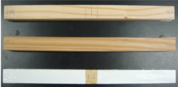
The deterioration in wood by the brown-rot fungus (Fomitopsispalustris) and the white-rot fungus (Trametesversicolor) were measured using ultrasonic velocity. Those were used for the decay exposure and 4 wood species of wood as the test specimens, Pinusdensiflora, Larixkaempferi, Pinuskoraiensis and Pinusrigida, were chosen with both the brown- and white-rot culture petridish during 12 weeks. After 12 weeks, the decrease rate of ultrasonic velocity was measured at 10

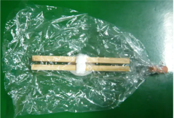
∼15%. In both brown- and white-rot exposure experiments, P. rigida showed significant decrease in ultrasonic velocity (20%), L. kaempferi on the other hand did not show decrease in ultrasonic velocity. After the fungal exposure experi- ment, the inside of specimens was investigated by computer tomography (C/T). After C/T investigation, bending tests were performed.

Keywords : Brown-rot fungus, White-rot fungus, Ultrasonic velocity, Computer tomography, Deterioration

1. INTRODUCTION 1)

There are many problems affecting wood in timber architecture and in wooden cultural pro- perties. These are caused by deterioration so that the performance is reduced due to the bio- logical and non-biological factors. The study on timber architecture and historic timber archi- tecture and deterioration is in progress. Kim et al. (2007) suggested that a pillar of historic tim- ber architecture tends to be susceptible to deteri- oration and there are many examples of decay of various types of wooden parts in the environ- ment around us. Kim et al. (2003) reported that

in deterioration evaluation of column members of ancient architecture through non-destructive inspection, deterioration of pillars at the section adjacent to the land and at the top of the upper part. Son et al. (2004) estimated the extent of decay of a Korean palace by nondestructive eva- luation (NDE). Deterioration of the wood threat- ens a building’s durability and internal stability.

Before assessing soundness of wood before it is used consideration of the systematic and his- torical importance of the wood is required and the method of measuring its internal state with avoiding direct destruction of wood is necessary.

Following these requirements among the meth- ods of evaluating wood soundness, the study of

1

Received March 21, 2013; Accepted March 19, 2014

2

Korea Forest Research Institute

Corresponding author : Won-Joung Hwang (e-mail: [email protected])

(2)

Fig. 1. The type of specimens.

the opposite side.

Park et al. (2008) tried to detect artificial flaws in wood using ultrasonic passing time. It is re- ported that there is a relationship between a lo- cation of the ultrasonic detector and the direc- tion of fracture. Furthermore, to detect internal deterioration of wood, an artificial defect is made and ultrasonic velocity is measured. The result showed that the larger the size of a de- fect, the less the ultrasonic velocity (Saeed K. et al., 2009).

Lee et al. (2008) investigated on how brown- rot fungus in wood is develops through meas- urement of mass loss rate, mechanical property and ultrasonic velocity with samples that have been exposed to brown-rot fungus in the wood The current study investigated the change in ul- trasonic transmission velocity caused by the de- cay period and the change in ultrasonic trans- mission velocity in decayed specimens using ul- trasonic transmission velocity about the absence of decay that is artificially produced through ul- trasonic measuring instrument, Wood Pole Tester.

2. MATERIALS AND METHOD

2.1. Fungal Culture

Fomitopsispalustris and Trametesversicolor were a specimen size of is 2 × 2 × 30 (cm). The test specimens were Pinusdensiflora, Larixleptolepis, Pinuskoraiensis and Pinusrigida. By taking heart- wood and sapwood samples from each species

of trees, atotal of eight test specimens was used in this study. A Wood Pole Tester (PROCEQ, CH/PILODYN, 6J), (77 kHz) was employed to measure the velocity of propagation of ultra sonic waves (Fig. 1). Computed tomography was undertaken using a TSX-0001A, (TOSHIBA corp), located in Korean Forest Research Insti- tute (KFRI), where it is employed to measure bending test of wood.

2.2. Experiment Method

2.2.1. Preparation of Test Specimens To find out the change in velocity of trans- mission of ultrasonic waves through wood caused by wood rot-fungieach test specimen (2 × 2 × 30 (cm)) was taken from the heartwood and sap- wood of the different wood species (Fig. 1).

The first condition is to make the space where it is both right and left side off 1cm from the center of the length direction of test specimens.

It is to protect the space from penetrating de-

caying rot-fungi when epoxy resin is sealed in

the marked space (HT1, ST1). For the second

condition, the method inoculating decaying rot-

fungi without epoxy resin sealed in the test speci-

mens were employed (HT2, ST2). Finally, a

control group of wood specimens which had not

been inoculated with wood rot fungi was included.

(3)

Fig. 2. Wood Pole tester. Fig. 3. Specimens under fungal exposure test.

2.2.2. Measuring the Velocity of Propagation of Ultra Sonic Waves

Measuring the velocity of propagation of ul- trasonic waves was conducted by measuring the change in the velocity of propagation of ultra sonic waves by rot-fungi inside wood at every 7 days from a week when the wood rot-fungi were infected by the Wood Pole Tester (77 kHz) (Fig. 2).

2.2.3. Experimental Construction

After pouring and solidifying 2% agar(which is without nutrition) with pressurization and sterilization in schale with 90∼100 diameter and 15 mm height, MRS agar is made including glucose 2.5%, peptone 0.5%, malt extract 1.0%, potassium dehydrogen phosphate 0.3% and mag- nesium sulfate 0.2%.

By positioning in the schalecentre of the length direction of a specimen, a mycelium was al- lowed to grow from the specimen’s centre. Sterile water was added to the specimen as required.

To protect the specimen from exterior fungi, test specimens were cultivated for 12 weeks in the sealed plastic bags, at a temperature of 26 ± 2°C and relative humidity greater than 70% (Fig. 3).

2.2.4. Computed Tomography

After the experiment on decay exposure, to investigate the internal shape of wood was com- puted tomography bya TSX-0001A with X-Rays for 5 seconds at 120 kV, 110 mA and set at 10 mm for intervals.

3. RESULTS AND DISCUSSION

3.1. Change in Ultrasonic Velocity

3.1.1. Influence of F. palustris

In the case of P. densiflora, ultrasonic veloc- ity in wood at the early stage was 4881.65 (m/s) for the heartwood and 4609.43 (m/s) for the sapwood and after 12 weeks it was recorded at 4026.50 (m/s) for the mean of heartwood and 4286.15 (m/s) for that of sapwood, respectively.

From this result, heartwood of P. densiflora de- creased by 17.51% and sapwood by 7.01%.

For P. koraiensis, ultrasonic velocity in wood

at the early stage was measured 4369.89 (m/s)

for the heartwood and 4466.01 (m/s) for the

sapwood and after 12 weeks it became 3891.82

(m/s) for the mean of heartwood and 4047.77

(m/s) for that of sapwood, indicating a decrease

in 10.94% for heartwood and 9.36% for sap-

(4)

Fig. 4. Computed tomography.

Fig. 5. Relationship between fungal exposure time and ultrasonic velocity (F.palustris).

wood compared with both means at the early stage, respectively (Fig. 4). Therefore the change in ultrasonic velocity in decayed heartwood changed more than for sapwood.

3.1.2. Impact of T. versicolor

Fig. 5 shows the relationship between F. pal-

After 12 weeks, the mean recordings for heart- wood and sapwood average were 3880.31 (m/s) and 4766.70 (m/s) respectively.

In L.kaempferi, one week after inoculation, the heartwood mean of ultrasonic velocity was 4670.53 (m/s) and the sapwood mean of that was 5057.41 (m/s). After 12 weeks, the each average of heartwood and sapwood was 4018.61 (m/s) and 4690.55 (m/s).

In the case of P.koraiensis, while mean ultra-

sonic velocity in wood at the early stage was

4812.75 (m/s) for the heartwood average and

5147.29 (m/s) for the sapwood average, after 12

weeks mean ultrasonic velocity in wood for the

(5)

Fig. 6. Relationship between fungal exposure time and ultrasonic velocity (T.versicolor).

heartwood and sapwood mean was 4444.44 (m/s) and 4737.10 (m/s).

In P.rigida, one week after inoculation, the heartwood was 4839.98 (m/s) and the sapwood 4614.77 (m/s) and after 12 weeks, both were measured in 3919.90 (m/s) and 3248.20 (m/s), respectively (Fig. 6).

3.1.3. Change Rate of Ultrasonic Velocity Following exposure to the brown-rot fungus exposure comparison of ultrasonic velocity for two different periods (before and after inocula- tion), the change rate of ultrasonic velocity for each specimen indicated a decrease of 19.19%

(P.densiflora’s sapwood), 8.27% (L.kaempferi’s sapwood), 13.95% (P.koraiensis’s sapwood) and 21.11% (P.rigida’s sapwood). Among 4 speci- mens, the rate of change in ultrasonic velocity for P.rigida’s sapwood was the most significant.

For T.versiclorthe ultrasonic velocity for the two different periods the rate of change in ultra- sonic velocity for each specimen was 12.98%

(P.densiflora’s sapwood), 13.95% (L.kaempferi’s sapwood), 7.65% (P.koraiensis’s sapwood) and 19% (P.rigida’s sapwood). Therefore for the 4 specimens, the rate of change for P.rigida’s sap- wood was again the most significant (Fig. 7).

Following decay F.palustris and T.versicolor over 12 weeks, computer tomography (C/T) was conducted. Fig. 7 shows the result of C/T fol- lowing decay exposure experiment. It is classi- fied by positioning that Type 2 was put above Type 1 and plastic plate is set in the middle.

Son et al. (2004) found that in the decay test the larger decrease in wood mass caused by the decay then the slower the ultrasonic velocity.

Furthermore the longer the wood exposure times

the slower the ultrasonic velocity and there is a

(6)

◆ P. densiflora heart wood * P. densiflora sap wood ■ L. leptolepis heart wood ●L. leptolepis sap wood

▲ P. koraiensis heart wood + P. koraiensis sap wood Х P. rigida heart wood -P. rigida sap wood

Fig. 7. Relationship between fungal exposure time and decreased rate of ultrasonic velocity (A: F. palustris, B:

T. versicolor).

difference in the extent of the decay and the rate of change in the ultrasonic velocity depend- ing on the properties of each specimen property.

Lee et al. (2008) reported on loss rate of mass and ultrasonic velocity through the test speci- men (10 × 3 × 10) that had been decayed by 2 types of brown-rot fungus. It was reported that at the early stage, the decay detection by meas- urement of ultrasonic velocity was possible and whereas qualitative evaluation was possible quantitative evaluation was unsatisfactory. Further- more during 8 weeks of decay activity intensity capacity fell 80% and ultrasonic velocity de- creased by around 30%. Compared with the findings of Lee et al. (2008), the current ex- perimental procedure used larger test specimens (2 × 2 × 30) and a longer incubation period (12

weeks). Considering the size of the test speci- mens and the difference between types of wood, the results of decrease in the rate of ultrasonic velocity were similar to those published by Lee et al. (2008).

3.2. Computer Tomography (C/T)

After the test specimens had been exposed to the decay fungi during 12 weeks period the in- ternal conditions of the wood samples were in- vestigated using computer tomography. In the test specimen of constant thickness and high density the decrease in X-ray energy was higher than that of the test specimen with low density.

Therefore, the brightness of a film is high due

to decreasing the sensitivity of the X-ray film.

(7)

Fig. 8. CT images of specimens after fungal exposure test (A: P.densiflora B: P.koraiensis C: P.densiflora D:

P.koraiensis) (A, B : decayed by F. palustris C,D : decayed by T. versicolor).

The greater the extent of the decay is, the lower the brightness is in the test specimen performed by C/T (Fig. 8). As Kim et al. (2006) reported that the brightness in the edge of the test speci- men is strongly observed and it is thought that the phenomenon results from higher the X-ray intensity reaching a film and the higher X-ray intensity the greater the brightness of a film.

4. CONCLUSION

This study using 4 different wood types which were separated into heartwood and sap- wood was undertaken to assess the extent of de- terioration and the change in ultrasonic velocity in the inside of wood following the decay peri- od from brown-rot and white-rot fungi.

The result showed that there is a significant decrease in ultrasonic velocity at the early stage of decay. Results for the 4 types of wood used indicated that the deterioration caused by the

brown-rot fungus in P.rigida (heartwood 21.11%, sapwood 18.09%) resulted in the largest change- L.kaempferi, the least change in ultrasonic velo- city. Moreover, comparing heartwood and sap- wood, heartwood decrease rate was higher than for sapwood.

Deterioration caused by the white-rot fungus was greatest in P.rigida (heartwood 19.00%, sapwood 29.61%). Like the brown-rot fungus the least change in the decrease in ultrasonic velocity was found to be for L.kaempferi. Except for P.rigida the remainder of the test specimens decrease in ultrasonic velocity was higher in- heartwood was higher than in sapwood.

From this study, in artificially produced de-

cay, qualitative evaluation is proved to be possi-

ble but additional research for quantitative eval-

uation is required

(8)

of the Korean Wood Science and Technology.

35(1): 51∼63.

2. Kim, Y. S. and S. M. Han. 2007. Deterioration and Preservation Technique of Wooden Cultural Properties (Part.Ⅱ)-Biodeterioration of Square Post to Support Wooden Printing Blocks Shelves, Janggengpanjeon. Journal of the Korean Wood Science and Technology. 35(1): 64∼72.

3. Korean Standards Association. 2004. Method of bending test for wood. KS F 2208.

4. Lee, J. J., G. M. Kim, and M. S. Bae. 2003. Trans- mission Process for Ultrasonic Wave in Wood.

Journal of the Korean Wood Science and Tech- nology. 31(2): 31∼37.

5. Lee, S. J., G. H. Kim, and J. J. Lee. 2008. Effect of brown-rotted wood on mechanical properties and ultrasonic velocity. Journal of the Korean Wood Science and Technology. 36(5): 24∼32.

6. Park, J. C. and S. I. Hong. 2008. Determination of Localized Defects in Wood by the Transfer Time of Ultrasonic Waves. Journal of the Korean Wood Science and Technology. 36(1): 61∼68.

7. Ross, R. J., L. A. Soltis, and P. Otton. 1998. Role of nondestructive evaluation in the inspection and

6 p.

9. Schad, K. C., D. L. Schmoldt, and R. J. Ross.

1996. Nondestructive methods for detecting de- fects in softwood logs. Res Pap.FPL-RP-546.

Madison, WL-U.S. Department of Agriculture, Forest Service, Forest Products Laboratory.13 p.

10. Saeed, Kazemi-Najafi., A. Shabafan, and G.

Ebrahimi. 2009. Intermal decay in standing beech trees using ultrasonic velocity measurement. Euro- pean Journal of Forest Ressearch. 128: 345∼350.

11. Son, D. W. and D. H. Lee. 2004. Wood Decay Detection by Non-destructive Methods. Journal of the Korean Wood Science and Technology.

32(4): 74 ∼81.

12. Son, D. W. and D. H. Lee. 2008. Evaluation on Termite Damage of the Traditional Wooden Building by Non-destructive Methods. Journal of the Korean Wood Science and Technology. 36(1):

21∼29.

13. Wang, X., R. J. Ross, J. A. Mattson, J. W. Fors-

man, E.A. Geske, and M. A. Wehr. 2002. Nonde-

structive evaluation techniques for assessing mod-

ulus of elasticity and stiffness of small-diameter

logs. Forest Prod. J. 52(2): 79 ∼85.

수치

Fig. 1. The type of specimens.
Fig. 2. Wood Pole tester. Fig. 3. Specimens under fungal exposure test.
Fig. 5. Relationship between fungal exposure time and ultrasonic velocity (F.palustris).
Fig. 6. Relationship between fungal exposure time and ultrasonic velocity (T.versicolor).
+3

참조

관련 문서

This study is based on the fact that demand for housing has been under the influence of change of population structure... people live in

Efficacy of syringe irrigation, RinsEndo and passive ultrasonic irrigation in removing debris from irregularities in root canals with different apical

A comparison of the cleaning efficacy of short-term sonic and ultrasonic passive irrigation after hand instrumentation in molar root canals.. Ultrasonic

To visualize the vari- ation in various transport characteristics (the non-dimen- sional axial velocity and temperature distributions, pressure gradient, rate of heat

The resolved-rate control method [9] is based on the manipulator velocity equation kX = kje where kj is the Jacobian matrix, e is the vector of relative joint rates, kX is the

✓ The rate of change of energy is equal to the sum of the rate of heat addition to and the rate of work done on a fluid particle.. ▪ First

 The rate of change of energy is equal to the sum of the rate of heat addition to and the rate of work done on a fluid particle..  First

1 John Owen, Justification by Faith Alone, in The Works of John Owen, ed. John Bolt, trans. Scott Clark, "Do This and Live: Christ's Active Obedience as the