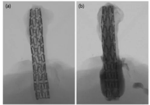
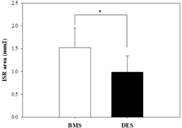
Ex vivo Morphometric Analysis of Coronary Stent using Micro-Computed Tomography
전체 글
수치

관련 문서
- "This work was supported by the Korea Foundation for the Advancement of Science and Creativity(KOFAC) grant funded by the Korea government(MOE)"... 스마트폰에는
- "This work was supported by the Korea Foundation for the Advancement of Science and Creativity(KOFAC) grant funded by the Korea government(MOE)"...
"This work was supported by the Korea Foundation for the Advancement of Science and Creativity(KOFAC) grant funded by the Korea government(MOE)"... 이러한 관점에서
- "This work was supported by the Korea Foundation for the Advancement of Science and Creativity(KOFAC) grant funded by
- "This work was supported by the Korea Foundation for the Advancement of Science and Creativity(KOFAC) grant funded by the Korea government(MOE)"... 다양한
- "This work was supported by the Korea Foundation for the Advancement of Science and Creativity(KOFAC) grant funded by the Korea government(MOE)"..
- "This work was supported by the Korea Foundation for the Advancement of Science and Creativity(KOFAC) grant funded by the Korea government(MOE)"...
This work was supported by the Korea Foundation for the Advancement of Science and Creativity(KOFAC) grant funded by the Korea government(MOE)... 과연 그들에게
