Hh-Ag1.5 처리가 돼지 체외수정란의 발육 및 세포사멸에 미치는 영향
권대진, 여재훈, 노원근, 곽태욱, 오건봉, 옥선아, 임석기, 박진기, 황성수*
농촌진흥청 국립축산과학원 동물바이오공학과
Effect of Hh-Ag1.5 Treatment on the In Vitro Development and Apoptosis of In Vitro Fertilized Embryos in Pigs
Dae-Jin Kwon, Jae-Hun Yeo, Won-Gun Noh, Tae-Uk Kwak, Keon Bong Oh, SunA Ock, Seok Ki Im, Jin-Ki Park and Seongsoo Hwang
*Animal Biotechnology Division, National Institute of Animal Science, RDA, Suwon 441-706, Korea
ABSTRACT
The present study was performed to investigate the effect of Hh-Ag1.5, a small-molecule chemical agonist of SMOothened receptor, on the in vitro maturation and development of in vitro fertilized (IVF) embryos in pigs. Oocytes or fertilized embryos were cultured in a maturation or embryo culture medium supplemented with 0 (control), 25, 50 or 100 nM of Hh-Ag1.5, respectively. Although the maturation rate were not different among treatment groups, the blastocyst formation rate in the group treated with 25 nM Hh-Ag1.5 was significantly increased compared to other groups (P<0.05). While the highest dose of Hh-Ag1.5 (100 nM) did negatively affect to the embryo development and cell number in blastocysts compared to other groups (P<0.05), the apoptotic cell index in blastocysts was significantly lower in 25 and 50 nM groups than in control and 100 nM groups (P<0.05). The mRNA expression of the pro- apoptotic gene Bax and the ratio of Bax/Bcl-XL decreased in among treatment groups compared to control (P<0.05).
The embryo quality related genes, Tert and Zfp42, were significantly decreased in 50 and 100 nM groups compared with control and 25 nM groups (P<0.05). In conclusion, the addition of 25 nM Hh-Ag1.5 to in vitro maturation and culture medium can enhance the developmental potential as well as quality of IVF embryos in pig.
(Key words : SMOothened, Hh-Ag1.5, apoptosis, embryo quality, pig)
†
본 연구는 어젠다 프로그램(PJ009457)과 2013년도 농촌진흥청 국립축산과학원 박사후 연수과정 지원사업에 의해 이루어짐.
*
Correspondence : E-mail : [email protected]
서 론
돼지는 이종 장기 생산 및 세포 치료를 위한 모델 동물로서 응용 및 연구 가치가 우수한 것으로 알려져 있다(Prather 등, 2003). 하지만 돼지를 이용한 연구에 있어서 양질의 수정란을 다수 생산하기 위한 많은 연구에도 불구하고, 현재까지 낮은 생산 효율은 해결해야 할 문제점으로 지적되고 있다.
체외 성숙에 이용되는 난자-난구세포 복합체(cumulus-oocyte complex, COC)는 성숙되는 동안 배발달에 필수적인 물질의 축적 및 세포질 환경을 구축하게 된다(Gandolfi와 Gandolfi, 2001; Sirard 등, 2006). 체내와 체외에서 성숙된 난자(meta- phase II, MII) 모두 수정되어 태아로 발달할 수 있는 능력을 가지고 있지만, 불완전한 체외 배양 체계는 비정상적인 염색 체 변이를 유도하기도 하며, 초기배 발육 동안 유전자 발현 양상의 변화를 야기시킬 수 있다(Carrell 등, 2005; Lonergan
등, 2006; Zheng과 Dean, 2007). 따라서 불완전한 체외 배양 조건은 체외에서 생산된 수정란과 체내수정란 간의 질적 차 이의 원인으로 여겨진다(Abeydeera 등, 1998; Kim 등, 2006).
Hedgehog(Hh) signaling pathway는 음성 조절자(negative regulator)인 Patched1(PTCH1)과 활성인자(activator)인 SMO- othened(SMO) 및 transcriptional effectors인 Gli family로 구성 되어 있으며, Hh signaling에 관련된 molecule들은 번식기계 (系)에 존재하며, 세포 증식과 steroid hormone 생산을 유도하 는 기능을 가지고 있다(Spicer 등, 2009). 마우스의 경우 Hh signaling은 초기배의 착상에 적합한 자궁 내 환경을 조절할 뿐만 아니라(Lee 등, 2006), 활성인자인 SMO의 활성을 직접적 으로 유도하였을 경우, 난소 내 협막(theca) 세포의 증식을 유 도하는 것으로 보아, Hh signaling이 난포란의 성숙에 관여하 고 있음이 시사되었다(Ren 등, 2009). 이후 Nguyen 등(2009)은 Hh signaling에 관련된 molecule(PTCH1, SMO, Gli1)들이 돼
지의 난소와 난자 모두에 존재하고 있음을 확인하였으며, 체 외 성숙 및 발육 배양액에 Hh 재조합 단백질을 첨가할 경우 단위 발생, 체외수정 및 복제란 생산 효율이 증가되는 것으로 보고하였다(Nguyen 등, 2010; Nguyen 등, 2011). 한편, Hh 재 조합 단백질을 첨가할 경우 PTCH1의 억제 효과로 인해 첨가 효과가 저해될 수 있기 때문에 Hh signaling의 활성을 효과적 으로 유도하기 위해 활성인자인 SMO를 직접적으로 활성화시 킨다면 그 효과를 증가시킬 수 있을 것이다. Hh-Ag1.5는 SMO 에 직접적으로 작용하는 Hh signaling의 저분자 agonist로서, Hh 재조합 단백질보다 활성이 높은 것으로 알려져 있다(Frank- Kamenetsky 등, 2002). 따라서 본 연구에서는 돼지 체외 성숙 (in vitro maturation, IVM) 및 체외 배양(in vitro culture, IVC) 배양액에 Hh-Ag1.5 첨가가 수정란의 질적 양적 생산 효율에 미치는 영향을 검토하였다.
재료 및 방법
1. 난포란의 채취 및 성숙 배양
도축장에서 도살된 암퇘지의 난소를 회수하여 25∼30℃의 생리식염수 용액에 넣어 실험실로 운반하여 직경이 3∼6 mm 의 난포로부터 18 gauge 주사 바늘이 부착된 주사기로 난포액 을 흡입하여 채란하고, 채취된 난자를 0.1%(w/v) polyvinyl alcohol(PVA)이 첨가된 Tyrode's lactate(TL)-Hepes액으로 3번 세척한 뒤, 실체 현미경 하에서 난구 세포가 균일하고 세포질 이 균질한 것만을 선별하여 난자 성숙에 공시하였다. 난포란 의 성숙배양을 위해 0.6 mM cysteine, 10 ng/mL epidermal growth factor(EGF), 10 IU/ml pregnant mare's serum gonado- tropin(PMSG), human chorionic gonadotropin(hCG), 10%(v/v) porcine follicular fluid(PFF)가 첨가된 TCM-199 배양액 500 μl 의 drop을 만들어 mineral oil로 피복하고, 성숙 배양 2∼3시간 전에 5% CO2, 39℃ 온도 조건 하에서 평형시킨 후, 각 drop당 100개의 난포란을 넣어 22시간 배양하였으며, 이후 호르몬이 첨가되지 않은 배양액으로 옮겨 20시간 동안 배양하였다. 일 부 난자는 성숙 배양 전 기간에 걸쳐 각각 25, 50, 100 nM의 Hh-Ag1.5를 첨가하였다.
2. 체외수정란의 생산
체외 성숙 배양 후 난포란을 0.1% hyaluronidase에 넣고 3 분간 교반(vortexing)하여 난구세포를 제거한 후, 체외수정에 이용하였다. 체외수정용 정액은 국립축산과학원에서 사육하 고 있는 일반 돼지로부터 채취하였으며, BTS 액으로 희석한 신선 정액을 이용하였다. 원심분리(500 × g) 후 상층액을 제거 하고 0.04% BSA가 첨가된 PBS 액으로 3회 세척한 후, mTBM 액으로 희석하여 체외수정용 배지에 5 × 10⁵ spermatozoa/ml가
되도록 정자를 주입하여 체외수정을 유도(6시간)하였다. 이후 PZM-3로 옮겨 5% CO2 및 39℃의 조건 하에서 6∼7일간 배 양하며, 분할율 및 배반포 형성율을 검사하였다. 일부 난자는 성숙 배양 시와 같은 농도(0∼100 nM)의 Hh-Ag1.5를 첨가하 였다.
3. 배반포의 Apoptotic Cell 검사
배반포의 정상성을 알아보기 위하여 DNA fragmentation을 검사하였다. 배양 6일째에 생산된 배반포를 실온에서 3.7%(w/v) paraformaldehyde가 첨가된 PBS로 40분간 처리하여 고정하였 다. 이후 0.5% Triton X-100과 3%(w/v) BSA가 첨가된 PBS로 37℃에서 30분간 처리하였다. Apoptotic cell를 검사하기 위해 TUNEL(In Situ Cell Death Detection Kit; Roche, Penzberg, Germany) 반응을 유도한 후, DAPI로 전체 세포의 핵을 염색 하였다. TUNEL 반응이 유도된 배반포를 slide glass로 옮겨 antifluorescence-fade 액으로 피복하고, cover glass로 덮어 표 본을 제작하였다. 제작된 표본은 Leica 형광 현미경(Leica Mic- rosystems, Mannheim, Germany)을 이용하여 전체 세포수와 apoptotic cell 수를 판단하였다.
4. Real-Time RT-PCR 분석
체외에서 성숙된 난자로부터 RNeasy Plus Mini Kit(Qiagen, Valencia, USA)을 이용하여 RNA 추출한 후, Agilent 2100 Bioanalyzer(Agilent Technologies, Santa Clara, USA)을 이용 하여 RNA의 integrity를 조정하였다. 이후 High-Capacity cDNA Reverse Transcription Kits(Life Technologies, Grand Island, USA)을 이용하여 제공된 사용법에 따라 cDNA를 생산하였 다. 배반포의 경우, FastLane Cell cDNA Kit(Qiagen, Valencia, USA)을 이용하여 제공된 방법에 따라 cDNA를 생산하였다.
Real-Time RT-PCR은 Rotor-Gene SYBR Green PCR Kit(Qia- gen, Valencia, USA) 및 7500 Fast Real-Time PCR System(Life Technologies, Grand Island, USA)를 이용하여 분석하였다. 분 석에 사용된 primer는 Table 1에 제시하였다. Real-Time RT- PCR의 증폭조건은 95℃에서 1분간 pre-incubation 과정을 거 친 후, 95℃에서 15초 동한 denaturation 후 60℃에서 1분간 annealing의 과정을 40 사이클 반복하도록 조작하였다.
5. 통계 처리
모든 처리에 대한 실험은 3회 이상 반복 실시하였으며, 난 자의 fragmentation, 발육율, 세포수 및 mRNA 발현에 대한 결 과들은 Statistical Analysis System(SAS Institute, Inc., Cary, USA)의 General Linear Models 절차를 이용한 Duncan's multiple range test로 유의성을 검토하였다. 유의차는 P<0.05 일 때 통계적 차이가 있는 것으로 판단하였다.
Table 1. Primer sets for Real Time RT-PCR
Gene Primer sequences(5’ to 3’) Product size(bps) References
Rnf24
F- CATGAGCTCTGTGCAGTGTG
185 XM_003134311.3
R- GGTCCACGGTCCTCCTTACT
Rab23
F- AGGCTGATGCTATGGGACAC
155 XM_001929636.1
R- TCAGCCACCACCTTCTCTCT
Zfp42
F- ACTTTTGAAGGATGCGGAAA
179 AM410991.1
R- CACTGATTTGTATTGGCCTTTG
Tert
F- CGCTCCTGAAAGCCAGAAAC
182 AY785158.1
R- CTGGCACAATCGCTCTCTG
Bax
F- CCTTTTGCTTCAGGGTTTCA
165 XM_003127290.2
R- ATCCTCTGCAGCTCCATGTT
Bcl-XL
F- CTGAATCAGAAGCGGAAACC
188 AF216205.1
R- CCTCCGGTACCTCAGTTCAA
Gapdh
F- CTGCGCTCTCTGCTCCTC
161 NM_001206359.1
R- ACAATGTCCACTTTGCCAGA
결과 및 고찰
본 연구에서는 SMO에 직접적으로 작용하는 Hh signaling 의 저분자 agonist인 Hh-Ag1.5가 돼지 체외성숙 및 체외 배발 달에 미치는 영향을 검토하였다. 체외성숙 배양액에 0, 25, 50, 100 nM의 농도로 첨가하였을 때 성숙율(83.0∼87.5%)에 는 처리구간 차이가 없었다(미제 시). 성숙된 난자에서 Hh signaling의 억제 유전자로 알려진 Rab23의 발현 정도를 검토 한 결과, 50 nM 이상 첨가하였을 경우에 발현이 증가하는 것 으로 나타났다(Fig. 1). 이는 일정 농도 이상의 Hh-Ag1.5 첨가 는 Hh signaling의 활성에 미치는 영향이 상대적으로 작다는 것을 시사한다.
Ring-finger protein 24(Rnf24)는 수정되지 않은 성숙 난자 에서 높은 수준으로 발현되며(Bobe 등, 2006), 수정 후 초기 분할 단계에서 급격히 저하되었다가 배반포 단계에서 최고 수준으로 발현된다(Malcuit 등, 2009). 따라서 Rnf24는 만능성 (pluripotency)과 관련이 있을 것으로 사료되며, 성숙란 및 배 반포에서의 발현 정도는 초기배 발달 능력을 평가하는 지표로 이용될 수 있을 것이다. 본 연구에서는 성숙란에서의 Rnf24 발현량을 검토한 결과, 50 nM Hh-Ag1.5 처리군에서 유의적 으로 높게 나타났다(P<0.05, Fig. 1).
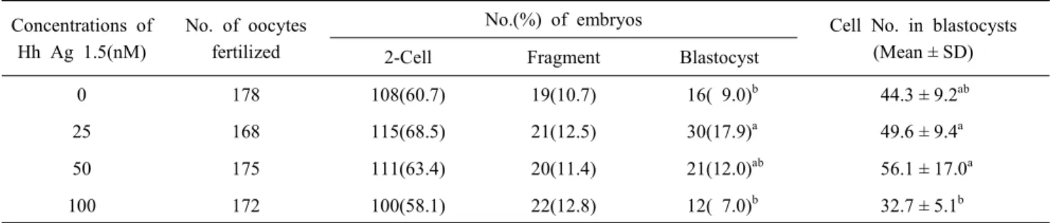
각 처리군에서 성숙된 난자는 체외수정에 이용하였으며, 이후 체외성숙 때와 동일한 농도의 Hh-Ag1.5를 첨가한 배양 액에서 발육시킨 결과를 Table 2에 나타내었다. 25 nM 처리
Fig. 1. Effect of Hh-Ag1.5 on Rab23 and Rnf24 mRNA expression of matured porcine oocytes in vitro. Cumulus-oocyte com- plexes(COCs) were cultured in a 500 μl drop of maturation medium supplemented with 0, 25, 50 or 100nM of Hh-Ag 1.5. Error bars indicate ± SD.
a∼c Within each gene, bars with different letters differ (P<0.05).
구에서 유의적으로 높은 배반포 발육율을 보였으며(P<0.05), 배반포의 세포 수에는 영향이 없는 것으로 나타났다. 하지만 100 nM로 첨가한 처리군에서 배반포 발육율과 세포수가 유
Table 2. Effects of Hh-Ag1.5 treatment during IVM on embryonic development after in vitro fertilization Concentrations of
Hh Ag 1.5(nM)
No. of oocytes fertilized
No.(%) of embryos Cell No. in blastocysts (Mean ± SD) 2-Cell Fragment Blastocyst
0 178 108(60.7) 19(10.7) 16( 9.0)b 44.3 ± 9.2ab
25 168 115(68.5) 21(12.5) 30(17.9)a 49.6 ± 9.4a
50 175 111(63.4) 20(11.4) 21(12.0)ab 56.1 ± 17.0a
100 172 100(58.1) 22(12.8) 12( 7.0)b 32.7 ± 5.1b
a,b Values with different superscripts are significantly different(P<0.05).
의적으로 낮게 나타나, 25 nM의 Hh-Ag1.5 첨가가 돼지 체외 수정란 생산에 효과적인 것으로 사료된다(Table 2, P<0.05).
체내에서 생산된 배반포라 할지라도 대부분의 배반포에서 적어도 1개 이상의 apoptotic cell이 관찰되지만, 일반적으로 apoptotic cell은 체외에서 생산된 배반포에서 유의적으로 많 은 수가 관찰되므로 난자의 질을 판단하는 지표로 이용된다 (Vandaele 등, 2007). 본 연구에서 생산된 배반포의 질적 양상 을 평가하기 위하여 TUNEL 분석을 통한 배반포의 apoptotic cell 수를 확인한 결과, 무처리군과 100 nM 첨가군에 비해 25 및 50 nM 첨가군에서 유의적으로 낮은 DNA fragmentation이 나타났다(Fig. 2, P<0.05). 또한 이러한 결과가 세포사멸 관련 유전자와 연관이 되는지를 확인하기 위하여 Real-time RT- PCR을 통한 세포사멸 관련 유전자인 Bax(pro-apoptotic gene) 및 Bcl-XL(anti-apoptotic gene) mRNA 발현 수준을 분석한 결 과, Bax의 경우 Hh-Ag1.5 처리군에 비하여 무처리군에서 가장
Fig. 2. Apoptotic cell index in blastocysts derived IVF embryos.
The apoptotic cell index(TUNEL-positive cells/total cells in blastocysts) at Day 7 after culture. The blastocysts were cultured in IVC medium supplemented with 0, 25, 50 or 100 nM of Hh-Ag1.5. Error bars indicate ± SD.
a,b Different letters indicate significant differences(P<0.05).
높게 발현되고 있는 것을 확인하였다(Fig. 3, P<0.05) 한 결과, Bax의 경우 Hh-Ag1.5 처리군에 비하여 무처리군에서 가장 높 게 발현되고 있는 것을 확인하였다(Fig. 3, P<0.05). 한편, Bcl- XL의 경우 25 nM 처리군에서 가장 높게 발현되었으며(Fig.
3, P<0.05), 전체적으로 Hh-Ag1.5 처리에 의해 배반포 내 Bax/Bcl-XL 비율이 감소되는 경향을 보였다. Bcl-2는 Shh-Gli1 signaling을 통해 발현이 직접적으로 활성화되며, 결과적으로 세포 내 Bax와 Bak1의 활성수준을 감소시키는 것으로 알려져 있어(Xu 등, 2009), 돼지 초기배 발달에 있어서 Shh-Gli1 signaling이 중요하게 작용하고 있는 것으로 확인되었다.
Telomerase는 염색체의 말단에 반복 염기서열 구조(5-TTAG GG-3)인 Telomere를 신장시키는 효소이며(Dahse 등, 1997), 소 수정란의 경우 수정 이후부터 배반포기까지 계속적으로 발현되며, 배반포기에 가장 높은 수준으로 발현되는 것으로 알려져 있다(Xu 와 Yang, 2001). 한편, Zfp42은 다양한 만능
Fig. 3. Expression of cell survival-related genes Bcl-XL and Bax in porcine blastocysts. IVF embryos were cultured in IVC medium supplemented with 0, 25, 50 or 100 nM of Hh-Ag 1.5. Error bars indicate ± SD.
a∼c Within each gene, bars with different letters differ (P<0.05).
세포주(pluripotent cell line)에서 발현되는 것으로 알려져 있 으며, 특히 마우스의 내부 세포괴(inner cell mass, ICM)에서 도 발현되기 때문에 줄기세포의 만능성을 판단하는 지표로 이용되고 있다(Rogers 등, 1991). 따라서 본 연구에서는 돼지 배반포에서 만능성과 관련된 유전자(telomerase reverse trans- criptase, Tert; zinc finger protein 42, Zfp42)들의 mRNA 발현 정도를 검토하여 배반포의 질을 판단하는 지표로 활용하였다 (Fig. 4). Tert의 발현량을 분석한 결과, 대조군와 25 nM 처리 군에서는 차이가 없었으나, 50 및 100 nM 처리군에서는 유의 적으로 낮게 발현되었다(Fig. 4A, P<0.05). Zfp42 발현은 25 nM 처리군에서 가장 높게 나타났으나, 대조군과의 유의적 차 이는 없었다. 따라서 높은 수준(50 nM 이상)의 Hh-Ag1.5 첨가 는 배반포의 질을 저하시키는 원인이 되는 것으로 사료된다.
이상의 결과를 종합해 보면, 돼지 체외 성숙 및 발육 배양액
(A)
(B)
Fig. 4. Expression of pluripotency-related genes Tert(A) and ZPF42 (B) in porcine blastocysts. IVF embryos were cultured in IVC medium supplemented with 0, 25, 50 or 100 nM of Hh-Ag1.5. Error bars indicate ± SD.
a,b,c
Within each gene, bars with different letters differ (P<0.05).
에 25 nM 농도의 Hh-Ag1.5를 첨가할 경우 배반포 형성율을 향상시킬 수 있을 뿐만 아니라, 질적으로 우수한 배반포를 생 산할 수 있어, 체외수정란 및 복제란 생산에 효과적으로 이용 할 수 있을 것으로 사료된다.
참 고 문 헌
Abeydeera LR, Wang WH, Cantley TC, Rieke A, Prather RS and Day BN. 1998. Presence of epidermal growth factor during in vitro maturation of pig oocytes and embryo culture can modulate blastocyst development after in vitro fertilization. Mol. Reprod. Dev. 51: 395-401.
Bobe J, Montfort J, Nguyen T and Fostier A. 2006. Identifica- tion of new participants in the rainbow trout (Oncorhynchus
mykiss) oocyte maturation and ovulation processes using
cDNA microarrays. Reprod. Biol. Endocrinol. 4: 39-54.Carrell DT, Liu L, Huang I and Peterson CM. 2005. Compa- rison of maturation, meiotic competence, and chromosome aneuploidy of oocytes derived from two protocols for in
vitro culture of mouse secondary follicles. J. Assist. Reprod.
Gen. 22: 347-354.
Dahse R, Fiedler W and Ernst G. 1997. Telomeres and telo- merase: biological and clinical importance. Clin. Chem. 43:
708-714.
Frank-Kamenetsky M, Zhang XM, Bottega S, Guicherit O, Wichterle H, Dudek H, Bumcrot D, Wang FY, Jones S, Shulok J, Rubin LL and Porter JA. 2002. Small-molecule modulators of Hedgehog signaling: identification and charac- terization of SMOothened agonists and antagonists. J. Biol.
1: 10-28.
Gandolfi TA and Gandolfi F. 2001. The maternal legacy to the embryo: cytoplasmic components and their effects on early development. Theriogenology 55: 1255-1276.
Kim S, Lee SH, Kim JH, Jeong YW, Hashem MA, Koo OJ, Park SM, Lee EG, Hossein MS, Kang SK, Lee BC and Hwang WS. 2006. Anti-apoptotic effect of insulin-like growth factor (IGF)-I and its receptor in porcine preimplantation embryos derived from in vitro fertilization and somatic cell nuclear transfer. Mol. Reprod. Dev. 73: 1523-1530.
Lee K, Jeong J, Kwak I, Yu CT, Lanske B, Soegiarto DW, Toftgard R, Tsai MJ, Tsai S, Lydon JP and DeMayo FJ.
2006. Indian hedgehog is a major mediator of progesterone signaling in the mouse uterus. Nat. Genet. 38: 1204-1209.
Lonergan P, Fair T, Corcoran D and Evans AC. 2006. Effect of culture environment on gene expression and develop-
mental characteristics in IVF-derived embryos. Theriogen- ology 65: 137-152.
Malcuit C, Trask MC, Santiago L, Beaudoin E, Tremblay KD and Mager J. 2009. Identification of novel oocyte and gra- nulosa cell markers. Gene Expr. Patterns. 9: 404-410.
Nguyen NT, Lin DPC, Yen SY, Tseng JK, Chuang JF, Chen BY, Lin TA, Chang HH and Ju JC. 2009. Sonic Hedgehog promotes porcine oocyte maturation and early embryo de- velopment. Reprod. Fertil. Dev. 21: 805-815.
Nguyen NT, Lin DP, Siriboon C, Lo NW and Ju JC. 2010.
Sonic Hedgehog improves in vitro development of porcine parthenotes and handmade cloned embryos. Theriogenology 74: 1149-1160.
Nguyen NT, Lo NW, Chuang SP, Jian YL and Ju JC. 2011.
Sonic Hedgehog supplementation of oocyte and embryo culture media enhances development of IVF porcine emb- ryos. Reproduction 142: 87-97.
Prather RS, Hawley RJ, Carter DB, Lai L and Greenstein JL.
2003. Transgenic swine for biomedicine and agriculture.
Theriogenology 59: 115-123.
Ren Y, Cowan RG, Harman RM and Quirk SM. 2009. Domi- nant activation of the Hedgehog signaling pathway in the ovary alters theca development and prevents ovulation. Mol.
Endocrinol. 23: 711-723.
Rogers MB, Hosler BA and Gudas LJ. 1991. Specific expre- ssion of a retinoic acid-regulated, zinc-finger gene, Rex-1,
in preimplantation embryos, trophoblast and spermatocytes.
Development 113: 815-824.
Sirard MA, Richard F, Blondin P and Robert C. 2006. Con- tribution of the oocyte to embryo quality. Theriogenology 65: 126-136.
Spicer LJ, Sudo S, Aad PY, Wang LS, Chun SY, Ben-Shlomo I, Klein C and Hsueh AJ. 2009. The Hedgehog-patched signaling pathway and function in the mammalian ovary: a novel role for Hedgehog proteins in stimulating prolifera- tion and steroidogenesis of theca cells. Reproduction 138:
329-339.
Vandaele L, Mateusen B, Maes DG, de Kruif A and Van Soom A. 2007. Temporal detection of caspase-3 and -7 in bovine in vitro produced embryos of different developmen- tal capacity. Reproduction 133: 709-718.
Xu J and Yang X. 2001. Telomerase activity in early bovine embryos derived from parthenogenetic activation and nuclear transfer. Biol. Reprod. 64: 770-774.
Xu XF, Guo CY, Liu J, Yang WJ, Xia YJ, Xu L, Yu YC and Wang XP. 2009. Gli1 maintains cell survival by up-regula- ting IGFBP6 and Bcl-2 through promoter regions in parallel manner in pancreatic cancer cells. J. Carcinog. 8: 13-37.
Zheng P and Dean J. 2007. Oocyte-specific genes affect folli- culogenesis, fertilization, and early development. Semin.
Reprod. Med. 25: 243-251.
(접수: 2013. 08. 22/ 심사: 2013. 08. 23/ 채택: 2013. 09. 10)