Drug-Eluting Stent Used to Treat a Case of Recurrent Right Coronary Artery In-Stent Restenoses often Accompanied by Acute Inferior Wall Myocardial Infarction
4
0
0
전체 글
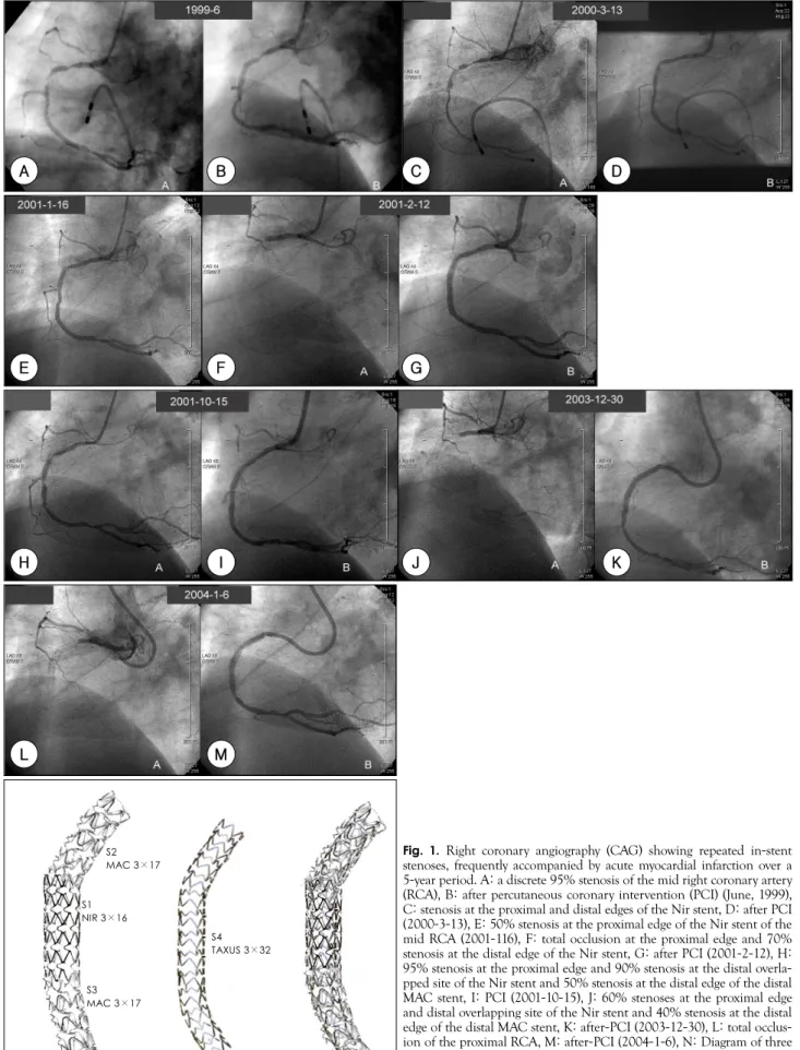
(2) Si-Hoon Park, et al:Drug-Eluting Stent in Recurrent In-Stent Restenosis·481. A. B. C. E. F. G. H. I. J. L. M. K. Fig. 1. Right coronary angiography (CAG) showing repeated in-stent. S2 MAC 3×17. S1 NIR 3×16 S4 TAXUS 3×32. S3 MAC 3×17. N. D. N S1+S2+S3+S4. stenoses, frequently accompanied by acute myocardial infarction over a 5-year period. A: a discrete 95% stenosis of the mid right coronary artery (RCA), B: after percutaneous coronary intervention (PCI) (June, 1999), C: stenosis at the proximal and distal edges of the Nir stent, D: after PCI (2000-3-13), E: 50% stenosis at the proximal edge of the Nir stent of the mid RCA (2001-116), F: total occlusion at the proximal edge and 70% stenosis at the distal edge of the Nir stent, G: after PCI (2001-2-12), H: 95% stenosis at the proximal edge and 90% stenosis at the distal overlapped site of the Nir stent and 50% stenosis at the distal edge of the distal MAC stent, I: PCI (2001-10-15), J: 60% stenoses at the proximal edge and distal overlapping site of the Nir stent and 40% stenosis at the distal edge of the distal MAC stent, K: after-PCI (2003-12-30), L: total occlusion of the proximal RCA, M: after-PCI (2004-1-6), N: Diagram of three bare metal stents and a Taxus stent within the bare metal stents in the RCA. RCA: right coronary artery, PCI: percutaneous coronary intervention, CAG: coronary artery angiography..
(3) 482·Korean Circulation J 2005;35:480-483. with a thrombolytic agent. He was discharged without rities(Fig. 1I). Two years and seven months later, the patient experienced an inferior AMI, which was treated a CAG. Seven months later, the patient complained of a chest pain, and CAG demonstrated total occlusion of the proximal MAC stent with grade II collaterals from the first RV wall branch, 60% stenoses at the proximal edge and the distal overlapping site of the Nir stent, and 40% stenosis at the distal edge of the distal MAC stent(Fig. 1J). Intervention was performed at the site, using a 3.0×32 mm Taxus under 14 atm, within three bare metal stents from the proximal RCA to the distal RCA(Fig. 1K, 1N). Seven days later, an inferior AMI developed. CAG showed total occlusion of the proximal RCA(Fig. 1L). The intravascular ultrasound(IVUS) findings revealed underexpansion of the stents: a stent area of 4.9 mm2 and a minimal luminal diameter(MLD) of 2.3 mm at the proximal MAC stent; a stent area of 3.3 mm2 and MLD of 2.0 mm2 at the proximal edge of the Nir stent, and a stent area of 5.3 mm2 and an MLD of 2.5 mm at the distal edge of the distal MAC stent. Intervention was successfully performed using a non-compliant balloon catheter (3.0×10 mm, 23 atm) at the proximal edge of the Nir stent(Fig. IM); the final minimal stent area was 5.1 mm2. The patient was prescribed low molecular heparin(LMH) for 4 weeks, at home. Antiplatelets, including aspirin, clopidogrel and cilostazol, were also prescribed. The patient declined a follow-up CAG because he remained asymptomatic throughout the 15 month follow-up period. A thallium scan showed normal findings, without perfusion defects. We concluded that his more than 5-year old RCA problem may have been resolved.. Discussion If we could have used DES under IVUS guidance from the onset in 1999, this patient would probably not have experienced a 5-year history of RCA. However, DES was not available in Korea until early 2003. If the bare metal stent(BMS) had initially been more fully available in 1999, and with the guidance of IVUS, could the ISR have been avoided? Possibly yes. In this patient, the bare metal stents appeared to be underexpanded at the proximal edge of the stent at the mid RCA, and this may have contributed to the ISR. The final stent area has been reported to be a predictor of the outcome, bur with inconsistent results.1-11) Therefore, is cutting balloon angioplasty better than conventional balloon angioplasty for ISR treatment? Although cutting balloon angioplasty is associated with some procedural advantages and a lower incidence of balloon slippage, it does not reduce recurrent ISR or major adverse cardiac events compared to conventional. balloon angioplasty.12) This case highlights DES as a promising tool for the treatment of recurrent ISR, and the importance of optimal stent expansion. Whether DES are effective at treating ISR is uncertain.13)14) Recently; however, the In-Stent Restenosis Registry produced favorable data at the 1 year follow-up, namely, one case of restenosis among 25 patients, without stent thrombosis(Brazilian data), and conversely, 2 deaths, 1 late thrombosis, 1 vessel occlusion and 2 in-lesion restenoses among 16 patients with more complex lesions, including a previous brachytherapy(Dutch data).15)16) These results imply that focal, less complex ISR is more responsive to DES than diffuse ISR. Our case showed multiple IRS, but focal lesions. The SECURE study after 6 months also demonstrated good interim results for DES in ISR, i.e., a low target lesion revascularization rate(11.6% in the radiation failure group vs. 5.4% in the non radiation failure group), with infrequent stent thrombosis (presented at TCT 2003). Optimal stent high pressure expansion, under IVUS guidance, warrants special attention for the treatment of ISR, even in the era of DES. Recent data17) has shown that stent underexpansion is associated with failure of the Sirolimus-eluting stent implantation for the treatment of ISR: 9 of 11 recurrences occurred in lesions with a mean stent area of <5.0 mm2, despite the use of a high inflation pressure(18±4 atm). As de-monstrated by our case, even higher pressure may be needed in ISR lesions to achieve an adequate stent area. Then, why did our patient show recurrent AMI? We assume that severe ISR, or residual stenosis, contributed to his developing AMI after the implantation of the BMS or DES. Finally, another key lesson is that adjunctive pharmacotherapy(unfractionated heparin, GPIIbIIIa inhibitors, and extended combination of antiplatelet therapy) should be optimized to prevent stent thrombosis in cases with complex/multiple stents and a high thrombus burden. We utilized low molecular heparin for 4 weeks, even after successful DES dilation. REFERENCES 1) Hoffmann R, Mintz GS, Mehran R, et al. Intravascular ultra-. sound predictors of angiographic restenosis in lesions treated with Palmaz-Schatz stents. J Am Coll Cardiol 1998;31:43-9. 2) Nakamura S, Colombo A, Gaglione A, et al. Intracoronary ultrasound observations during stent implantation. Circulation 1994;89:2026-34. 3) de Feyter PJ, Kay P, Disco C, Serruys PW. Reference chart derived from post-stent-implantation intravascular ultrasound predictors of 6-month expected restenosis on quantitative coronary angiography. Circulation 1999;100:1777-83. 4) Hong MK, Park SW, Mintz GS, et al. Intravascular ultrasonic predictors of angiographic restenosis after long coronary stenting. Am J Cardiol 2000;85:441-5..
(4) Si-Hoon Park, et al:Drug-Eluting Stent in Recurrent In-Stent Restenosis·483. 5) de Jaegere P, Mudra H, Figulla H, et al. Intravascular ultra-. 11) Reiber JH, van der Zwet PM, Koning G, et al. Accuracy and. sound-guided optimized stent deployment: immediate and 6 months clinical and angiographic results from the Multicenter Ultrasound Stenting in Coronaries Study (MUSIC Study). Eur Heart J 1998;19:1214-23. 6) Berry E, Kelly S, Hutton J, et al. Intravascular ultrasoundguided interventions in coronary artery disease: a systematic literature review, with decision-analytic modelling, of outcomes and cost-effectiveness. Health Technol Assess 2000;4:1-117. 7) Fitzgerald PJ, Oshima A, Hayase M, et al. Final results of the Can Routine Ultrasound Influence Stent Expansion (CRUISE) study. Circulation 2000;102:523-30. 8) Mudra H, di Mario C, de Jaegere P, et al. Randomized comparison of coronary stent implantation under ultrasound or angiographic guidance to reduce stent restenosis (OPTICUS Study). Circulation 2001;104:1343-9. 9) Schiele F, Meneveau N, Vuillemenot A, et al. Impact of intravascular ultrasound guidance in stent deployment on 6-month restenosis rate: a multicenter, randomized study comparing two strategies-with and without intravascular ultrasound guidance. J Am Coll Cardiol 1998;32:320-8. 10) Nissen SE, Yock P. Intravascular ultrasound: novel pathophysiological insights and current clinical applications. Circulation 2001;103:604-16.. precision of quantitative digital coronary arteriography: observer-, short-, and medium-term variabilities. Cathet Cardiovasc Diagn 1993;28:187-98. 12) Albiero R, Silber S, di Mario C, et al. Cutting balloon versus conventional balloon angioplasty for the treatment of in-stent restenosis: results of the restenosis cutting balloon evaluation trial (RESCUT). J Am Coll Cardiol 2004;43:943-9. 13) Degertekin M, Regar E, Tanabe K, et al. Sirolimuseluting stent for treatment of complex in-stent restenosis: the first clinical experience. J Am Coll Cardiol 2003;41:184-9. 14) Saia F, Lemos PA, Sianos G, et al. Effectiveness of sirolimuseluting stent implantation for recurrent in-stent restenosis after brachytherapy. Am J Cardiol 2003;92:200-3. 15) Sousa JE, Serruys PW, Costa MA. New frontiers in cardiology: drug-eluting stents: part I. Circulation 2003;107:2274-9. 16) Sousa JE, Costa MA, Abizaid A, et al. Sirolimuseluting stent for the treatment of in-stent restenosis: a quantitative coronary angiography and three-dimensional intravascular ultrasound study. Circulation 2003;107:24-7. 17) Fujii K, Mintz GS, Kobayashi Y, et al. Contribution of stent underexpansion to recurrence after sirolimus-eluting stent implantation for in-stent restenosis. Circulation 2004;109:1085-8..
(5)
수치
관련 문서
The positive change pattern of CK-MB according to time on acute myocardiac infarction patient.. The positive change pattern of troponin-T according to time on
Utility of T1-and T2-weighted high-resolution vessel wall imaging for the diagnosis and follow up of isolated posterior inferior cerebellar artery dissection with
Approved clinical use of bone marrow stem cells for myocardial infarction treatment... Cardiac
(Background) Gallbladder wall thickening(GWT) and gallbladder contraction are often observed in patients with acute hepatitis.. The incidence of acute hepatitis A
The solid-state structure of crystals often have less clearly defined bonds, so a simpler model is used, in which the atoms are represented by touching spheres. In this model the
최대정수함수가 극댓값을 갖는 점과 극솟값을 갖는 점을 모두 구하고, 그 점에서 극값을 구하 시오.. 함수가 미분 가능하면 극값을
그러므로 이러한 표현은 함수의 이름과 미분계수를 구하는 점이 명확하여 혼동할 염려가 없을
최대정수함수가 극댓값을 갖는 점과 극솟값을 갖는 점을 모두 구하고, 그 점에서
