Surgery for Cerebral Stroke 26 : 158•`163, 1998
Original
Recurrent Hemorrhage in Patients with Previous Surgery for Cerebral Aneurysms
Man-Bin YIM, M. D., Ph. D., Woon-Ill BAEK, M. D., Chang-Young LEE, M. D., Jang-Chull LEE, M. D., Ph. D., Eun-Ik SON, M. D., Ph. D., Dong-Won KIM, M. D., Ph. D., and In-Hong KIM, M. D.
Summary: Among 875 patients with intracranial aneurysm operated on during the past 14 years, the authors encountered eleven who had experienced recurrent hemorrhage caused by the rupture of aneurysms which had not been noticed at the time of the initial operation. The age at the time of initial hemorrhage was relatively young (average 43.7 years), and the interval between initial and recurrent hemorrhage varied between 4 and 16 years. Multiple aneurysms occurred in four cases and hypertension in four others.
Clinical grades at the time of the second admission were relatively poor, and in eight patients there were complications with intracerebral hematomas, intraventricular hemorrhages or acute subdural hematoma. Retrospective evaluation of the first angiograms disclosed tiny aneurysms in five cases, and these grew and ruptured at recurrent hemorrhage. In eight patients, the outcome was good; one remained moderately disabled, and two died.
We conclude that the possibility of recurrent hemorrhage after the clipping of a ruptured aneurysm should be considered in all aneurysmal patients, especially in those who are young or have multiple aneurysms or hypertension. Their angiograms should be investigated detailly to determine whether or not suspicious tiny aneurysms are present. In addition, late postoperative follow-up angiography to determine the growth or development of another aneurysm might also be needed.
Key words:
・cerebral aneurysm
・growth
・de novo
・recurrent hemorrhage
・surgery
Surg Cereb Stroke (Jpn)26:158-163,1998
Introduction
The rupture of an intracranial aneurysm is a very dan- gerous event and it can be prevented by the surgical repair.
Once an aneurysm has been eliminated by a clipping com- pletely, the patient is thought to be cured. Late postoper- ative follow-up angiography to detect the development of a new aneurysm is usually not performed. However, sev- eral authors report9)11)14) the formation of new aneurysms (de novo aneurysm) on arteries which were found normal on previous angiogram in patients who received surgery
for ruptured aneurysms. Therefore, it is necessary to make some guidelines for follow-up angiography after surgical management of the ruptured aneurysms, especially for young patients.9)
On the other hand, some controversy exists in the management of tiny suspicious aneurysmal lesions which are discovered incidentally at the time of investigation of subarachnoid hemorrhage from another source. Most neu- rosurgeons hesitate to operate unless an aneurysm is clear- ly verified by angiography, and some authors19)2°) do not recommend surgical treatment for such lesions because
Department of Neurosurgery, School of Medicine, Keimyung University (Received July 28, 1997) [Mailing address: Man-Bin Yim, M. D., Department of Neurosurgery, Dong San Medical Center, Keimyung University, 194 Dong-San Dong, Taegu 700-712, Korea]
the probability of subsequent rupture of these lesions is very low. However, Schievink et al.,15) and Solomon and Corre1116) recommended surgical repair for these lesions by reporting the cases with previously documented asymp- tomatic intact small aneurysms that subsequently ruptured.
We now report our experience with eleven patients who had a recurrent hemorrhage caused by the rupture of an aneurysm which had not been noticed at the time of initial operation for ruptured one. We attempt to find which patients after clipping of a aneurysm should be needed postoperative follow-up angiography to detect the development of another aneurysm. We also discuss about the management of tiny suspicious aneurysmal lesions which are discovered incidently.
Materials and Methods
Among 875 patients who underwent surgery for the rupture of an aneurysm from Sept. 1982 to Sept. 1996, recurrent hemorrhage caused by the rupture of the second aneurysm which had not been noticed at the time of the initial operation were developed in 11 patients. The med- ical records of these 11 patients were reviewed, and re- examined the previous angiograms which were taken at the time of the initial operation to find any evidence of the second aneurysms. Clinical characteristics of these
patients, such as patients ages, previous history of hyper- tension, clinical grades on admission and outcomes, and the intervals between the initial and the recurrent hemor- rhage were investigated.
Results
1. Clinical summary at the time of the initial surgery There were three men and eight women. The ages of patients were relatively young, ranging from 26 to 55 years
(mean age, 43.7 years). The clinical grades6) on admission were grade I in one case, II in seven, IV in one, and unknown owing to the initial operation was done at other hospitals in two.
The sites of aneurysms were posterior communicating artery (Pcom) in four cases, middle cerebral artery (MCA) in three, anterior choroidal artery (Acho) and anterior com- municating artery (Acom) in one of each. There were two cases of multiple aneurysms such as internal carotid artery bifurcation (ICA Bif) and distal anterior cerebral artery (A2) in one, and ophthalmic artery (Oph) and MCA in one.
Retrospective detailed re-examination of the previous angiographic films which were taken at the time of the ini- tial operation could be performed in six patients. Five of them had a tiny microaneurysm approximately 1 mm in size that was not noticed at the time of the initial opera-
Fig. 1 Growing aneurysm of the patient 8. Left vertebral angiogram performed at the time of the first admission shows a suspicious tiny aneurysm at the junction of the vertebral artery (VA) and PICA (arrow). The left MCA aneurysm which ruptured at that time was clipped. Because of recurrent subarachnoid hemor- rhage, a further left vertebral angiogram (B) was obtained five years later, a definite aneurysm is seen at the VA-PICA junction (arrow). Postoperative left ver- tebral angiogram (C) shows complete obliteration of that aneurysm.
tion. It eventually became an apparent aneurysm at the time of the recurrent hemorrhage (Fig. 1 and 2). In one patient, no aneurysmal shadow was noticed on the right posterior communicating artery where the second aneurysm developed (Fig. 3). In the remaining five patients, retrospective detailed re-examination of the pre- vious angiographic films could not be performed due to several reasons (Table 1).
2. Clinical summary at the time of the second surgery The intervals between initial and recurrent hemor- rhage varied between 4 to 16 years (average 7.82 years), and hypertension history was noticed in four patients. The clinical grades on admission were grade II in two cases, III in one, IV in seven and V in one.
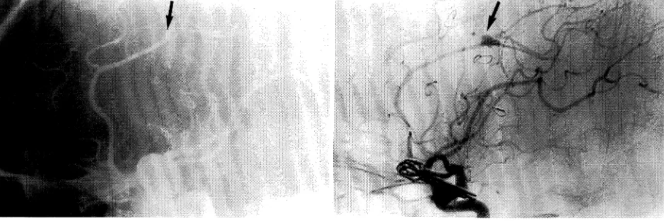
The sites of the second aneurysms were Pcom in four cases, MCA in three, P1-P2 junction of the posterior cere- bral artery (PCA), posterior inferior cerebellar artery Fig. 2 Growing aneurysm of the patient 10. Left carotid angiogram performed at the time of the
first admission shows a large ophthalmic aneurysm and a suspicious tiny aneurysm in the distal anterior cerebral artery (ACA) (arrow) (A). Because of recurrent hemorrhage, a further left carotid angiogram (B) was obtained five years later; a definite aneurysm is seen at the distal ACA (arrow).
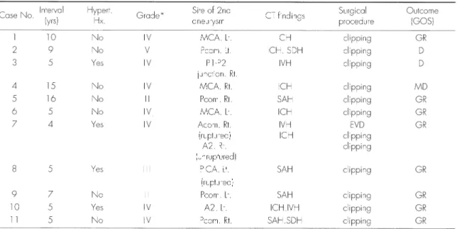
Table 1 Clinical summary at the time of the initial surgery#
#Abbreviations: GOS=Glasgow Outcome Scale8); GR=good recovery; Acom=anterior communicating artery; Al =horizontal portion of the anterior cerebral artery; Pcom=posterior communicating artery; ICA bif=internal carotid artery bifurcation; A2=distal anterior cerebral artery;
Pcom=posterior communicating artery; MCA=middle cerebral artery; Oph=ophthalmic artery; Acho=anterior choroidal artery
*Hunt & Hess grade()) on admission.
**The first operation was performed at other hospital.
***Retrospective re-examination of previous angiographic films was impossible due to loss of those films.
****On the basis of the results of a unilateral carotid angiogram
, an emergency operation was performed. The patient declined postoperative four-vessel angiography.
160 脳 卒 中 の外 科26:1998
(PICA) and distal anterior cerebral artery (A2) in one of each. There was one case of multiple aneurysms at Acorn and A2. The brain computed tomographic scan, taken at second admission, showed a pure subarachnoid hemor- rhage (SAH) in only three cases. In other eight cases, intracerebral hemorrhage (ICH), intraventricular hemor- rhage (NH) or acute subdural hematoma were complicat- ed. In eight patients, the outcome after surgical manage- ment was good; one remained moderately disabled, and two died (Table 2).
Discussion
The recurrent hemorrhage in patient who received previous surgical obliteration of ruptured cerebral aneurysm is rare.1)7)9)-14)17)18)The hemorrhage may be originated from the residual neck of the previous clipped aneurysmal sac,1)7)10) growing aneurysm from the previous angiographically occult microaneurysm at the time of the first angiography18) and de novo aneurysm which devel- oped after surgery9)11)-14)18). Many authors reported devel-
opment of de novo aneurysms in patients who had been received surgery for ruptured aneurysms9)11)-14)18) and in patients who had been done therapeutic carotid ligation
for the treatment of cerebral aneurysms.2)3) Drapkin and Rose2) reported a case of de novo aneurysm following com- mon carotid ligation for the management of the right ICA bifurcation aneurysm. Fujiwara et al.,3) also described two cases of de novo aneurysm formation following therapeu- tic carotid occlusion for intracranial ICA aneurysms.
They2)3) suggest that the hemodynamic changes caused by carotid ligation might be the underlying basis for the for- mation of de novo aneurysms.
Miller et al.,12) on the other hand, reported seven cases of de novo aneurysm among 620 patients who received clipping of initial intracranial aneurysm. The interval from the clipping of initial aneurysms and rupture of de novo aneurysms were three to twenty years. Marchel et al.,11) also reported five cases of formation of new aneurysms among 964 patients treated for intracranial aneurysms.
Among these five cases, three cases had multiple Fig. 3 Suspect de novo aneurysm of the patient 1 1 . Right carotid angiograms (A and B) taken
at the time of the first admission shows no aneurysm at the junction of the internal carotid (ICA) and posterior communicating artery (Pcom) (arrows). The left anterior choroidal artery aneurysm, which ruptured at that time, was clipped. Because of recurrent hemorrhage, fur- ther right carotid angiogram (C and D) were obtained five year later. They show a new aneurysm at the ICA-Pcom junction (arrows).
aneurysms and, in 4 cases, the aneurysms were formed within 3 to 6 years after treatment of aneurysms. They stressed that the possibility of formation of new aneurysms after the cure of a demonstrable one should be considered in all aneurysmal patients, especially in multiple cases.
Rinne and Hernesniemi13) estimated that the incidence of de novo aneurysm formation and rupture was 63 per 100,000 per year in patients known to have a subarachnoid hemorrhage. They recommended long-term neuroradiolog- ical follow-up in young patients with surgical treatment of
cerebral aneurysm. Sakaki et al.,14) also reported nine patients who had de novo cerebral aneurysms which rup- tured at intervals of 4 to 7.5 years after clipping of an ini- tial aneurysm among 986 patients with cerebral aneurysms.
They regard the hypertension as an important predispos- ing factor for the development of de novo aneurysm and recommended meticulous control of blood pressure for the prevention of development of new aneurysm.
Although previously mentioned authors11)12)14) regard- ed the all aneurysms which were found at the site of arter- ies normal on previous angiograms at the time of initial surgery as de novo aneurysm, some authors17)18) thought that some of these aneurysms might not be true de novo aneurysms. Wakai18) described that he could find a tiny blister approximately 1 mm in size that was not noticed at the time of the initial operation by the retrospective re- examination of the first angiograms in the second
aneurysm that developed several years after the initial aneurysm operation. He described that the incidence of true de novo aneurysm in his series would be 0.2%.
In this study, we had 11 patients who had recurrent hemorrhage caused by the rupture of the second aneurysm among 875 patients with intracranial aneurysm operated
on. Its incidence was 1.26%. Among these 11 patients, we could find a tiny microaneurysm in 5 of 6 patients who could be re-examined the previous angiographic films ret- rospectively. We suspect de novo aneurysm was only one in our cases. Therefore, we agreed with the opinion of the Wakai18) that the all of the de novo aneurysms which were reported in the literature previously might not true de novo aneurysm. Some of them might be grown aneurysm from the microaneurysm which were not detected definitely in the previous angiogram.
There exist some controversy in the management of a suspicious microaneurysm which is discovered incidently at the time of investigation of subarachnoid hemorrhage from another source. Most neurosurgeons agreed that all suspicious lesions that can be reached using the same approach for treatment of the ruptured aneurysm should be confirmed and managed surgically if those are aneurysms.5) However, in the lesions which were required the second operation to confirm and manage that lesion, controversy exists. Most neurosurgeons hesitate to do operation unless an aneurysm is clearly verified by angio-
#Abbreviations: Hyped. Hx.=hypertension history; CT=computed tomography; GOS=Glasgow Outcome Scale; GR=good recovery; MD=moderate disability; D=death; MCA=middle cerebral artery; Pcom=posterior communicating artery; Acom=anterior communicating artery; A2=distal anterior cerebral artery; PICA=posterior inferior cerebellar artery; ICH=intracerebral hematoma;
SDH=subdural hematoma; IVH=intraventricular hemorrhage; SAH=subarachnoid hemorrhage
*Hunt & Hess grade6 on admission
Table 2 Clinical summary at the time of the second operation#
162 脳 卒 中 の外 科26:1998
graphy. Furthermore, some authors do not recommend surgical treatment for such lesions because the probabili- ty of subsequent rupture of these lesions are very low.19)20) Wiebers et al.,20) reported none of the 102 unruptured aneurysms smaller than 10 mm in diameter ruptured dur- ing a mean follow-up period of 8.3 years. The authors rec- ommended that intact aneurysms smaller than 10 mm should not be surgical intervention.
On the other hand, Schievink et al.,15) reported three patients with previously documented asymptomatic intact saccular intracranial aneurysms smaller than 5 mm in diameter that subsequently ruptured. They recommended surgical repair for those lesions because they are not innocuous. Solomon and Correll16) also reported a case which had a small asymptomatic aneurysm that was left untreated and subsequently ruptured.
We also recommend prophylactic surgical repair for those lesions because, in our series, five tiny microa- neurysms that were left untreated grew and ruptured sub- sequently. In terms of follow-up angiogram in patients who received previous surgery for ruptured aneurysms, several authors4)9)11)-15) recommended postoperative follow-up angiogram in relatively young patients,4)9)13) multiple aneurysm patient,9)11) and patients with hypertension,14) oral contraceptive use,14) and hemodynamic effects.") The interval at which follow-up angiography should be per- formed is not clearly defined. Rinne and Hernesniemi13) and Koeleveld et al.,9) recommended every 5 years after first SAH. They9)13) presumed that magnetic resonance angiography may make a role in this fields in the future.
In our series, the ages of our patients at the time of initial hemorrhage were also relatively young and hyper- tension history was noticed in four patients. The interval from initial hemorrhage to recurrent hemorrhage was 4 to
16 years. Therefore, we also suggest that patients who have the aneurysm surgery at relatively young age and patients with hypertension might be benefited from follow-up angiographic study 4 or more years after their first SAH.
Conclusion
The authors conclude that the possibility of recurrent hemorrhage after the clipping of a ruptured aneurysm should be considered in all aneurysmal patients, especial- ly in those who are young or have multiple aneurysms or hypertension. Their angiograms should be investigated detailly to determine whether or not suspicious tiny aneurysms are present. In addition, late postoperative fol- low-up angiography to determine growth or development of another aneurysm might be also needed.
References
1) Drake CG, Friedman MI, Peerless SJ: Failed aneurysm surgery. Reoperation in 115 cases. J Neurosurg 61: 848-856, 1984
2) Drapkin AJ, Rose WS: Serial development of 'de novo' aneurysms after carotid ligation: case report. Surg Neurol 38: 302-308, 1992
3) Fujiwara S, Fujii K, Fukui M: De novo aneurysm formation and aneurysm growth following therapeutic carotid occlu- sion for intracranial internal carotid artery (ICA) aneurysms.
Acta Neurochir (Wien) 120: 20-25, 1993
4) Heiskanen O, Marttila I: Risk of rupture of a second aneurysm in patients with multiple aneurysms. J Neurosurg 32: 295-299, 1970
5) Heiskanen O: Risk of bleeding from unruptured aneurysms in cases with multiple intracranial aneurysms. J Neurosurg 55: 524-526, 1981
6) Hunt WE, Hess RM: Surgical risk as related to time of inter- vention in the repair of intracranial aneurysms. J Neurosurg 28: 14-20, 1968
7) Jafar JJ, Weiner HL: Surgery for angiographically occult cere- bral aneurysms. J Neurosurg 79: 674-679, 1993
8) Jennett B, Bond M: Assessment of outcome after severe brain damage. A practical scale. The Lancet 1: 480-484, 1975 9) Koeleveleld RF, Heilman CB, Klucznik RP, et al: De novo development of an aneurysm: case report. Neurosurgery 29:
756-759, 1991
10) Lin T, Fox AJ, Drake CG: Regrowth of aneurysm sacs from residual neck following aneurysm clipping. J Neurosurg 70:
556-560, 1989
11) Marchel A, Bidzinski J, Bojarski P: Formation of new aneurysms. Report of five cases. Acta Neurochir (Wien) 112:
96-99, 1991
12) Miller CA, Hill SA, Hunt WE: "De novo" aneurysms. A clini- cal review. Surg Neurol 24: 173-180, 1985
13) Rinne JK, Hernesniemi JA: De novo aneurysms: special mul- tiple intracranial aneurysms. Neurosurgery 33: 981-985, 1993 14) Sakaki T, Tominaga M, Miyamoto K, et al: Clinical studies
of de novo aneurysms. Neurosurgery 32: 512-517, 1993 15) Schievink WI, Piepgras DG, Wirth FP: Rupture of previous-
ly documented small asymptomatic saccular intracranial aneurysms. Report of three cases. J Neurosurg 76: 1019- 1024, 1992
16) Solomon RA, Correll JW: Rupture of a previously docu- mented asymptomatic aneurysm enhances the argument for prophylactic surgical intervention. Surg Neurol 30: 321-331, 1988
17) Solomon RA: Clinical studies of de novo aneurysms.
Neurosurgery 32: 517, 1993 (comment)
18) Wakai S: Clinical studies of de novo aneurysms.
Neurosurgery 34: 1102, 1994 (correspondence)
19) Wiebers DO, Whisnant JP, O'Fallon WM: The natural histo- ry of unruptured intracranial aneurysms. N Engl J Med 304:
696-698, 1981
20) Wiebers DO, Whisnant JP, Sundt TM, et al: The significance of unruptured intracranial saccular aneurysms. J Neurosurg 66: 23-29, 1987