© 2016 The Korean Ophthalmological Society
This is an Open Access article distributed under the terms of the Creative Commons Attribution Non-Commercial License (http://creativecommons.org/licenses /by-nc/3.0/) which permits unrestricted non-commercial use, distribution, and reproduction in any medium, provided the original work is properly cited.
Original Article
Obstructive Sleep Apnea in Patients with Branch Retinal Vein Occlusion: A Preliminary Study
Hee Jung Kwon
1, Eui Chun Kang
2, Junwon Lee
2, Jinu Han
2, Won Kyung Song
11
Department of Ophthalmology, CHA Bundang Medical Center, CHA University, Seongnam, Korea
2
Institute of Vision Research, Department of Ophthalmology, Yonsei University College of Medicine, Seoul, Korea
Purpose: Our study aimed to determine whether obstructive sleep apnea (OSA) is common among branch retinal vein occlusion (BRVO) patients without systemic risk factors using a Watch PAT-100 portable monitoring device.
Methods: The study participants included consecutive patients with BRVO of less than 3 months duration with- out any risk factors known to be associated with OSA (diabetes, coronary artery disease, stroke, hematologic diseases, autoimmune disease, etc.) except for hypertension. All patients underwent full-night unattended polysomnography by means of a portable monitor Watch PAT-100 device. The apnea-hypopnea index (AHI) was calculated as the average number of apnea and hypopnea events per hour of sleep, and an AHI score of five or more events was diagnosed as OSA.
Results: Among 19 patients (6 males and 13 females), 42.1% (8 of 19) had an AHI reflective of OSA. In the 13 patients who had no concurrent illness, including hypertension, 30.8% (4 of 13) had positive test results for OSA; three of these patients were ranked as mild OSA, while one had moderate OSA. The OSA group had an average AHI of 12.3 ± 7.8, and the average AHI was 2.0 ± 0.9 in the non-OSA group. Although it was not sta- tistically proven, we found that OSA patients experienced a more severe form of BRVO.
Conclusions: We found a higher than expected rate of OSA in BRVO patients lacking concomitant diseases typically associated with OSA. Our findings suggest that OSA could be an additional risk factor in the patho- genesis of BRVO or at least a frequently associated condition that could function as a triggering factor.
Key Words: Branch retinal vein occlusion, Hypertension, Obstructive sleep apnea
Retinal vein occlusion (RVO) is the second most com- mon retinal vascular disease after diabetic retinopathy and is an important cause of blindness and visual morbidity [1].
This condition affects males and females equally and oc- curs most frequently between the ages of 60 and 70. The
prevalence of RVO is 0.7%, and the 5-year incidence is 0.8% [2]. The most common risk factors associated with branch retinal vein occlusion (BRVO) are systemic hyper- tension, diabetes, hyperlipidemia, smoking, and age-relat- ed atherosclerosis [3]. The interruption of venous flow in the eyes of BRVO patients afflicted with the aforemen- tioned risk factors almost always occurs at a retinal arte- riovenous intersection, where a retinal artery crosses a ret- inal vein [4].
Obstructive sleep apnea (OSA) is the most common sleep-related breathing disorder, with an estimated preva-
Received: February 27, 2015 Accepted: May 21, 2015
Corresponding Author: Won Kyung Song, MD, PhD. Department of
Ophthalmology, CHA Bundang Medical Center, CHA University, #59
Yatap-ro, Bundang-gu, Seongnam 13496, Korea. Tel: 82-31-780-5330,
Fax: 82-31-780-5333, E-mail: [email protected]
lence of 2% for women and 4% for men in the middle-aged population [5]. It is characterized by the repetitive com- plete or partial collapse of the upper airway during sleep [6], which causes the cessation (obstructive apnea) or sig- nificant reduction (obstructive hypopnea) of airflow. These respiratory events result in intermittent hypoxemia and hypercapnia, cortical arousals and surges of sympathetic activity [7]. OSA is an insidious disorder, and most patients are unaware of its symptoms; it is becoming increasingly recognized as an important cause of medical morbidity and mortality. The occurrence of OSA has been linked to increased cardiovascular morbidity and mortality, hyper- tension, diabetes mellitus, atherosclerosis, and stroke, most of which are also common risk factors for BRVO [7-10].
Previous studies have shown an association between RVO and OSA [11,12]. However, these studies included both branch and central RVO patients, and there were not any exclusion criteria. In the present study, we aimed to determine whether OSA is common among patients with BRVO who do not have any systemic risk factors.
Materials and Methods
This study was carried out in the ophthalmology clinic of the hospital of the Yonsei University College of Medi- cine in Seoul, Korea. The institutional review board ap- proved the ethical and methodological aspects of the inves- tigation, and patients were required to provide signed written informed consent as a prerequisite for their partici- pation. This study is also registered at ClinicalTrials.gov, number NCT01291862. Patients with BRVO of less than 3 months duration were consecutively enrolled between April 2010 and March 2011. BRVO was assessed with an indirect ophthalmoscope by a retinal specialist and con- firmed by fluorescein angiography (FA). Patients with in- sulin-dependent diabetes, insulin-independent diabetes, coronary artery disease, stroke, hematologic diseases, au- toimmune diseases, or any other condition reported to be associated with OSA were excluded, with the exception of patients with well-controlled hypertension. Further, any ocular disease other than retinal vascular occlusion and conditions compromising the capability of patients to un- derstand or participate in the study were considered exclu- sionary.
All patients underwent full-night unattended level III
polysomnography by means of a portable monitor Watch PAT-100 device (WP100; Itamar Medical, Caesarea, Israel).
The WP100 is a Food and Drug Administration-approved portable diagnostic device for sleep apnea and is easy to use because the testing is done in the patient’s own bed- room. This device is typically worn on the patient’s wrist, and there are two non-invasive, finger-mounted probes: a peripheral arterial tone (PAT) probe and a pulse oximeter (http://www.itamar-medical.com/). This device continu- ously records on four channels: the PAT signal via a fin- ger-mounted PAT probe, arterial oxygen saturation via fin- ger-mounted pulse oximeter, sleep-wake state from the build-in actigraphic signal, and heart rate (derived from the PAT signal) [13]. The PAT signal measures the arterial pulsatile volume changes in the finger, which are regulated by ɑ-adrenergic innervation of the smooth muscles of its vasculature, and thus reflects sympathetic nervous system activity [13]. Because discrete obstructive airway events cause arousal from sleep and sympathetic activation, these events are associated with attenuation of the PAT signal [14]. On the following day, patients returned the WP100 device, and the overnight polysomnographic data were up- loaded and automatically analyzed using commercial soft- ware (zzzPAT ver. 44, Itamar Medical). Despite the gold standard for an OSA diagnosis being the in-laboratory ap- proach using level I polysomnography, the use of the WP100 has been validated to have reasonable reliability and specificity [13-15].
In addition to analyzing the PAT, heart rate and pulse oximetry data, the WP100 calculates three indices accord- ing to the number of events per hour of sleep: the ap- nea-hypopnea index (AHI), respiratory disturbance index (RDI) and oxygen desaturation index (ODI). The AHI is calculated as the average number of apnea and hypopnea events per hour of sleep; the RDI is determined based on the total number of apneas, hypopneas and respiratory ef- fort-related arousals per hour of sleep, while the ODI is calculated as the number of oxygen desaturations of at least 4% from baseline per hour of sleep. An AHI of five or more events confirmed a positive OSA diagnosis. Cut- off points of 5, 15 and 30 were used to indicate mild, mod- erate and severe levels of OSA, respectively [16]. An RDI of five or more events was considered abnormally elevated.
Records were accepted when patients had spent at least 6
hours in bed and when good to excellent recordings of ar-
terial oxygen saturation and respiration were achieved.
We also evaluated the participants’ ophthalmic results, including the initial best-corrected visual acuity and the presence of a non-perfusion area larger than five disc di- ameters on FA performed at least 6 months after the diag- nosis of the BRVO. Moreover, we assessed patients’ snor- ing behavior and incidence of daytime sleepiness or fatigue using the Berlin questionnaire.
Statistical analyses were performed using the SPSS ver.
17.0 (SPSS Inc., Chicago, IL, USA). We compared patients both with and without OSA using the Fisher’s exact test for categorical data and the Mann-Whitney U-test for numeri- cal data.
Results
This study included a total of 19 patients (6 males and 13 females). Among these, 13 had no concurrent illness, in- cluding hypertension, while six participants had well-con- trolled hypertension. None of these patients had taken medication except for hypertension medication. The mean age of patients was 59.6 ± 9.4 years (range, 46 to 78 years).
None of the patients were obese, and they all had a body mass index (BMI) of <30. Ten patients disclosed on the questionnaire that they snored during sleep, and four pa- tients complained of excessive daytime sleepiness. Overall, 42.1% of our patients (8 of 19) were found to have AHI values reflective of OSA, and 57.9% (11 of 19) were diag- nosed with an abnormally elevated RDI (Table 1). Among the 13 patients who had no concurrent illness (including hypertension), the average AHI was 4.4 ± 5.9, with 30.8%
of the patients (4 of 13) testing positive for OSA. Three of these patients were ranked as mild OSA, and one had mod- erate OSA. For the patients with hypertension, the average AHI was found to be 10.4 ± 8.4, with 66.7% of the patients (4 of 6) testing positive for OSA: three with mild OSA and one with moderate OSA.
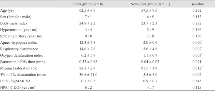
Table 2 shows the results of a comparison within the groups both with and without OSA. There were no differ- ences in age, sex, BMI, hypertension history, smoking his- tory, and time (minutes) spent at less than 90% oxygen sat- uration in the OSA and non-OSA groups. The OSA group had an average AHI of 12.3 ± 7.8, of which seven patients were ranked as mild OSA and two as moderate OSA. In the non-OSA group, the average AHI was found to be 2.0
± 0.9. For all patients, AHI showed a positive correlation
with RDI (γ = 0.587, p = 0.000), ODI (γ = 0.700, p = 0.000), minimal oxygen saturation (γ = -0.488, p = 0.005), and 4%
to 9% desaturation times (γ = 0.752, p = 0.000). However, AHI had no significant correlation with patient age, weight, height, BMI, and time (minutes) spent at less than 90% oxygen saturation.
We also analyzed the differences in ophthalmic exam- inations within the OSA group and the non-OSA group.
The initial visual acuity was not statistically different be- tween the two groups. Although it was not statistically dif- ferent, the OSA group contained relatively more patients with non-perfusion areas larger than five disc diameters on FA compared to the non-OSA group (75.0% vs. 36.4%).
Discussion
Several mechanisms by which OSA evokes RVO have been proposed. First, the change in retinal microcirculation caused by sleep apnea is related to the occurrence of RVO [11]. Respiratory events caused by OSA result in hypoxia, hypercapnia and activation of the sympathetic nervous system. Hypoxia-induced vasodilation of the central retinal artery can compress the adjacent central retinal vein and interfere with retinal blood flow. Hypercapnia-induced cerebral vasodilation can increase the intracranial and ce- rebral spinal fluid pressure, inducing papilledema and ele- vated venous pressure in the optic nerve head. This vaso- dilation also has the effect of reducing the rate of retinal circulation. Moreover, activated sympathetic tone can also stimulate increases in arterial blood pressure. These three factors all impact hemodynamic changes in the retinal ar- tery, which contribute to the occlusion of the retinal vein located in the same adventitial sheath and may help ex- plain why RVO increases the incidence of OSA [11].
Second, the thrombogenicity of sleep apnea patients is
another probable mechanism for the promotion of RVO in
OSA [12]. Intermittent hypoxia produces reactive oxygen
species and inflammatory cytokines (interleukin-6 and tu-
mor necrosis factor-ɑ), which increase the expression and
release of tissue factors of vascular endothelial cells and
trigger the extrinsic coagulation pathway and platelet ag-
gregation. Reactive oxygen species and inflammatory cy-
tokines also impair the repair capacity of endothelial cells
and suppress fibrinolysis. All these factors may contribute
to the hypercoagulable state frequently present in OSA
Table 1. Demographic characteristics and polysomnographic results of enrolled patients Sex / age BMI HTN Smoking history AHI OSA
severity RDI ODI <90%
saturation time (min)
Minimal saturation
(%)
4% to 9%
desaturation times
Initial VA (logMAR)
>5 DD NPA on FA
F / 46 26.4 Abs Abs 2.08 - 3.13 2.43 0.1 89 7 0.50 Pre
M / 76 22.7 Abs Abs 1.61 - 2.76 0.92 0.1 89 4 0.70 Pre
F / 65 21.2 Abs Abs 2.62 - 2.76 1.38 0 93 10 1.00 Abs
F / 53 22.7 Abs Abs 1.97 - 4.22 0.28 0 93 2 0.10 Abs
F / 51 23.5 Abs Abs 1.55 - 3.1 0.69 0 92 4 0.30 Abs
M / 50 26.8 Abs Pre 2.57 - 18.17 2.06 0.2 89 12 0.40 Abs
M / 50 25.3 Abs Pre 0.97 - 3.5 0.12 0 92 1 2.00 Pre
M / 54 21.8 Abs Abs 0.2 - 4.9 0 0 94 0 0.20 Abs
F / 68 25.9 Abs Abs 2.4 - 3.4 0 0 93 0 2.00 Pre
F / 68 23.7 Abs Abs 5.71 Mild 7.7 3.42 0.1 89 12 1.50 Pre
F / 64 24.3 Abs Abs 5.63 Mild 8.09 2.11 0 91 6 0.30 Pre
F / 66 21.0 Abs Abs 7.7 Mild 11.6 3 0 91 11 1.00 Abs
F / 60 22.9 Abs Abs 22.82 Moderate 23.04 9.13 0.8 85 40 1.00 Pre
F / 53 25.0 Pre Abs 2.86 - 9.09 1.56 0 92 6 1.70 Abs
M / 66 19.9 Pre Pre 3.33 - 5.36 2.17 0 90 15 1.00 Abs
F / 60 27.3 Pre Abs 6.25 Mild 7.4 2.68 0.4 89 21 0.20 Abs
F / 56 26.6 Pre Abs 10.45 Mild 11.43 6.2 1 83 34 0.20 Pre
M / 48 27.0 Pre Abs 14.8 Mild 21.5 6.52 0 90 37 0.40 Pre
F / 78 25.6 Pre Abs 25 Moderate 25.7 19.2 1.9 87 133 0.70 Pre
BMI = body mass index; HTN = hypertension; AHI = apnea-hypopnea index; OSA = obstructive sleep apnea; RDI = respiratory distur- bance index; ODI = oxygen desaturation index; VA = visual acuity; logMAR = logarithm of the minimum angle of resolution; NPA = non-perfusion area; DD = disc diameter; FA = fluorescein angiography; Abs = absence; Pre = presence.
Table 2. Demographic data for patients with OSA and for those without OSA
OSA group (n = 8) Non-OSA group (n = 11) p-value
Age (yr) 62.5 ± 8.9 57.5 ± 9.6 0.272
Sex (female : male) 7 : 1 6 : 5 0.153
Body mass index 24.8 ± 2.2 23.7 ± 2.3 0.272
Hypertension (yes : no) 4 : 4 2 : 9 0.166
Smoking history (yes : no) 0 : 8 3 : 8 0.170
Apnea-hypopnea index 12.3 ± 7.8 2.0 ± 0.9 0.000
*Respiratory disturbance 14.6 ± 7.6 5.6 ± 4.8 0.002
*Oxygen desaturation index 6.2 ± 5.9 1.1 ± 0.9 0.003
*Saturation <90% time (min) 0.53 ± 0.68 0.04 ± 0.07 0.091
Minimal saturation (%) 88.1 ± 2.9 91.5 ± 1.9 0.012
*4% to 9% desaturation times 36.8 ± 41.0 5.5 ± 5.0 0.002
*Initial logMAR VA 0.7 ± 0.5 0.9 ± 0.7 0.545
NPA >5 DD (yes : no) 6 : 2 4 : 7 0.115
OSA = obstructive sleep apnea; logMAR = logarithm of the minimum angle of resolution; VA = visual acuity; NPA = non-perfusion area; DD = disc diameter.
*