Korean J Gastroenterol Vol. 73 No. 3, 182-185 https://doi.org/10.4166/kjg.2019.73.3.182 pISSN 1598-9992 eISSN 2233-6869
CASE REPORT
Korean J Gastroenterol, Vol. 73 No. 3, March 2019 www.kjg.or.kr
췌장으로 전이한 자궁경부선암 1예
김도준, 박진명, 김지현, 남길우, 강창돈, 이성준, 이경열1, 전용환2
강원대학교 의학전문대학원 강원대학교병원 내과, 병리과1, 영상의학과2
Pancreatic Metastasis from Adenocarcinoma of the Uterine Cervix
Do Jun Kim, Jin Myung Park, Ji Hyun Kim, Kilwoo Nam, Chang Don Kang, Sung Joon Lee, Kyoungyul Lee1 and Yong Hwan Jeon2 Departments of Internal Medicine, Anatomic Pathology1 and Radiology2, Kangwon National University Hospital, Kangwon National University School of Medicine, Chuncheon, Korea
Pancreatic metastasis from cervical cancer is extremely rare. We report a case of metastatic adenocarcinoma of the pancreas from uterine cervical cancer. A 70-year-old woman was referred because of a pancreatic mass detected by CT. She had been diagnosed with uterine cervical adenocarcinoma 20 months previously. After concurrent chemoradiotherapy, CT showed no evidence of the cer- vical mass, and follow-up showed no evidence of recurrence. Endoscopic ultrasound-guided fine needle aspiration biopsy of the pan- creatic mass resulted in a diagnosis of metastatic adenocarcinoma from uterine cervix. (Korean J Gastroenterol 2019;73:182-185) Key Words: Pancreas; Endoscopic ultrasound-guided fine needle aspiration; Adenocarcinoma; Uterine cervical neoplasms
Received June 2, 2018. Revised July 16, 2018. Accepted August 9, 2018.
CC This is an open access article distributed under the terms of the Creative Commons Attribution Non-Commercial License (http://creativecommons.org/licenses/
by-nc/4.0) which permits unrestricted non-commercial use, distribution, and reproduction in any medium, provided the original work is properly cited.
Copyright © 2019. Korean Society of Gastroenterology.
교신저자: 박진명, 24289, 춘천시 백령로 156, 강원대학교 의학전문대학원 강원대학교병원 내과
Correspondence to: Jin Myung Park, Department of Internal Medicine, Kangwon National University Hospital, Kangwon National University School of Medicine, 156 Baengnyeong-ro, Chuncheon 24289, Korea. Tel: +82-33-258-9235, Fax: +82-33-258-2404, E-mail: [email protected], ORCID: https://orcid.org/0000-0002-8798-0587 Financial support: None. Conflict of interest: None.
INTRODUCTION
Pancreatic metastasis from carcinoma of the uterine cervix is rare, and the condition has only been reported on a small number of occasions.1-5 Here, we report a case of pancreatic metastasis from adenocarcinoma of the cervix confirmed by endoscopic ultrasound (EUS)-guided fine needle aspiration biopsy.
CASE REPORT
A 70-year-old woman with a prior diagnosis of uterine cer- vical adenocarcinoma 20 months previously was referred be- cause of a pancreatic mass. MRI revealed a 3.4 cm sized cervical mass that had invaded posterior fornix and both para-
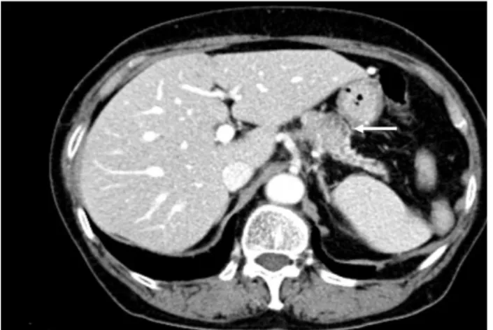
metria with suspected metastasis to bilateral internal iliac lymph nodes (stage IIB). The patient received six cycles of concurrent chemoradiotherapy (cisplatin, 54.0 Gy) and intra- cavitary radiotherapy (24.0 Gy) over nine weeks, and 4 months after completing treatment, follow-up CT showed no cervical mass or enlarged lymph nodes in the pelvic cavity. Eighteen months after treatment completion, abdominal CT revealed a 3.5 cm sized mass at the pancreatic body (Fig. 1), but no mass in the cervix, and as a result she was referred for further evaluation.
At this time the patient complained of low back pain of three weeks duration. Abdominal pain and weight loss were not obvious. Vital signs were stable, and physical examination revealed no specific abnormality. Laboratory test results were as follows: white blood count 6,300/mm3 (neutrophils 77%),
Kim DJ, et al. Pancreatic Metastasis from Adenocarcinoma of the Uterine Cervix 183
Vol. 73 No. 3, March 2019 Fig. 1. Abdominal computed tomography image showing a
heterogeneously enhanced mass in the pancreatic body (white arrow).
Fig. 2. Magnetic resonance T2-weighted image showing a 3.5 cm mass in the pancreatic body (white dashed arrow).
Fig. 3. FDG PET/CT revealed abnormal FDG uptake in the pancreatic body (SUV max 5.9). FDG PET/CT, fluorodeoxyglucose positron emission tomography/computed tomography; SUV, standardized uptake value.
Fig. 4. Endoscopic ultrasound showed a hypoechoic mass in the pancreatic body; fine needle aspiration bioppsy was performed.
Fig. 5. Pathology of the pancreatic tumor showing atypical neoplastic glands with cellular crowding, stratification, and hyperchromatic nuclei (H&E, ×200).
Fig. 6. Tumor cells were positive for p16 by immunohistochemical staining (original magnification, ×200).
hemoglobin 12.5 g/dL, platelets 236,000/mm3, BUN 15.1 mg/dL, creatinine 1.0 mg/dL, sodium 144 mmol/L, potassium 4.7 mmol/L, chloride 108 mmol/L, total protein 7.2 g/dL, albu- min 4.7 g/dL, AST 29 U/L, alanine transaminase 26 U/L, ALP 97 U/L, total bilirubin 0.5 mg/dL, amylase 53 U/L, lipase
184 김도준 등. 췌장으로 전이한 자궁경부선암 1예
The Korean Journal of Gastroenterology
43 U/L, and CA 19-9 19.2 U/mL (normal, <37.0 U/mL).
Pancreatic MRI revealed a 3.5 cm mass in the body and irregular dilation of the upstream main pancreatic duct in an atrophic pancreas tail (Fig. 2). Whole body PET/CT showed intense 18-fluorodeoxyglucose uptake in the pancreatic mass (Fig. 3) and mild uptake in multiple tiny lung nodules. EUS showed an ill-defined hypoechoic mass in the pancreatic body. Fine needle aspiration biopsy was performed using 22-gauge needle (Procore; Cook Medical, Bloomington, IN, USA) (Fig. 4), and results revealed adeno- carcinoma with atypical neoplastic glands, cellular crowding, stratification, and hyperchromatic nuclei (Fig. 5). Although considered pancreatic ad- enocarcinoma, immunohistochemical staining was performed given the history of endocervical type adenocarcinoma. The tumor was found to be positive for cytokeratin 7, p16 (Fig.
6), and caudal-related homeobox transcription factor 2, which matched results for cervical cancer tissue collected by cervical biopsy 20 months previously. Human papillomavirus (HPV) gen- otyping (GeneFinder TM HPV Liquid Bead MicroArray Genotype kit; Infopia Co., Ltd., Anyang, Korea) revealed the presence of the HPV type 18 genome, which was also detected in cervical cancer tissue. The final diagnosis was pancreatic metastasis from cervical adenocarcinoma. The patient was discharged from hospital to receive treatment at another institution.
DISCUSSION
Metastatic cancer of the pancreas is rare with a reported frequency of 2-5% among all malignant pancreatic tumors.4 Renal cell carcinoma is the most common solid tumor that metastasizes to the pancreas,6 whereas pancreatic meta- stasis from cervical cancer is extremely rare.1-5
The histopathology of cervical cancer is usually squamous cell carcinoma, but the prevalence of adenocarcinoma is increasing. Uterine cervical adenocarcinoma comprises nearly 20-25% of all cervical malignancies in developed countries, and more aggressive biological behavior has been reported in patients with intermediate and high-risk factors after surgery. Furthermore, in patients with advanced stage disease (over III), radiotherapy alone and concurrent cisplatin-based chemo-radiotherapy have been reported to be ineffective.7
In a case report of pancreatic metastasis from cervical adenocarcinoma in a 57-year-old woman with a history of stage IIIA adenocarcinoma of cervix,8 PET/CT revealed focal
pancreatic uptake, and EUS-guided fine needle aspiration of the pancreatic lesion revealed adenocarcinoma matching the histopathologic findings of the lesion at the primary site.
To the best of our knowledge, the described case is only the second reported case of pancreatic metastasis from cervical adenocarcinoma.
Pancreatic metastasis has been diagnosed based on the findings of imaging modalities such as ultrasonography, EUS, CT, MRI, or PET, and the recent increased usage of EUS-guided fine needle aspiration has made it possible to achieve histo- pathological diagnoses. Differentiating primary and metastatic pancreatic cancer is not straightforward. In CT images, primary pancreatic cancer and metastatic cancer have similar en- hancement patterns, except for metastatic cancer from renal cell carcinoma. In a recent study, it was observed that pancre- atic metastasis showed persistent low attenuation in 75% of cases in non-renal cell carcinoma by multidetector-row CT.9 In the described case, we concluded pancreatic metastasis originated from cervical cancer because the patient had a history of cervical adenocarcinoma, which had been diagnosed 20 months previously and because the HPV type 18 genome was detected in pancreatic and cervical specimens.
Furthermore, immunostaining results for pancreatic and cer- vical tumor specimens for CK7, p16 and CDX2 were identical.
Primary and metastatic pancreatic cancer are known to ex- press CK7 and CDX2, although in previous studies only a mi- nority of cases exhibited CDX2 positivity.10,11 Therefore, im- munohistochemical results for CK7 and CDX2 are not sufficient to differentiate these tumors. Diffuse positive immunostaining for p16 is a good surrogate marker of high-risk HPV infection in uterine cervical cancer.12 However, it has been reported diffuse positive staining for p16 is also possible in pancreatic cancer,13 and thus, p16 positivity does not in itself indicate metastatic cervical cancer. Accordingly, we performed HPV genotyping. Type 18 is the second-most common HPV after type 16 in cervical adenocarcinoma and is more specific for adenocarcinoma than squamous cell carcinoma.14 High-risk HPV genomes have not been reported in primary pancreatic cancer except in one study, in which HPV type 16 was detected in a patient with a borderline pancreatic mucinous cystic neoplasm.15 Accordingly, confirmation of the presence of high-risk HPV genomes is important for determining whether a pancreatic tumor has metastasized from HPV-associated pri- mary cancer elsewhere in the body, such as the uterine cervix.
Kim DJ, et al. Pancreatic Metastasis from Adenocarcinoma of the Uterine Cervix 185
Vol. 73 No. 3, March 2019
In the described case, HPV type 18 was detected in pancreatic and cervical cancer specimens, which favored metastasis from cervical cancer rather than primary pancreatic cancer.
We report a rare case of pancreatic metastasis from cer- vical adenocarcinoma. Metastatic pancreatic tumors should be considered in patients presenting with a pancreatic mass, particularly those with a history of malignancy.
REFERENCES
1. Kuwatani M, Kawakami H, Asaka M, Marukawa K, Matsuno Y, Hosaka M. Pancreatic metastasis from small cell carcinoma of the uterine cervix demonstrated by endoscopic ultrasonography- guided fine needle aspiration. Diagn Cytopathol 2008;36:
840-842.
2. Mackay B, Osborne BM, Wharton JT. Small cell tumor of cervix with neuroepithelial features: ultrastructural observations in two cases. Cancer 1979;43:1138-1145.
3. Ogawa H, Tsujie M, Miyamoto A, et al. Isolated pancreatic meta- stasis from uterine cervical cancer: a case report. Pancreas 2011;40:797-798.
4. Nishimura C, Naoe H, Hashigo S, et al. Pancreatic metastasis from mixed adenoneuroendocrine carcinoma of the uterine cer- vix: a case report. Case Rep Oncol 2013;6:256-262.
5. Wastell C. A solitary secondary deposit in the pancreas from a carcinoma of the cervix. Postgrad Med J 1966;42:59-61.
6. Ballarin R, Spaggiari M, Cautero N, et al. Pancreatic metastases from renal cell carcinoma: the state of the art. World J Gastroenterol 2011;17:4747-4756.
7. Takeuchi S. Biology and treatment of cervical adenocarcinoma.
Chin J Cancer Res 2016;28:254-262.
8. Mahajan S, Pandit-Taskar N. Uncommon metastasis to the pan- creas from adenocarcinoma of the cervix detected on surveil- lance 18F-FDG PET/CT Imaging. Clin Nucl Med 2017;42:
e511-e512.
9. Choi TW, Kim SH, Shin CI, Han JK, Choi BI. MDCT findings of pan- creatic metastases according to primary tumors. Abdom Imaging 2015;40:1595-1607.
10. Chu PG, Schwarz RE, Lau SK, Yen Y, Weiss LM. Immuno- histochemical staining in the diagnosis of pancreatobiliary and ampulla of Vater adenocarcinoma: application of CDX2, CK17, MUC1, and MUC2. Am J Surg Pathol 2005;29: 359-367.
11. McCluggage WG, Shah R, Connolly LE, McBride HA. Intestinal- type cervical adenocarcinoma in situ and adenocarcinoma ex- hibit a partial enteric immunophenotype with consistent ex- pression of CDX2. Int J Gynecol Pathol 2008;27:92-100.
12. Ohtsubo K, Watanabe H, Yamaguchi Y, et al. Abnormalities of tu- mor suppressor gene p16 in pancreatic carcinoma: im- munohistochemical and genetic findings compared with clin- icopathological parameters. J Gastroenterol 2003;38:663-671.
13. Doxtader EE, Katzenstein AL. The relationship between p16 ex- pression and high-risk human papillomavirus infection in squ- amous cell carcinomas from sites other than uterine cervix: a study of 137 cases. Hum Pathol 2012;43:327-332.
14. Tong TR, Chan A, Lai TW, et al. Identification of HPV-16 in border- line mucinous cystic neoplasm of pancreas. Int J Biomed Sci 2007;3:72-75.
15. de Sanjose S, Quint WG, Alemany L, et al. Human papillomavirus genotype attribution in invasive cervical cancer: a retrospective cross-sectional worldwide study. Lancet Oncol 2010;11:1048-1056.