Effect of Snake Venom Toxin on Inhibition of Colorectal Cancer HT29 Cells Growth via Death
Receptors Mediated Apoptosis
※Yoon Seop Shim and Ho Sueb Song*
Department of Acupuncture & Moxibustion Medicine, College of Orinetal Medicine, Gachon University
[Abstract]
Objectives : We investigated whether snake venom toxin(SVT) from Vipera lebetina turanica sensitizes HT29 human epithelial colorectal cancer cells to tumor necrosis factor(TNF)-related apoptosis-inducing ligand(TRAIL) induced apoptosis in cancer cells.
Methods : Cell viability assay was used to assess the inhibitory effect of TRAIL on cell growth of HT29 human colorectal cancer cells. And 6-diamidino-2-phenylindole(DAPI), terminal deoxynucleotidyl transferase mediated dUTP nick end labeling assay(TUNEL) staining assay were used to evaluate cell-apoptosis. Western blot analysis were conducted to observe apoptosis related proteins and death receptor. To assess whether the synergized inhibitory effect of SVT and TRAIL on reactive oxygen species(ROS) generation was reversed by strong anti-oxidative agent.
Results : SVT with TRAIL inhibited HT29 cell growth different from TRAIL alone. Consistent with cell growth inhibition, the expression of TRAIL receptors; Expression of death receptor(DR)4 and DR5 was significantly increased and intrinsic pro-apoptotic cleaved caspase-3, -9 was subsequently increased together with increase of Bax/Bcl-2 ratio and extrinsic pro-apototic caspase-8 was also activated. In addition, the expression of anti-apoptotic survival proteins, a marker of TRAIL resistance(eg, cFLIP, survivin, X-linked inhibitor of apoptosis protein(XIAP) and Bcl-2) was suppressed by the combination treatment of SVT and TRAIL. Pretreatment with the ROS scavenger N-acetylcysteine abolished the SVT and TRAIL-induced upregulation of DR4 and DR5 expression and expression of the intrinsic pro-apoptotic caspase-3 and-9.
Conclusion : The collective results suggest that SVT facilitates TRAIL-induced apoptosis in HT29
human epithelial colorectal cancer cells through up-regulation of the TRAIL receptors; DR4 and DR5 and consecutive induction of bilateral apoptosis via regulating apoptosis related proteins.
Key words : Snake venom toxin;
Colorectal cancer;
TRAIL;
DR4;
DR5;
Caspase;
Apoptosis;
Survival protein
Received : 2014. 05. 14.
Revised : 2014. 05. 14.
Accepted : 2014. 05.22.
On-line : 2014. 06.20.
✱ Corresponding author : Department of Acupuncture & Moxibustion Medicine, Gil Oriental Medicine Hospital of Gachon Univercity, 12, Dokjeom-ro 29beon-gil, Namdong-gu, Incheon, 405-760, Republic of Korea
Tel : +82-70-7120-5012 Email : [email protected]
This is an Open-Access article distributed under the terms of the Creative Commons Attribution Non-Commercial License (http://creativecommons.org/licenses/by-nc/3.0) which permits unrestricted non-commercial use, distribution, and reproduction in any medium, provided the original work is properly cited.
The Acupuncture is the Journal of Korean Acupuncture & Moxibustion Medicine Society. (http://www.TheAcupuncture.org) Copyright © 2014 KAMMS. Korean Acupuncture & Moxibustion Medicine Society. All rights reserved.
Ⅰ. Introduction
Colorectal carcinoma is mostly common and fatal, even holding a third rank in males and a second in females in the causes of cancer-related death worldwide1,2), due to current medical limitations such as incomplete visualization of its cells3), relatively rare, highly chemo-resistance4,5) and its quiescent or slow proliferation6).
A single layer of epithelial cells on colorectum play a major role in maintaining a protective barrier to various immune cells between the lumen and the lamina propria7,8) and preserving homeostasis between cell proliferation and apoptosis. Frequent exposure to imbalance of their dynamic equilibrium leads to inflammatory condition9) and subsequently results in fatal colorectal cancer10).
Therefore, a novel therapeutic strategy and agent is urgently needed to cope with this cancer overcoming the tough challenge on the basis of better understanding the molecular mechanisms of apoptosis. Apoptosis, a programmed cell death can be triggered by two major pathways including the mitochondria- dependent intrinsic pathway and the death receptor (DR)-related extrinsic pathway11,12).
Recently, more and more attention is paid to the latter for a promising therapeutic approach of various cancers including colorectal cancer because of the well recognized attributes that DR signaling apoptosis is so selective and specific as to do little harm to normal cells and to effectively induce intrinsic apoptosis as well as extrinsic one13,14).
Of the above DR associated apoptotic pathway, DR4 and DR5 draws researchers attraction as represen- tatives, which exists on the cell surface of Tumors and binds to its ligand Tumor necrosis factor(TNF)- α-related apoptosis-inducing ligand(TRAIL), a member of the TNF ligand super family, and sub- sequently induces apoptosis through up-regulation of pro-apoptotic caspases via activation of caspase- 815,16) and concomitant initiation of mitochondria regulated apoptosis17-21). According to previous studies22-27), agents such as curcumin, baicalein,
ursolic acid, gossypol and 15-deoxy-delta- prostaglandin J2 actually up-regulated the DR4 and DR5 TRAIL receptors in various cancer cells.
However, another challenge, resistance of DR4 and DR5 to TRAIL makes clinicians or researchers not eagerly use TRAIL alone but decide to use it with other chemotherapeutic agents together, for they synergistically sensitize cancer cells through enhancement of DR4 and DR5 expressions via overcoming the TRAIL resistance28-31).
Natural snake venom toxin(SVT) from Vipera lebetina turanica was previously demonstrated as a promising chemotherapeutic agent against the growth of human prostate cancer cell and neuroblastoma cell through induction of apoptotic cell death that is mediated by the modulated expression of apoptosis regulatory proteins32-34).
In this study, I evaluated whether SVT from VVipera lebetina turanica combined with TRAIL represents anticancer effects in HT29 epithelial human colorectal cancer cells, and how it overcomes TRAIL resistance and restore the sensitivity in them.
Ⅱ. Materials and methods
A. Materials
SVT from Vipera lebetina turanica, N-acetycys- teine and SP600125 were purchased from Sigma.
Soluble Recombinant human Apo2L/TRAIL was purchased from Peprotech(Rocky Hill, NJ). All of the secondary antibodies such as Bax, Bcl-2, survivin, c-FLIP, XIAP, caspase-3, -9, -8, cleaved caspase-3, -9, -8, DR4, DR5 used in Western blot analysis were purchased from Santa Cruz Biotechnology(Santa Cruz, CA). T4 polynucleotide kinase was obtained from Promega(Madison, WI). Poly(dI-dC), horseradish peroxidase-labeled donkey anti-rabbit secondary antibody, and ECL detection reagent were obtained from Amersham Pharmacia Biotech(Piscataway, NJ).
All other reagents were purchased from Sigma unless otherwise stated.
B. Cell culture
The human colorectal cancer HT29 cell lines were purchased from the American Type Culture Collection (Manassas, VA). They were cultured in RPMI-1640 medium supplemented with 10 % fetal bovine serum (FBS) and penicillin/streptomycin(100 U/㎖). Cell cultures were then maintained at 37 ℃ in a humidified atmosphere with 5 % CO2.
C. Cell viability assay
To determine the cell number, human colorectal cancer HT29 cells were plated in 24 well plates(5×10⁴ cells/well) with or without TRAIL, and subconfluent cells were subsequently treated with SVT(0.1, 0.5, 1, 2 and 5 ㎍/㎖) for 24 hr. After treatment, cells were trypsinized and pelleted by centrifugation for 5 min at 1,500 rpm, resuspended in 5 ㎖ of phosphate-buffered saline(PBS), and 0.1 ㎖ of 0.2 % trypan blue was added to the cancer cell suspension in each of the solutions (0.9 ㎖ each). Subsequently, a drop of suspension was placed into a Neubauer chamber and the living cancer cells were counted. Cells that showed signs of staining were considered to be dead, whereas those that excluded trypan blue were considered viable. Each assay was carried out in triplicate.
D. Measurement of ROS
Generation of ROS was assessed by 2,7- dichlorofluorescein diacetate(DCFH-DA, Sigma Aldrich, StLouis, MO, USA), anoxidation-sensitive fluorescent probe. Intracellular H2O2 or low-molecular-weight peroxides can oxidize 2,7-dichlorofluorescein diacetate to the highly fluorescent compound dichlorofluorescein (DCF). Briefly, human colorectal cancer HT29 cells were plated in 6 well plates(5×10⁴ cells/well), and subconfluent cells were subsequently treated with SVT and TRAIL for 30 min. After the cells were trypsinized, the 1×10⁴ cells were plated in black 96 well plate and incubated with 10 µM DCFH-DA at 37 ℃ for 4 hr. The
fluorescence intensity of DCF was measured in a microplate-reader at an excitation wave length of 485
㎚ and an emission wave length of 538 ㎚.
E. Western blot analysis
Cells were homogenized with lysis buffer(50 mM Tris, pH 8.0, 150 mM NaCl, 0.02 % NaN3, 0.2 % SDS, 1 mM phenylmethylsulfonyl fluoride, 10 ㎕/㎖ aprotinin, 1 % igapel 630 [Sigma], 10 mM NaF, 0.5 mM EDTA, 0.1 mM EGTA, and 0.5 % sodium deoxycholate) and centrifuged at 23,000 g for 1 hr. Equal amounts of proteins(80 ㎍) were separated on SDS/12 %-polyacrylamide gels and then transferred to a nitrocellulose membrane(Hybond ECL; Amersham Pharmacia Biotech). Blots were blocked for 2 hr at room temperature with 5 %(w/v) non-fat dried milk in tris buffered saline(10 mM Tris, pH 8.0, 150 mM NaCl) containing 0.05 % Tween-20.
The membrane was incubated for 5 hr at room temperature with following specific antibodies: mouse monoclonal antibodies directed against cleaved caspase-8(1:1,000 dilutions; Cell Signaling Technology, Beverly, MA), against Bax(1:500 dilutions; Santa Cruz Biotechnology), and against XIAP, survivin, bcl-2, cleaved caspase-3, -9 and c-FLIP(1:1,000 dilutions;
Cell Signaling Technology, Beverly, MA). The blot was then incubated with the corresponding anti-rabbit/goat immunoglobulin G-horseradish peroxidase-conjugated secondary antibody(Santa Cruz Biotechnology Inc).
Immunoreactive proteins were detected with the Enhanced Chemiluminescence Western blotting detection system(Amersham Pharmacia Biotech Inc, Buckingham shire, United Kingdom).
F. Apoptosis evaluation
Human colorectal HT29 cells(2.5×10⁵ cells/well) were cultured on 8-chamber slides. The cells were treated with SVT(0.5 ㎍/㎖). The cells were washed twice with PBS and fixed by incubation in 4 % paraformaldehyde in PBS for 1 hr at room temperature. Membrane was permeabilized by exposure to 0.1 % Triton X-100 in
phosphate-buffered saline for 5 min at room temperature. TdT-mediated dUTP nick and labeling (TUNEL) assays were performed by using the in situ Cell Death Detection Kit(Roche Diagnostics GmbH, Mannheim, Germany) according to the manufacturer’s instructions. For 4'-6-Diamidino-2-phenyl indole (DAPI) staining, slides were incubated for 15 min at room temperature in the dark with mounting medium for fluorescence containing DAPI(Vector Laboratories Inc, Burlingame, CA). The cells were then observed through a fluorescence microscope(Leica Microsystems AG, Wetzlar, Germany).
G. Statistical analysis
The data were analyzed using the GraphPad Prism 4 ver. 4.03 software(GraphPad Software, La Jolla, CA).
Data are presented as mean± SD. The differences in all data were assessed by one-way analysis of variance (ANOVA). When the p value in the ANOVA test indicated statistical significance, the differences were assessed by the Dunnetts test. A value of p<0.05 was considered to be statistically significant.
Ⅲ. Results
A. Effect of snake venom toxin on cell growth in HT29 human colorectal cancer cells
To assess the inhibitory effect of SVT on cell growth of HT29 human colorectal cancer cells, I analyzed cell viability by direct cell counting. The cells were treated with several concentrations of SVT(0.1, 0.5, 1, 2, and 5
㎍/㎖) for 24 hr. As shown in Fig. 1, SVT significantly inhibited cell proliferation of HT29 human colorectal cancer cells over 0.5 ㎍/㎖ of it in a concent- ration-dependent manner with IC50 value of 1.24 ㎍/㎖ (Fig. 1).
Fig. 1. Effect of snake venom toxin on cell viability in HT29 human colorectal cancer cells The results were expressed as a percentage of viable cells.
Columns, means of three experiments, with triplicates of each experiment; bars, SD.
*, p<0.05, significantly different from untreated control cells.
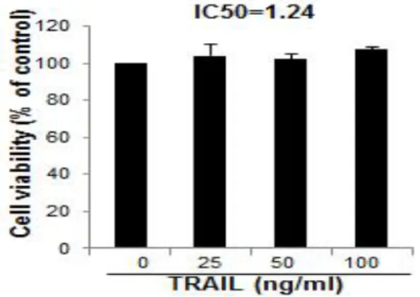
B. Effect of TRAIL on cell growth in HT29 human colorectal cancer cells
To assess the inhibitory effect of TRAIL on cell growth of HT29 human colorectal cancer cells, I analyzed cell viability by direct cell counting. The cells were treated with several concentrations of TRAIL(25, 50 and 100 ng/㎖) for 24 hr. As shown in Fig. 2, TRAIL had little influence upon cell viability of HT29 human colorectal cancer cells with IC50 value of 1.24 ㎍/㎖(Fig. 2).
Fig. 2. Effect of TRAIL on cell viability in HT29 human colorectal cancer cells
The results were expressed as a percentage of viable cells.
Columns, means of three experiments, with triplicates of each experiment; bars, SD.
C. Synergic effect of snake venom toxin and TRAIL on cell growth in HT29 human colorectal cancer cells
To assess the inhibitory effect of SVT and TRAIL on cell growth of HT29 human colorectal cancer cells, I analyzed cell viability by direct cell counting. The cells were treated with TRAIL(50 ng/㎖) or SVT(0.5 ㎍/㎖) or TRAIL(50 ng/㎖) plus SVT(0.5 ㎍/㎖) for 24 hr. As shown in Fig. 3, SVT(0.5 ㎍/㎖) significantly inhibited cell proliferation of HT29 human colorectal cancer cells, compared to control, whereas TRAIL(50 ng/㎖) alone did show little antiproliferative effect. TRAIL(50 ng/㎖) plus SVT(0.5 ㎍/㎖) synergistically and more significantly hindered cell growth of HT29 human colorectal cancer cells with IC50 value of 1.24 ㎍/㎖, compared to SVT(0.5 ㎍/㎖) alone(Fig. 3).
Fig. 3. Snake venom toxin enhanced TRAIL- induced cytotoxicity in HT29 human colorectal cancer
The results were expressed as a percentage of viable cells.
Columns, means of three experiments, with triplicates of each experiment; bars, SD.
*, p<0.05, significantly different from untreated control cells.
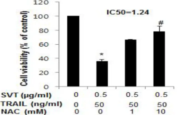
D. Reverse effect of NAC on synergized proliferation of snake venom toxin-combined TRAIL in HT29 human colorectal cancer cells
To assess whether the synergized inhibitory effect of SVT combined TRAIL on cell growth of HT29 human
Fig. 4. Snake venom toxin-combined TRAIL- induced antiproliferative activity was reversed in HT29 human colorectal cancer cells by anti- oxidative NAC
The results were expressed as a percentage of viable cells.
Columns, means of three experiments, with triplicates of each experiment; bars, SD.
*, p<0.05, significantly different from untreated control cells.
#. p<0.05, significantly different from TRAIL(50 ng/㎖) plus SVT(0.5 ㎍/㎖).
colorectal cells was reversed by strong anti-oxidative agent, NAC(1 or 10 mM), I analyzed cell viability by direct cell counting. The cells were treated with TRAIL(50 ng/
㎖) plus SVT(0.5 ㎍/㎖) for 24 hr with or without NAC(1 and 10 mM). As shown in Fig. 4, TRAIL(50 ng/㎖) plus SVT(0.5 ㎍/㎖) significantly inhibited cell proliferation of HT29 human colorectal cancer cells, compared to control.
After treatment of NAC(1 or 10 mM), growth of HT29 human colorectal cancer cells was conversely increased concentration- dependently. Moreover, NAC(10 mM) significantly reversed the synergized inhibitory effect of SVT and TRAIL with IC50 value of 1.24 ㎍/㎖, compared to TRAIL(50 ng/㎖) plus SVT(0.5 ㎍/㎖)(Fig. 4).
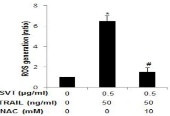
E. Reverse effect of NAC on snake venom toxin-combined TRAIL induced ROS generation in HT29 human colorectal cancer cells
To assess whether the synergized inhibitory effect of SVT and TRAIL on ROS generation was reversed by strong anti-oxidative agent, NAC(10 mM) on cell growth of HT29 human colorectal cancer cells, I analyzed ROS measurement as described in materials
Fig. 5. ROS dependent snake venom toxin- combined TRAIL-induced apoptosis was reversed by anti-oxidative NAC
Columns, means of three experiments, with triplicates of each experiment; bars, SD.
*, p<0.05, significantly different from untreated control cells.
#. p<0.05, significantly different from TRAIL(50 ng/㎖) plus SVT(0.5 ㎍/㎖).
and method. The cells were treated with TRAIL(50 ng/㎖) plus SVT(0.5 ㎍/㎖) for 30 min with(10 mM) or without NAC. As shown in Fig. 5, TRAIL(50 ng/㎖) plus SVT(0.5
㎍/㎖) significantly increased ROS generation, compared to control. However, they conversely and significantly decreased it following NAC(10 mM) treatment, compared to TRAIL(50 ng/㎖) plus SVT(0.5 ㎍/㎖)(Fig. 5).
F. Synergic effect of snake venom toxin and TRAIL on apoptosis in HT29 human colorectal cancer cells
To determine whether SVT enhances TRAIL-induced apoptotic cell death of TRAIL-insensitive HT29 human colorectal cancer cells. I found that SVT alone increased about 39 % of caspase active cells, compared to control, 2.6 %, inducing 41.6 % apoptosis in HT29 human colorectal cancer cells, where TRAIL, 2.8 % represented little increase of them in contrast.
However, combination treatment with SVT and TRAIL synergistically enhanced apoptotic cell death to 68.2 % in HT29 human colorectal cancer cells, compared to SVT alone(Fig. 6).
Fig. 6. Snake venom toxin enhanced TRAIL- induced apoptosis in HT29 human colorectal cancer cells
The green color in the fixed cells marks TUNEL-labeled cells.
Data means± SD expressed as percentage of control value, which is set to 100 %.
At least three independent experiments were carried out in triplicate.
*, p<0.05, significantly different from control cells.
G. Effect of snake venom toxin on the expression of death receptors 4 and 5 in HT29 human
colorectal cancer cells
To ascertain the underlying mechanism that may be responsible for enhancement of TRAIL-induced apoptotic cell death by SVT, I examined the effect of SVT on the expression of death receptors. SVT increased the expressions of both DR4 and DR5 in TRAIL-insensitive HT29 cells(Fig. 7). Especially, it represented dose dependent enhancement in DR5 expression(Fig. 7). Contrary to SVT, TRAIL exerted little influence on the expression of DR4 and DR5 in TRAIL-insensitive HT29 human colorectal cancer cells(Fig. 7). However, expression of DR4 and DR5 was synergistically further increased by the combined treatment of SVT and TRAIL(Fig. 7).
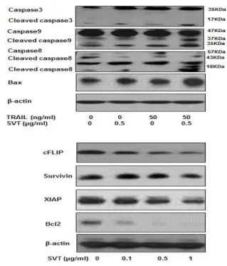
H. Effect of snake venom toxin on TRAIL-induced expression of apoptosis related proteins
I selected HT29 human colorectal cancer cell line to confirm its sensitivity to TRAIL and to ascertain the
Fig. 7. Snake venom toxin enhanced the expression of DR4 and DR5 in the HT29 human colorectal cancer cells
Expression of DR4, DR5 and β-actin was detected by western blotting using specific antibodies.
effect of SVT and/or TRAIL on the activation of caspase-8, caspase-3, caspase-9 cleavage and the expression of Bax in it. Although TRAIL alone had little effect on the activation of caspases cleavage and Bax expression, SVT increased the their expression, compared to control. Moreover, combination treatment of SVT and TRAIL significantly increased expression of the pro-apoptotic proteins(Fig. 8). On the basis of the previous reports that various anti-apoptotic proteins including survivin, Bcl-2, XIAP and cFLIP have been shown to induce resistance to TRAIL-induced apoptosis35,36). I investigate whether SVT sensitized HT29 cells to TRAIL through down-regulation of the expression of these anti-apoptotic proteins irrespective of their TRAIL resistance. As shown in Fig. 8, SVT decreased expression of XIAP, survivin, bcl-2 and cFLIP concentration dependently. These results indicated that the
SVT enhances TRAIL-induced
Fig. 8. Snake venom toxin enhanced TRAIL- induced apoptotic proteins expressions in HT29 human colorectal cancer cells
Expression of cFLIP, survivin, XIAP, Bcl-2, bax and β-actin was detected by western blotting using specific antibodies.
β-actin protein was used an internal control.
Each band is representative for three experiments.
apoptotic cell death through the over-expression of DR4 and DR5, as well as the down-regulation of anti-apoptotic protein expression.
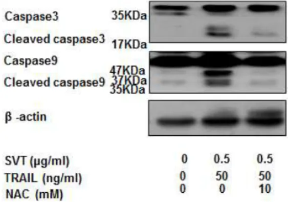
I. Reversal of synergistic effect of snake venom toxin-combined TRAIL on pro-apoptotic proteins in HT29 human colorectal cancer cells
HT29 cells were pretreated with SVT for 24 hr and washed out. After that the cells were treated with TRAIL for 24 hr, and whole cell extracts were analyzed by western blotting using antibodies against caspase-3, caspase-9, β-actin. β-actin protein was used an internal control. Each band is representative for three experiments. I found that pretreatment(1 hr) of ROS scavenger N-acetylcysteine(NAC, 10 mM) reversed the activation of cleavaged caspase-3 and -9
Fig. 9. Increased expressions of snake venom tToxin-combined TRAIL-induced apoptotic protein was reversed by anti-oxidative NAC
Whole cell extracts were analyzed by western blotting using the antibodies against caspase-3, caspase-9 and β-actin.
β-actin protein was used an internal control.
Each band is representative for three experiments.
by the combination treatment of SVT and TRAIL, suggesting TRAIL should be a promising target of apoptosis in HT29 cells(Fig. 9).
J. Reversal of synergistic effect of snake venom toxin-combined TRAIL on expression of DR4 and DR5 in HT29 human colorectal cancer cells
HT29 cells were pretreated with SVT for 24 hr and
Fig. 10. The activation of DR4 and DR5 was reversed by anti-oxidative NAC
Whole cell extracts were analyzed by western blotting using the antibodies against DR4, DR5 and β-actin.
β-actin protein was used an internal control.
Each band is representative for three experiments.
washed out. After that the cells were treated with TRAIL for 24 hr, and whole cell extracts were analyzed by western blotting using antibodies against DR4, DR5 and β -actin. β-actin protein was used an internal control. Each band is representative for three experiments. I found that pretreatment(1 hr) of ROS scavenger N-acetylcysteine(NAC, 10 mM) reversed the increased activation of DR4 and DR5 by the combination treatment of SVT and TRAIL(Fig. 10).
Ⅳ. Discussion
Apoptosis is a form of programmed cell death, which can be triggered by two major pathways including mitochondria-dependent intrinsic pathway and DR- associated extrinsic pathway11,12,37,38).
In the intrinsic apoptotic pathway, ROS stimuli exerts influence upon intrinsic mitochondrial regulated apoptotic pathway, translocating a truncated form of Bid(tBid) into mitochondria and consecutively making a change of the mitochondrial outer membrane potential due to increased Bax/Bcl-2 ratio, and subsequently resulting in mitochondrial intrinsic apoptosis through activation of caspase-9 and caspase-3 via cytochrome C release, whereas it consecutively let DR4 and DR5 binding to its ligand, subsequently causing apoptosis through caspase-8-mediated rapid activation of caspase cascades in the extrinsic apoptotic pathway39-43).
Therefore, this implicate that ROS generation could be involved in extrinsic apoptosis as well as intrinsic one, that is substantiated by previous reports25,44).
In this study, it was found that Vipera lebetina turanica snake venom toxin inhibited cell growth of HT29 human epithelial colorectal cancer cells through induction of both intrinsic, mitochondria-mediated, and extrinsic DR-mediated apoptosis via increased binding of DR4, DR5 with TRAIL, and that the combination of SVT and TRAIL increased apoptotic cell death synergistically in spite of TRAIL insensitivity to them, representing SVT overcame TRAIL resistance and let the apoptosis going through enhancement of expression of proapoptotic proteins and control of
anti-apoptotic ones simultaneously.
That is, SVT significantly inhibited cell proliferation of HT29 human colorectal cancer cells(Fig. 1). However, inconsistent with a previous report45), TRAIL had little influence upon cell viability of them(Fig. 2). However, TRAIL(50 ng/㎖) plus SVT(0.5 ㎍/㎖) synergistically and more significantly hindered cell growth of them, compared to SVT(0.5 ㎍/㎖) alone(Fig. 3). After treatment of strong anti-oxidant, N-acetyl-cysteine, NAC(1 or 10 mM), antiproliferation of HT29 human colorectal cancer cells and increase of ROS generation by combined SVT and TRAIL was abolished dose-dependently, suggesting inhibition of the cell growth was related with bilateral apoptosis induced by SVT together with TRAIL. I reconfirmed the above in the TUNEL assay that combination treatment with SVT and TRAIL synergistically increased apoptotic cell death to 68.2 % in HT29 human colorectal cancer cells, compared to SVT alone or TRAIL alone(Fig. 6). TRAIL mediates apoptotic cell death through enhanced expression of two death receptors, DR4 and DR5 which are expressed on the surface of cancer cells46,47). The binding of TRAIL to DR4 and DR5 result in DR mediated extrinsic apoptosis48,49).
Expression of DR4 and DR5 was also synergistically further increased by the combined treatment of SVT and TRAIL than SVT or TRAIL alone(Fig. 7). Taken together, it was suggested that SVT should play a major role in inducing DR mediated extrinsic apoptosis through compensating TRAIL resistance in HT29 human epithelial colorectal cancer cells.
Caspases, a family of cysteine aspartyl-specific proteases, control the triggering and executing apoptosis in both pathways including mitochondria dependent intrinsic apoptotic and DR mediated extrinsic apoptotic pathway50). In the activation of caspase cascade, of three main pathways including DR-ligand complex-activated caspase-8 pathway, the mitochondria dependent pathway characterized by mitochondrial membrane permeability change according to pro-apototic Bax/anti-apoptotic Bcl-2 ratio, consecutive cytochrome c release and subsequent activation of apoptosome-associated caspases-9 and -3 and the endoplasmic reticulum(ER)-specific
apoptotic pathway involving caspase-12 activation, former two pathways are more noteworthy regarding both apoptotic pathways in various cancer cells and considered a potential target of developing new anticancer drugs51-53) where induction of apoptosis critically depends on upregulation of proapoptotic caspases known as executioners of apoptosis54-56).
In the expression of apoptosis related proteins, SVT with TRAIL in HT29 cells represented increased Bax/Bcl-2 ratio, subsequently upregulated intrinsic apoptotic pathway related cleaved caspase-9, -3 as well as activated extrinsic DR-mediated apoptotic pathway related caspase-8, suggesting SVT exerts substantial influence upon bilateral apoptosis in them nevertheless of TRAIL resistance(Fig. 8).
Several studies have reported that resistance of cancer cells to TRAIL-induced apoptosis is known to be related to intracellular levels of anti-apoptotic proteins and that DR up-regulation and cell survival protein down regulation may be a promising strategy for sensitizing TRAIL-resistant cancer cells to TRAIL-induced apoptotic cell death57-61).
Coincident to the above, SVT decreased expression of anti-apoptotic XIAP, survivin, bcl-2 and cFLIP in addition to DR4, 5 enhancement in the HT29 human epithelial colorectal cancer cells, indicating that the SVT strengthened TRAIL-induced apoptotic cell death through the over-expression of DR4 and DR5, as well as the down-regulation of anti-apoptotic protein expression.
These results also reconfirmed through experiment to ascertain whether pretreatment(1 hr) of ROS scavenger N-acetylcysteine(NAC, 10 mM) abolish the activation of DR4,5 and pro-apoptotic cleavaged caspase-3 and -9 induced by the combination treatment of SVT and TRAIL(Fig. 9, 10)
In conclusion, this study shows that SVT strengthen sensitizing HT29 human epithelial colorectal cancer cells to the TRAIL induced apoptosis, and that the combination treatment of SVT and TRAIL could get over TRAIL resistance. The results suggest that SVT may be a potent agent for the treatment or prevention of colorectal cancer.
V. Conclusion
The collective results suggest that SVT facilitates TRAIL-induced apoptosis in HT29 human epithelial colorectal cancer cells through up-regulation of the TRAIL receptors; DR4 and DR5 and consecutive induction of bilateral apoptosis via regulating apoptosis related proteins.
Ⅵ. References
1. Yu Z, Li W. Induction of apoptosis by puerarin in colorectal cancer HT29 cells. Cancer Lett. 2006 ; 238(1) : 53-60.
2. Jemal A, Bray F, Center MM, Ferlay J, Ward E, Forman D. Global cancer statistics. CA Cancer J Clin. 2011 ; 61(2) : 69–90.
3. Tomida A, Tsuruo T. Drug resistance mediated by cellular stress response to the microenvironment of solid tumors. Anticancer Drug Des. 1999 ; 14(2) : 169-77.
4. Goldberg RM, Sargent DJ, Morton RF et al. A ran- domized controlled trial of fluorouracil plus leucovorin, irinotecan, and oxaliplatin combi- nations in patients with previously untreated metastatic colorectal cancer. J Clin Oncol. 2004 ; 22(1) : 23–30.
5. Kamb A, Wee S, Lengauer C. Why is cancer drug discovery so difficult? Nature Rev Drug Discovery.
2007 ; 6(2) : 115-20.
6. Dalerba P, Cho RW, Clarke MF. Cancer stem cells:
models and concepts. Annual Rev Med. 2007 ; 58(1) : 267-84.
7. de Santa Barbara P, van den Brink GR, Roberts DJ. Development and differentiation of the intestinal epithelium. Cell Mol Life Sci. 2003 ; 60(7) : 1322–32.
8. Sansonetti PJ. War and peace at mucosal sur- faces. Nat Rev Immunol. 2004 ; 4(12) : 953–64.
9. Cho JH. The genetics and immunopathogenesis of inflammatory bowel disease. Nat Rev Immunol.
2008 ; 8(6) : 458–66.
10. van den Brink GR, Hardwick JC. Hedgehog inter- action in colorectal cancer. Gut. 2006 ; 55(7) : 912–4.
11. Adams JM. Ways of dying: multiple pathways to apoptosis. Genes Dev. 2003 ; 17(20) : 2481-95.
12. Shi Y. Mechanical aspects of apoptosome assembly.
Curr Opin Cell Biol. 2006 ; 18(6) : 677-84.
13. Koschny R, Walczak H, Ganten TM. The promise of TRAIL. Potential and risks of a novel anticancer therapy. J Mol Med. 2007 ; 85(9) : 923-35.
14. Johnstone RW, Frew AJ, Smyth MJ. The TRAIL apoptotic pathway in cancer onset, progression and therapy. Nat Rev Cancer. 2008 ; 8(10) : 782-98.
15. Ashkenazi A, Dixit VM. Death receptors: signaling and modulation. Science. 1998 ; 281(5381) : 1305-8.
16. Wajant H, Gerspach J, Pfizenmaier K, Tumor thera- peutics by design: targeting and activation of death receptors. Cytokine Growth Factor Rev.
2005 ; 16(1) : 55-76.
17. Sprick MR, Weigand MA, Rieser E, et al. FADD/
MORT1 and caspase-8 are recruited to TRAIL receptors 1 and 2 and are essential for apoptosis mediated by TRAIL receptor 2. Immunity. 2000 ; 12(6) : 599-609.
18. Scaffidi C, Fulda S, Srinivasan A et al. Two CD95 (APO-1/Fas) signaling pathways. EMBO J. 1998 ; 17(6) : 1675-87.
19. Luo X, Budihardjo I, Zou H, Slaughter C, Wang X.
Bid, a Bcl-2 interacting protein, mediates cyto- chrome c release from mitochondria in response to activation of cell surface death receptors. Cell.
1998 ; 94(4) : 481-90.
20. Chai J, Du C, Wu JW, et al. Structural and bio- chemical basis of apoptotic activation by Smac/
DIABLO. Nature. 2000 ; 406(6798) : 855-62.
21. Wei MC, Zong WX, Cheng EH et al. Proapoptotic BAX and BAK: a requisite gateway to mitochon- drial dysfunction and death. Science. 2001 ; 292(5517) : 727-30.
22. Prasad S, Yadav VR, Kannappan R, Aggarwal BB.
Ursolic acid, a pentacyclin triterpene, potentiates TRAIL-induced apoptosis through p53-indepen-
dent upregulation of death receptors. Evidence for the role of reactive oxygen species and JNK. J Biol Chem. 2001 ; 286(7) : 5546-57.
23. Prasad S, Yadav VR, Ravindran J, Aggarwal BB.
Cardamonin sensitizes tumour cells to TRAIL through ROS- and CHOP-mediated upregulation of death receptors and down-regulation of survival proteins. Br J Pharmacol. 2012 ; 165(3) : 741-53 24. Sung B, Ravindran J, Prasad S, Pandey MK,
Aggarwal BB. Gossypol induces death receptor-5 through activation of the ROS-ERK-CHOP path- way and sensitizes colorectal cancer cells to TRAIL. J Biol Chem. 2010 ; 285(46) : 35418-27.
25. Jung EM, Lim JH, Lee TJ, Park JW, Choi KS, Kwon TK. Curcumin sensitizes tumor necrosis factor-related apoptosis-inducing ligand(TRAIL)- induced apoptosis through reactive oxygen species-mediated upregulation of death receptor 5(DR5). Carcinogenesis. 2005 ; 26(11) : 1905-13.
26. Taniguchi H, Yoshida T, Horinaka M et al. Bai- calein overcomes tumor necrosis factor- related apoptosis-inducing ligand resistance via two di- fferent cell-specific pathways in cancer cells but not in normal cells. Cancer Res. 2008 ; 68(21) : 8918-27.
27. Su RY, Chi KH, Huang DY, Tai MH, Lin WW. 15 - deoxy-Delta12, 14-prostaglandin J2 up- regulates death receptor 5 gene expression in HCT116 cells:
Involvement of reactive oxygen species and C/EBP homologous transcription factor gene transcrip- tion. Mol Cancer Ther. 2008 ; 7(10) : 3429-40.
28. Bin L, Thorburn J, Thomas LR, Clark PE, Humphreys R, Thorburn A. Tumor-derived mutations in the TRAIL receptor DR5 inhibit TRAIL signaling through the DR4 receptor by competing for ligand binding. J Biol Chem. 2007 ; 282(38) : 28189-94.
29. Lee SH, Shin MS, Kim HS et al. Somatic mutations of TRAIL-receptor 1 and TRAIL- receptor 2 genes in non-Hodgkin’s lymphoma. Oncogene. 2001 ; 20(3) : 399-403.
30. Keane MM, Ettenberg SA, Nau MM, Russell EK, Lipkowitz S. Chemotherapy augments TRAIL-induced apoptosis in breast cell lines. Cancer Res. 1999 ; 59(3) : 734-41.
31. Lacour S, Hammann A, Wotawa A, Corcos L, Solary E, Dimanche-Boitrel MT. Anticancer agents sensitize tumor cells to tumor necrosis factor- related apoptosis-inducing ligand-mediated caspase-8 activation and apoptosis. Cancer Res. 2001 ; 61(4) : 1645-51.
32. Son DJ, Park MH, Chae SJ et al. Inhibitory effect of SVT from Vipera lebetina turanica on hormone-refractory human prostate cancer cell growth: Induction of apoptosis through inacti- vation of nuclear factor kappaB. Mol Cancer Ther. 2007 ; 6(2) : 675-83.
33. Park MH, Son DJ, Kwak DH et al. Snake venom toxin inhibits cell growth through induction of apoptosis in neuroblastoma cells. Arch Pharm Res. 2009 ; 32(11) : 1545-54.
34. Oh MG, Song HS. Inhibitory effect of snake venom on colon cancer cell growth through induction of death receptor dependent apoptosis. J of Korean Acupuncture & Moxibustion Medicine Society.
2012 ; 29(1) : 25-35.
35. Rauert H, Stuhmer T, Bargou R, Wajant H, Sieg- mund D. TNFR1 and TNFR2 regulate the extrinsic apoptotic pathway in myeloma cells by multiple mechanisms. Cell Death Dis. 2011 ; 2(1) : e194.
36. Yadav VR, Prasad S, Aggarwal BB. Cardamonin sensitizes tumor cells to TRAIL through ROS- and CHOP-mediated upregulation of death receptors and downregulation of survival proteins. Br J Pharmacol. 2012 ; 165(3) : 741-53.
37. Das GC, Holiday D, Gallardo R, Haas C. Taxol- induced cell cycle arrest and apoptosis: dose- response relationship in lung cancer cells of different wildtype p53 status and under isogenic condition. Cancer Lett. 2001 ; 165(2) : 147-53.
38. Debatin KM. Activation of apoptosis pathways by anticancer treatment. Toxicol Lett. 2000 ; 111-112(1) : 41-8.
39. Pulido MD, Parrish AR. Metal-induced apoptosis:
Mechanisms. Mutat Res. 2003 ; 533(1-2) : 227-41.
40. Selimovic D, Hassan M, Haike Y, Hengge UR. Taxol- induced mitochondrial stress in melanoma cells is mediated by activation of c-Jun N-terminal kinase
(JNK) and p38 pathways via uncoupling protein 2. Cell Signal. 2008 ; 20(2) : 311–22.
41. Barbu A, Welsh N, Saldeen J. Cytokine-induced apoptosis and necrosis are preceded by disruption of the mitochondrial membrane potential(Δψm) in pancreatic RINm5F cells: Prevention by Bcl-2.
Mol Cell Endocrinol. 2002 ; 190(1) : 75-82.
42. Reed JC. Apoptosis-regulating proteins as targets for drug discovery. Trends Mol Med. 2001 ; 7(7) : 314–9.
43. Li JY, Xu ZJ, Tan MY, Su WK, Gong XG. 3- (4- (Benzo[d]thiazol-2-yl) -1-phenyl-1Hpyrazol- 3-yl) phenyl acetate induced HepG2 cell apoptosis through a ROS-mediated pathway. Chem Biol Interact. 2010 ; 183(3) : 341–8.
44. Kim H, Kim EH, Eom YW et al. Sulforaphane sen- sitizes tumor necrosis factor-related apoptosis- inducing ligand(TRAIL)-resistant hepatoma cells to TRAIL-induced apoptosis through reactive oxygen species-mediated up-regulation of DR5.
Cancer Res. 2006 ; 66(3) : 1740-50.
45. Kim KT, Song HS. Inhibitory effect of snake venom toxin on colorectal cancer HCT116 cells growth through induction of intrinsic or extrinsic apoptosis.
Journal of Korean Acupuncture & Moxibustion Medicine Society. 2013 ; 30(1) : 43-55.
46. Ashkenazi A, Herbst RS. To kill a tumor cell: the potential of proapoptotic receptor agonists. J Clin Invest. 2008 ; 118(6) : 1979-90.
47. Eberle J, Fecker LF, Forschner T, Ulrich C, Röwert-Huber J, Stockfleth E. Apoptosis pathways as promising targets for skin cancer therapy. Br J Dermatol. 2007 ; 156(3) : 18-24.
48. Pennarun B, Meijer A, de Vries EG, Kleibeuker JH, Kruyt F, de Jong S. Playing the DISC: turning on TRAIL death receptor-mediated apoptosis in cancer. Biochim Biophys Acta. 2010 ; 1805(2) : 123-40.
49. Jung YH, Heo J, Lee YL, Kwon TK, Kim YH.
Quercetin enhances TRAIL-induced apoptosis in prostate cancer cells via increased protein stability of death receptor 5. Life Science. 2010 ; 86(9-10) : 351-7.
50. Earnshaw WC. Apoptosis. A cellular poison cupboard.
Nature. 1999 ; 397(6718) : 387-9.
51. Yoneda T, Imaizumi K, Oono K et al. Activation of caspase-12, an endoplastic reticulum(ER) resident caspase, through tumor necrosis factor receptor- associated factor 2-dependent mechanism in response to the ER stress. J Biol Chem. 2001 ; 276(17) : 13935-40.
52. Zhang L, Xing D, Chen M. Bim(L) displacing Bcl- x(L) promotes Bax translocation during TNFalpha- induced apoptosis. Apoptosis. 2008 ; 13(7) : 950-8.
53. Cohen GM. Caspases. The executioners of apoptosis.
Biochem J. 1997 ; 326(pt 1) : 1-16.
54. Chai WS, Zhu XM, Li SH, Fan JX, Chen BY. Role of Bcl-2 family members in caspase-3/9-dependent apoptosis during Pseudomonas aeruginosa infection in U937 cells. Apoptosis. 2008 ; 13(6) : 833-43.
55. Radha V, Sudhakar C, Ray P, Swarup G. Induc- tion of cytochrome c release and apoptosis by Hck-SH3 domain-mediated signalling requires caspase-3. Apoptosis. 2002 ; 7(3) : 195-207.
56. Nicholson DW. Caspase structure, proteolytic sub- strates, and function during apoptotic cell death.
Cell Death Differ. 1999 ; 6(11) : 1028-42.
57. Wang S, El-Deiry WS. TRAIL and apoptosis in- duction by TNF-family death receptors. Oncogene.
2003 ; 22(53) : 8628-33.
58. Jäättelä M. Escaping cell death: Survival proteins in cancer. Exp Cell Res. 1999 ; 248(1) : 30-43.
59. Kim YH, Lee DH, Jeong JH, Guo ZS, Lee YJ.
Quercetin augments TRAIL-induced apoptotic death: involvement of the ERK signal trans- duction pathway. Biochem Pharmacol. 2008 ; 75(10) : 1946-58.
60. Psahoulia FH, Drosopoulos KG, Doubravska L, Andera L, Pintzas A. Quercetin enhances TRAIL- mediated apoptosis in colorectal cancer cells by inducing the accumulation of death receptors in lipid rafts. Mol Cancer Ther. 2007 ; 6(9) : 2591-9.
61. Sung B, Park B, Yadav VR, Aggarwal BB. Celastrol, a triterpene, enhances TRAIL-induced apoptosis through the down-regulation of cell survival proteins and up-regulation of death receptors. J Biol Chem. 2010 ; 285(15) : 11498-507.