• 검색 결과가 없습니다.

N/A
N/A
Protected

Academic year: 2021

Share ""

Copied!
5
0
0

로드 중.... (전체 텍스트 보기)

전체 글

(1)

접수일 : 2015 년 8 월 6 일 , 게재승인일 : 2015 년 10 월 28 일 책임저자 : 박중현 , 서울시 강남구 언주로 211

󰂕 06273, 강남세브란스병원 재활의학과

Tel: 02-2019-3491, Fax: 02-3463-7585 E-mail: [email protected]

고주파 열치료를 이용한 선택적 신경박리술 후 발생한 깊은 종아리 신경 손상

󰠏 증례 보고 󰠏

연세대학교 의과대학 재활의학교실 및 희귀난치성 신경근육병 재활연구소

장철훈ㆍ조한얼ㆍ이진형ㆍ박중현

Deep Peroneal Nerve Injury Induced by Radiofrequency Selective Neurolysis

󰠏 A Case Report 󰠏

Chul Hoon Jang, M.D., Han Eol Cho, M.D., Jin Hyung Lee, M.D. and Jung Hyun Park, M.D., Ph.D.

Department of Rehabilitation Medicine and Rehabilitation Institute of Neuromuscular Disease, Yonsei University College of Medicine, Seoul, Korea

Selective neurolysis is widely performed in private local plastic surgery clinics to give the leg a thinner shape by inducing semi-permanent nerve damage. However, it can cause long-lasting injury to nerves near the target if the procedure is not performed precisely. This case report ad- dresses a rare complication of selective neurolysis for cos- metic calf reduction. A 32 year-old woman suffered from right foot drop after a radiofrequency selective neurolysis procedure on both gastrocnemius muscles. Magnetic reso- nance image of the calf revealed peripheral nerve injury of the right common peroneal nerve. An electrodiagnostic study also revealed injury of the right deep peroneal nerve.

There was no improvement in her ankle dorsiflexion or toe extension on the electrodiagnostic studies at her 6 month follow-up. To avoid such a catastrophic outcome, well- trained techniques, comprehensive knowledge of anatomy and supplementary device for procedure would be essential.

(Clinical Pain 2015;14:93-97)

Key Words: Peroneal neuropathy, Neurolysis, Drop foot

INTRODUCTION

Cosmetic calf reduction surgery is widely performed in

private plastic surgery clinics. Reduction surgery process includes liposuction, gastrocnemius(GCM) tenotomy, parti- al resection of GCM muscle and selective neurolysis of the GCM.

1

Selective neurolysis damages motor nerves in- nervating the GCM muscle, which makes the muscle para- lyzed and dystrophic. The radiofrequency selective neu- rolysis has become a common procedure among the plastic clinics because it requires simpler preparation and also guarantees semi-permanent effects on the target muscles if using radiofrequency ablations.

The radiofrequency selective neurolysis blocks all tibial nerve branches which innervate the GCM muscle. However, the neurolysis could cause injury on other nerves near the target such as sural nerve, soleus muscle branch of tibial nerve and peroneal nerve if it is not performed with ex- treme precision.

2

Previous article reported several compli- cations of selective neurolysis such as temporary walking discomfort and transient numbness of calf.

3

However, the severe complication of the unwanted nerve injury has not been commonly reported until now.

Here we present a complication case of radiofrequency selective neurolysis; severe deep peroneal nerve injury.

Although the nerve injury seems to be rather prevalent in

local cosmetic clinics, there are very few case reports on

this side effect. And even in the reports, they just com-

mented mild and transient complications in the aspect of

clinical symptoms such as foot drop or ankle instability.

3,4

There is few case reports with anatomical and functional

analysis which are accompanied by magnetic resonance im-

age (MRI) images and electrodiagnostic studies to verify

the nerve injuries. Therefore, it could be meaningful to re-

view the case with complications using calf MRI and elec-

trodiagnostic studies.

(2)

Table 1. Results of Motor Nerve Conduction Study

Nerve Stimulation Site Recording Site Latency (ms) Amplitude (mV) Velocity (m/s)

Rt. Common Peroneal Ankle EDB No response

Lt. Common Peroneal Ankle EDB 3.25 4.4 51.6

Rt. Tibial Ankle AH 3.35 21.9 43.1

Lt. Tibial Ankle AH 2.85 19.8 45.5

EDB: extensor digitorum brevis, AH: abductor hallucis.

Fig. 1. Calf MRI (10 days after the procedure) – signal change in common peroneal nerve. (A) Multifocal nodular lesions at medial and lateral gastrocnemius muscle heads (white arrow). Signal change around the common peroneal nerve at the level of fibular head in T2 axial MRI image is observed (in the white circle). (B) Imaginary picture for the anatomical location of the nerve around knee and calf in coronal MRI image. White circle is the nerve branch to lateral GCM which was the main target for neurolysis. Common peroneal nerve (red circle) is suspected to be damaged during neurolysis due to close location.

CASE REPORT

A 32-year-old female visited our hospital for right foot drop. She received selective neurolysis using radiofre- quency on both calves for cosmetic purposes a month ago at a private plastic surgery clinic. Incision was done by be- hind the knee and radiofrequency probe was inserted.

Multiple radiofrequency nerve ablations were performed around the medial and lateral head of both GCM muscles to block tibial nerve branches. The probe delivers thermal effects that inhibit the tibial nerve and produce conduction block. After the procedure, she had no obvious muscle weakness in her legs. However, 4 days later, she visited to the clinic again for her right foot drop. She was unable to dorsiflex her right ankle and extend all five toes, and

the manual muscle test (MMT) grade showed zero grade in right ankle-dorsi flexor muscle and right toe extensor muscles. MMT of ankle eversion was normal and sensory test of both lower extremity was also normal.

Ten days after injury, calf MRI was taken at other clinic.

In the image, multifocal nodular lesions (up to 1.7 cm) with denervation-related edema at medial and lateral gastro- cnemius, soleus and peroneus longus muscles was ob- served. Signal change around the common peroneal nerve at the level of fibular head with swelling of the common peroneal nerve was also seen (Fig. 1).

Electrodiagnostic examination was done three weeks af-

ter surgery. There was no response at compound muscle

action potential (CMAP) of right deep peroneal nerve con-

duction study (Table 1). Acceptable latency and amplitude

of sensory nerve action potential (SNAP) at superficial per-

(3)

Table 2. Results of Sensory Nerve Conduction Study

Nerve Stimulation Site Recording Site Latency (ms) Amplitude (mV)

Rt. Superficial Peroneal Leg Ankle 2.95 15.8

Lt. Superficial Peroneal Leg Ankle 2.95 17.9

Rt. Tibial Leg Ankle 2.20 25.1

Lt. Tibial Leg Ankle 2.05 24.8

Table 3. Results of Motor Nerve Conduction Study by Time after Injury

Three weeks after injury Two months after injury Four months after injury CMAP of Peroneal n.

innervation muscles

No response in TA, EDB No response in EDB and Low amplitude in TA

Low amplitude in TA and Prolonged latency & low amplitude in EDB EDB: extensor digitorum brevis, TA: tibialis anterior, CMAP: compound muscle action potential.

oneal nerve and sural nerve were observed (Table 2).

In needle electromyographic examination study, abnor- mal spontaneous activities such as positive sharp wave and fibrillation potential and decreased volitional activities was observed on right tibialis anterior, extensor digitorum brevis and extensor digitorum longus muscles. Motor unit action potential was not found in the muscles innervated by right deep peroneal nerve such as the right tibialis anterior, ex- tensor digitorum brevis and extensor digitorum longus (Table 3). These finding were suggestive of right deep per- oneal nerve injury. It implied that iatrogenic injuries on oth- er nerves near the target happened after the procedure. After the patient was diagnosed with deep peroneal nerve injury, she was equipped with ankle orthosis to prevent foot drop during gait, and received rehabilitation treatment such as gait training and electrical stimulation therapy on right ti- bialis muscle. When she first visited our hospital after a month later from the injury, her MMT grades for ankle dor- siflexion was changed from zero to trace. After the first vis- it, she received strengthening exercises and electrical stim- ulation therapy on the right ankle several times in our clinic.

After a month from the initial visit, the MMT of ankle dor- siflexion had not changed from trace grade. Follow-up elec- trodiagnostic studies were taken after 2 months and after 4 months from the injury. CMAP of Peroneal nerve in TA muscle with surface electrode had been changed from no response to low amplitude and low latency after 4 month from the injury. But it still showed no motor unit action potential of the right TA and EHL muscles at 2 months

and 4 months after the injury (Table 3).

DISCUSSION

The rate of foot drop after cosmetic calf reduction is not reported before, however foot drop could be a critical com- plication after cosmetic calf reduction. Eventually, foot drop could cause ankle plantar flexion contracture and equinus gait pattern. However, the prognosis of foot drop has not been reported up until now. Our report deals with a complication which can occur in selective neurolysis of the lateral head of GCM; severe deep peroneal nerve injury with serial electrodiagnostic studies and calf MRI image.

The mechanism of foot drop could be deep peroneal nerve injury near medial tibial nerve branch innervated at GCM during radiofrequency selective neurolysis. In this case, ankle dorsiflexion weakness was caused by muscle weakness innervated by damaged peroneal nerve such as tibialis anterior muscle. The deep peroneal nerve runs be- side to the fibula bone which could be more susceptible to mass effect by swelling and the swelling is suspected to be aggravated for 4 days. This could explain the reason why foot drop happened 4 days after injury. As the patient ambulated after radiofrequency selective neurolysis we could exclude the mechanism of nerve compression due to bed rest status.

To the best of our knowledge, there are no case reports

of foot drop after calf reduction neurolysis diagnosed by

MRI image. Foot drop after radiofrequency ablation for

(4)

varicose veins were reported before.

5

Furthermore no fur- ther study such as electrodiagnostic studies was performed to verify the cause of foot drop. So, there is no reliable consensus to predict the prognosis of peroneal nerve injury caused by cosmetic calf reduction neurolysis. However, we can presume the prognosis of our patient with generalized MRI interpretation and serial electrodiagnostic studies.

In the calf MRI, signal change around the common per- oneal nerve and swelling of the nerve were seen. Acute axonal nerve lesions caused hyperintense signal on T2-weighted MRI at and distal to the nerve lesion site. It is well-known that MRI signal alterations occur as early as 24 hours after an axonal nerve lesion and correlate with nerve fiber degeneration, and later with nerve edema on histology. Nerves and muscle normally exhibit an inter- mediate signal on MRI image, but they produce a hyper- intense signal that is, they appear bright following nerve injury and denervation.

4

The increased nerve signal shows regression in the period of two weeks after the onset of nerve regeneration.

6

In our study, a calf MRI during the acute stage (3 weeks later) was taken, so we could con- clude that acute axonal nerve damage happened at the right common peroneal nerve with the MRI image and the nerve regeneration had not been observed for that time. To be certain, follow-up MRI images to check nerve regeneration with changes in the T2 intensity should be needed.

In the electrodiagnostic study of peroneal nerve injury, any CMAP response recorded from the TA or EDB mus- cles at baseline were associated with good prognosis.

7

In our patient, there were no CMAP responses recorded from TA and EDB in initial electrodiagnostic study and Nerve conduction of tibial nerve study was normal (Table 1).

Peroneal muscle showed no abnormal spontaneous activity on electrodiagnostic study. Therefore, based on the pa- tient’s results, sufficient motor recovery in ankle dorsi- flexion for gait would not be expected. To summarize our case, there was a signal change in the right common per- oneal nerve with swelling seen on the MRI image and ab- normal spontaneous activities in the muscles innervated by the deep peroneal nerve such as right TA, EDB and EDL, confirming that the neurolysis procedure had damaged the right deep peroneal nerve.

During neurolysis of the lateral GCM nerve branch, the deep peroneal nerve could be injured because it runs very close to the tibial nerve in the popliteal region beside fibula

bone (Fig. 1). Anatomically, it is known that the nearest point between the peroneal nerve and the lateral GCM nerve branch is 2.5 cm.

1

Lesions of the deep peroneal nerve could cause weakness in foot eversion, ankle dorsiflexion or sensory loss on the lower extremity. If the damage on deep peroneal nerve portion is dominant like our case, weakness of ankle dorsiflexion and toe extension could be obvious. The neurolysis procedure using radiofrequency ablation must be done with great caution because it causes long lasting impairment to other nerves near the target due to the semi-permanent effect of the technique. The well- trained techniques and comprehensive knowledge of anat- omy in calf could enhance the accuracy of procedures and reduce complications. Even with expert hands, supple- mentary device in the procedure to enhance technique is essential for better output.

8

Introducing the technique to lo- calize the target nerve by using both direct ultrasono- graphic visualization and electric nerve stimulation could be helpful to confirm the target nerve. Well-trained techni- ques, comprehensive knowledge of anatomy and supple- mentary device are essential to avoid unwanted nerve injury.

REFERENCES

1. Han SH, Chung NS, Park DY. Ankle Plantar-Flexion Contracture Complication After Aesthetic Calf Volume Reduction Procedure. Ann Plast Surg 2015; 75: 19-23 2. Kim SC, Kang MH, Ock JJ. Calf-contouring surgery of gas-

trocnemius hypertrophy: selective neurectomy of the sural nerve. Aesthetic Plast Surg 2008; 32: 889-893

3. Tsai FC, Mardini S, Fong TH, Kang JH, Chou CM.

Selective neurectomy of the gastrocnemius and soleus mus- cles for calf hypertrophy: an anatomical study and 700 clin- ical cases. Plast Reconstr Surg 2008; 122: 178-187 4. Bendszus M, Stoll G. Technology insight: visualizing pe-

ripheral nerve injury using MRI. Nat Clin Pract Neurol 2005; 1: 45-53

5. Kumar RS, Gopinath M. A rare cause of foot drop after ra- diofrequency ablation for varicose veins: case report and re- view of the literature. Neurol India. 2010; 58: 303-305 6. Park YJ, Jo YW, Bang SI, Kim HJ, Lim SY, Mun GH, et

al. Radiofrequency volume reduction of gastrocnemius mus- cle hypertrophy for cosmetic purposes. Aesthetic Plast Surg 2007; 31: 53-61

7. Hwang K, Kim YJ, Chung IH, Won HS, Tanaka S, Lee SI.

Innervation of calf muscles in relation to calf reduction. Ann

(5)

Plast Surg 2003; 50: 517-522

8. Derr JJ, Micklesen PJ, Robinson LR. Predicting recovery af-

ter fibular nerve injury: which electrodiagnostic features are

most useful? Am J Phys Med Rehabil 2009; 88: 547-553

수치

Fig.  1.  Calf  MRI  (10  days  after  the  procedure)  – signal  change  in  common  peroneal  nerve
Table  2.  Results  of  Sensory  Nerve  Conduction  Study

참조

관련 문서

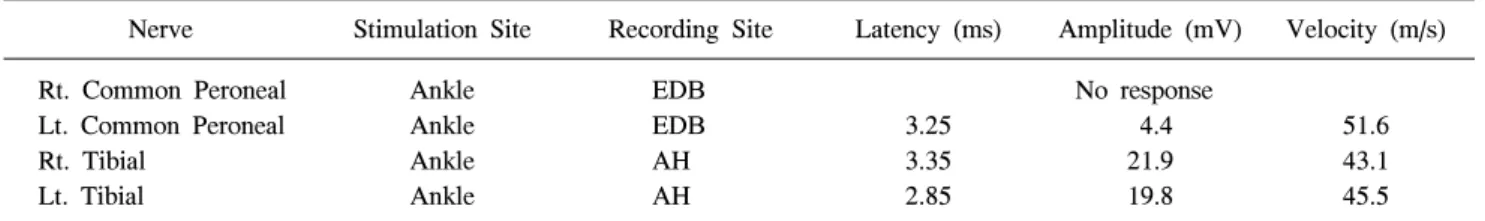
신경전도검사에서 왼쪽 정강신경(tibial nerve)과 종아리신 경(peroneal nerve)의 말단잠복기(terminal latency)와 F 파가 연장되어 있었고 종아리신경의

Key words: Microneurosurgery, Nerve extractor, Peripheral nerve injury, Sural nerve graft, Trigeminal nerve.. 서

However, in this case, we performed a sural nerve cable graft on the dam- aged temporal branch of a facial nerve on a male patient one month after a nerve injury.. We evaluated

A case of isolated injury of the lateral dorsal cutaneous branch of the sural nerve is reported. It was thought to be a direct result of compression to the distal sural nerve

Methods : Measurements were made from median nerve, ulnar nerve, deep peroneal nerve, tibial nerve of 44 upper extremities and 38 lower extremities with Excel

In fact, diverse pathologic conditions affecting small branch- es of the posterior tibial nerve or lateral and medial plantar nerve can cause symptoms of tarsal tunnel syndrome..

In this case, the intraneural ganglion cyst was found to track distally within for the nerve sheath along the common peroneal nerve to the recurrent anterior tibial branch for about

The medial sural cutaneous nerve (MSCN) and peroneal communicating nerve (PCN) conjoin in the calf area to form the sural nerve (SN).. In previous anatomic studies, there was