Address for correspondence;
Sun-Young Kim
Department of Neurology, Ulsan University Hospital, 877 Bangeojinsunhwando-ro, Dong-gu, Ulsan 682-714, Korea Tel: +82-52-250-8860 Fax: +82-52-250-7089 E-mail: [email protected]
Case Report
에틸린 글리콜 중독 이후 나타난 지연성 다발성 뇌신경병 및 다발신경뿌리신경병
울산대학교 의과대학 울산대학교병원 신경과1, 내과2
김민수
1․김선영
1․권지현
1․김욱주
1․정현철
2Multiple Cranial Neuropathy and Polyradiculoneuropathy as Delayed Sequelae of Ethylene Glycol Intoxication
Min-Soo Kim
1, Sun-Young Kim
1, Jee-Hyun Kwon
1, Wook-Joo Kim
1, Hyun-Chul Jung
21Department of Neurology, Ulsan University Hospital, Ulsan University College of Medicine, Ulsan; 2Department of Internal Medicine, Ulsan University Hospital, Ulsan University College of Medicine, Ulsan, Korea
Multiple cranial and peripheral neuropathies as a delayed sequellae of ethylene glycol poisoning is a less well known clinical entity and its information about long-term electrophysiological and clinical outcomes is limited. We report a 45-year-old male who presented with acute renal failure and subsequently developed multiple cranial neuropathy, respi- ratory failure, and flaccid tetraparesis. Through sequential electrophysiological studies, we would like suggest that the main pathophysiology of ethylene glycol-related neuropathy is a demyelinating polyradiculoneuropathy with secondary axonal degeneration.
Key Words: Ethylene glycol, Multiple cranial neuropathy, Polyneuropathy
Received 3 May 2013; received in revised form 12 July 2013; accepted 24 October 2013.
Introduction
Ethylene glycol (EG), a solvent found in products from anti- freeze fluid, is a rare cause of peripheral neuropathy.1 Multiple bilateral cranial nerve impairment is a delayed sequelae of ethylene glycol intoxication and usually develops 5 to 10 days after ingestion. It is often associated with limb weakness,
areflexia, and CSF albuminologic dissociation, mimicking Guillian- Barre Syndrome (GBS).2-5 However, the main pathophysiology of EG-related neuropathy is uncertain because few studies regarding sequential electrophysiological study or postmortem pathologic study for EG-related neuropathy have been performed.
Previous electrophysiological studies have shown predominantly axonopathy6or polyradiculoneuropathy.2,5 Here, we report a patient who attempted suicide by the ingesting EG and developed a delayed neurological deficit mimicking fulminant GBS. Serial electrophysiological evaluation and clinical outcomes are described to speculate the main pathophysiology of EG-related neuropathy.
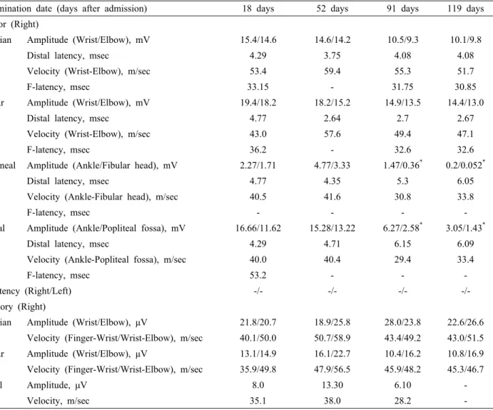
Table 1. Sequential electrophysiological study of the patient
Examination date (days after admission) 18 days 52 days 91 days 119 days
Motor (Right)
Median Amplitude (Wrist/Elbow), mV 15.4/14.6 14.6/14.2 10.5/9.3 10.1/9.8
Distal latency, msec 4.29 3.75 4.08 4.08
Velocity (Wrist-Elbow), m/sec 53.4 59.4 55.3 51.7
F-latency, msec 33.15 - 31.75 30.85
Ulnar Amplitude (Wrist/Elbow), mV 19.4/18.2 18.2/15.2 14.9/13.5 14.4/13.0
Distal latency, msec 4.77 2.64 2.7 2.67
Velocity (Wrist-Elbow), m/sec 43.0 57.6 49.4 47.1
F-latency, msec 36.2 - 32.6 32.6
Peroneal Amplitude (Ankle/Fibular head), mV 2.27/1.71 4.77/3.33 1.47/0.36* 0.2/0.052*
Distal latency, msec 4.77 4.35 5.3 6.05
Velocity (Ankle-Fibular head), m/sec 40.5 41.6 30.8 33.8
F-latency, msec - - - -
Tibial Amplitude (Ankle/Popliteal fossa), mV 16.66/11.62 15.28/13.22 6.27/2.58* 3.05/1.43*
Distal latency, msec 4.29 4.71 6.15 6.09
Velocity (Ankle-Popliteal fossa), m/sec 40.0 40.4 29.4 33.4
F-latency, msec 53.2 - - -
H-latency (Right/Left) -/- -/- -/- -/-
Sensory (Right)
Median Amplitude (Wrist/Elbow), μV 21.8/20.7 18.9/25.8 28.0/23.8 22.6/26.6
Velocity (Finger-Wrist/Wrist-Elbow), m/sec 40.1/50.0 50.7/58.9 43.4/49.2 43.0/51.5
Ulnar Amplitude (Wrist/Elbow), μV 13.1/14.9 16.1/22.7 10.4/16.2 10.8/16.9
Velocity (Finger-Wrist/Wrist-Elbow), m/sec 35.9/49.8 47.9/56.5 45.9/48.2 45.3/46.7
Sural Amplitude, μV 8.0 13.30 6.10 -
Velocity, m/sec 35.1 38.0 28.2 -
*conduction block was seen.
Case Report
A 45-year-old man visited the emergency room of our hospital with a confused mental state and abdominal pain after ingestion of anti-freeze fluid for a suicidal attempt. Initial assessments revealed hypertension (blood pressure: 187/94 mmHg), tachy- cardia (pulse: 125 beats/min), and tachypnea (respiratory rate: 27 breaths/min). The physical examination was unremarkable except for the drowsy mental status without any focal neurological deficit. Arterial blood gas analysis revealed a metabolic acidosis (pH 7.110, PO2 102.1 mmHg, PCO2 11.9 mmHg, bicarbonate 3.7 mmol/L). Serum biochemistry showed normal sodium (145 mmol/L), potassium (5.6 mmol/L), blood urea nitrogen (7.5 mg/dL) and creatinine (1.67 mg/dL) levels. The complete blood count indicated a mild leukocytosis. Liver function tests,
serum glucose, lactate, coagulation profile, chest radiogram, and electrocardiogram were normal. Urinalysis showed a mild pro- teinuria.
The patient was transferred to the intensive care unit, and he- modialysis was started due to anuria and severe metabolic acidosis. He improved quickly and became alert with stable vital signs. Eight days after admission, he developed a bilateral facial muscle weakness and became easily fatigued, and the patient was referred to us for neurological consultation of his bifacial weakness. On neurological examination, he showed bilateral peripheral type facial nerve palsy without limb weakness. Deep tendon reflexes were lost in his knees and ankles. Brain MRI with gadolinium enhancement was unremarkable, and cerebrospinal fluid (CSF) analysis revealed an elevated protein level (109.9 mg/dL) with normal glucose (65 mg/dL) and cell counts (4 white
blood cells/mm3). Tests for hepatitis-associated antigen, anti- nuclear antibody, serum VDRL, viral antibodies, serum protein electrophoresis and urine porphyrin screen came back as negative. Serum vitamin B12 and folate levels were within normal limits. Anti-ganglioside antibodies against GM1 and GD1 were also negative. Serum mercury and lead levels were normal.
The serum EG level at 7 days after hemodialysis was withn normal limits.
Since then, the patient started to develop progressive hearing impairment, swallowing difficulty and voice change over 10 days.
Examination revealed bilateral sensorineural hearing loss, diminished gag reflex, and vocal cord paralysis. The patient showed a progressive worsening of mentality, bilateral 7th, 8th, 9th, 10th, and 12th cranial nerve palsies, and lower limb predom- inant quadriparesis, and finally developed a respiratory failure at 11 days after admission. The patient was artificially ventilated with a tracheostomy and treated with a hemodialysis. Fifty days after admission, the patient showed some neurological improve- ment with an improved mentality and limb muscle weakness, and recovery of sensorineural hearing loss. Dysphagia, hoarseness, bilateral facial muscle weakness, and lower limb paralysis (ankle flexion and extension weakness grade 1/5) were still evident.
During admission, he stayed at the intensive care unit for 61 days and was dialyzed for 40 days. Three months after admission, his upper limb muscle power became normalized with a marked improvement of bifacial weakness and dysphagia, but with a per- sisting lower extremity weakness (grade 3/5 in proximal legs and grade 1/5 in distal legs). The patient also complained of distal limb paresthesia, especially in both feet.
Electrophysiological examination was performed four times during his admission, and the results are summarized in Table 1.
The first nerve conduction study at 18 days after admission showed a loss of F-waves of bilateral peroneal nerves, gastro- cnemius-soleus H-reflexes, and bilateral facial compound muscle action potentials (CMAPs). Blink responses were also lost bilaterally. On brainstem auditory evoked potential (BAEP) study, none of the wave was evoked, indicating severe bilateral end-organ damage. Nerve conduction studies were repeated 52 days after admission and showed a persistent loss of F-waves from all tested nerves and gastrocnemius-soleus H-reflexes. A third electrophysiological examination was performed 91 days af- ter admission. Motor nerve conduction studies in the lower limbs showed reduced CMAPs and prolonged terminal latencies with
probable conduction blocks. Needle electromyogram showed moderate abnormal spontaneous activities in the distal lower legs with reduced recruitment patterns. A sensory nerve conduction study in the lower limbs showed low reduced compound nerve action potentials. F-waves and gastrocnemius-soleus H-reflexes of the lower limbs remained lost. The final electrophysiological examination (at 119 days after admission) showed a further re- duction of CMAPs and CNAPs of motor and sensory nerves of the lower limbs. Probable conduction blocks and loss of F-waves and gastrocnemius-soleus H-reflexes of the lower limbs persisted.
The BAEP returned to normal. After 119 days of hospitalization, the patient was able to walk by himself but with using an ankle orthosis because of persistent foot drop. He still had mild bilateral facial weakness, as well as numbness and paresthesias in both feet.
Discussion
EG is a common constituent of antifreeze, paint, and other de- tergents, and is responsible for both inadvertent and intentional poisoning, with a reported incidence of exposure in the United States of almost 5,000 episodes annually.1 EG itself is a non-toxic agent but its metabolites, such as oxalic acid, glycolate and glyox- ylate are toxic. Metabolic acidosis and oxalate crystals deposition produce variable clinical manifestations. Symptoms and signs of EG poisoning are traditionally divided into three chronological stages.3 (1) CNS depression, ranging from stupor to coma (3-12 hours after ingestion), (2) possible cardiopulmonary failure (within 72 hours); and (3) renal failure, ranging from mild azote- mia to frank anuria. Multiple cranial neuropathies including facial paralysis develop very rarely, occurring 6-15 days after ingestion of EG.2-5 As a delayed neurological syndrome of EG poisoning, facial diplegia is a classic finding in addition to multiple cranial nerve deficits, limb weakness, areflexia, and CSF albuminocyto- logic dissociation.2,5-7
With an exclusion of other toxic or viral neuropathies, a diag- nosis of EG-related toxic neuropathy was made in our patient, al- though he did not show toxic levels of EG in the serum, which is probably due to the delayed sampling after several cycles of dialysis. Although the definitive laboratory test for EG poisoning is the elevated serum concentration of EG, it often shows poor correlation with an ingested dose and is difficult to define toxic serum level exactly.8 Because our patient had a clear history of
Figure 1. Serial motor-conduction study findings for the peroneal and tibial nerves in the patient. (A, C) Conduction block is present between the ankle and knee. (B, D) Follow-up study suggested a secondary axonal degeneration.
EG ingestion with an elevated anion gap at presentation, which was presumed to be secondary to the renal failure, it is almost cer- tain that EG intoxication is responsible for the clinical features of the patient. Furthermore, all the clinical findings are compatible with a delayed neurological syndrome of EG-poisoning as pre- viously reported by others.2,5-7 All the previously reported cases of delayed sequellae of EG-poisoning are characterized by develop- ment of multiple cranial neuropathies associated with severe limb weakness, which is worse in lower extremities.
Serial neurological examination and neurophysiological as- sessment was available in our case. The initial nerve conduction study only showed a loss of F-waves and H-reflex, which strongly suggested a lumbosacral polyradiculopathy. However, subsequent nerve conduction study began to suggest an evidence of demyeli- nating neuropathy, such as probable conduction blocks and de- layed distal latencies with slowing of nerve conduction velocities in lower extremities. Finally, a progressive reduction of CMAPs, loss of sural nerve CNAPs, and appearance of moderate abnormal spontaneous activites on needle EMG in subsequent studies in- dicates that secondary axonal degeneration had been followed by
segmental demyelination. Thus, considering the serial neuro- physiological findings and high CSF protein level observed in this patient, the overall feature suggested a demyelinating poly- radiculoneuropathy with a secondary axonal degeneration.
Alzouebi et al5 described sequential neurophysiological exami- nation of EG-related acute polyradiculoneuropathy, and reported reversible conduction blocks with a predominant axonal degener- ation in the distal legs. In their report, conduction block was reversed within a few months without additional features of demyelinating neuropathy. In the present case, the reduction of CMAPs more than 50% was observed between distal and prox- imal stimulation sites in peroneal and posterior tibial nerves, which is compatible with conduction block (Figure 1). In addi- tion, our case also showed a prolonged terminal latencies and slowing of nerve conduction velocities. Therefore, compared with the case of Alzouebi et al,5 the neurophysiological pattern observed in our case is more close to that of Guillain-Barré syn- drome where conduction blocks associated with slowing of nerve conduction velocities and prolonged distal latencies are fre- quently observed after several days to week after the onset of
A B
C D
symptoms.
In contrast to our case, EG-related neuropathy has been known to take a form of primary axonal polyneuropathy or polyradiculopathy.2,5,6 The reason why the same chemical could produce different pathophysiological feature is unclear, but this might be related to the severity and stage of the nerve injury caused by EG. The doses of toxic material and host factors, such as individual variability in the hepatic P450 system, may also play a role in determining severity and nature of the neuropathy. While the pathogenesis of severe polyradiculopathy observed in EG in- toxication is also unclear, deposits of calcium oxalate in the lep- tomeninges or its blood vessels and in the subarachnoid space of cranial nerves have been reported at postmortem examinations.9 Thus, the deposition of oxalate crystals in the meninges may di- rectly damage the proximal nerve roots and secondary axonal de- generation could take place in peripheral nerves as suggested in our case. This feature could be regarded as a unique feature of EG-related neuropathy because many other toxic neuropathies are characterized by dying back axonal degeneration.10
In conclusion, this case illustrates that EG-related neuropathy could show features of demyelinating including conduction blocks, and can mimic Guillain-Barré syndrome. EG poisoning should be considered in patients with rapidly progressive neuro- pathy who are associated with acute renal failure and high anion gap.
REFERENCES
1. Pellegrino B, Parravani A, Cook L, Mackay K. Ethylene gly- col intoxication: Disparate findings of immediate versus de- layed presentation. W V Med J 2006;102:32-34.
2. Zhou L, Zabad R, Lewis RA. Ethylene glycol intoxication: elec- trophysiological studies suggest a polyradiculopathy. Neurology 2002;59:1809-1810.
3. Lewis LD, Smith BW, Mamourian AC. Delayed sequelae after acute overdoses or poisonings: cranial neuropathy related to ethylene glycol ingestion. Clin Pharmacol Ther 1997;61:692- 699.
4. Hasbani MJ, Sansing LH, Perrone J, Asbury AK, Bird SJ.
Encephalopathy and peripheral neuropathy following diethylene glycol ingestion. Neurology 2005;64:1273-1275.
5. Alzouebi M, Sarrigiannis PG, Hadjivassiliou M. Acute poly- radiculoneuropathy with renal failure: mind the anion gap. J Neurol Neurosurg Psychiatry 2008;79:842-844.
6. Tobe TJ, Braam GB, Meulenbelt J, van Dijk GW. Ethylene glycol poisoning mimicking Snow White. Lancet 2002;359:
444-445.
7. Baldwin F, Sran H. Delayed ethylene glycol poisoning present- ing with abdominal pain and multiple cranial and peripheral neuropathies: a case report. J Med Case Reports 2010;4:220.
8. Barceloux DG, Krenzelok EP, Olson K, Watson W. American academy of clinical toxicology practice guidelines on the treat- ment of ethylene glycol poisoning. Ad Hoc Committee. J Toxicol Clin Toxicol 1999;37:537-560.
9. Froberg K, Dorion RP, McMartin KE. The role of calcium oxalate crystal deposition in cerebral vessels during ethylene glycol poisoning. Clin Toxicol (Phila) 2006;44:315-318.
10. Le Quesne PM. Electrophysiological investigation of toxic neuropathies. Acta Neurol Scand Suppl 1982;92:75-87.