INTRODUCTION
The tarsal tunnel is a fibro-osseous space located posterior to the medial malleolus. It consists of an upper and lower com- partment, and is bound superficially by the flexor retinaculum and the medial surfaces of the talus and calcaneus on its deep surface (1, 2). Several anatomic structures pass through the tar- sal tunnel, including the posterior tibial nerve, the posterior tibial artery and vein, and three medial tendons of the flexor hallucis longus, flexor digitorum longus, and the posterior tibi-
alis muscle (Fig. 1).
Tarsal tunnel syndrome is an entrapment neuropathy of the posterior tibial nerve or one of its branches within the tarsal tunnel (2-4), where its symptoms and signs include pain and paresthesia in the toes, sole, or heel. To the best of our knowl- edge, in up to 50% of the cases, potential causes of tarsal tunnel syndrome could be identified and included intrinsic lesions, such as accessory muscles, ganglion cysts, neurogenic tumors, varicose veins, lipomas, synovial hypertrophy, and scar tissue.
Otherwise, various extrinsic conditions, such as foot deformi-
J Korean Soc Radiol 2012;66(2):183-192
Received August 9, 2011; Accepted November 8, 2011 Corresponding author: Sheen-Woo Lee, MD Department of Radiology, Gil Hospital, Gachon University of Medicine and Science, 1198 Guwol-dong, Namdong-gu, Incheon 405-760, Korea.
Tel. 82-32-460-3060 Fax. 82-32-460-3065 E-mail: [email protected]
The study was partly supported by the Gachon University New Investigator Fund.
Copyrights © 2012 The Korean Society of Radiology
Purpose: The purpose of this study was to access the diverse conditions that lead to the clinical manifestations of tarsal tunnel syndrome and evaluate the usefulness of magnetic resonance imaging (MRI) in preoperative evaluation.
Materials and Methods: Thirty-three patients who underwent ankle MRI and sur- gery under the impression of tarsal tunnel syndrome were retrospectively analyzed.
The findings on ankle MRI were categorized into space occupying lesions within the tarsal tunnel, space occupying lesions of the tunnel wall, and non-space occupying le- sions. Associated plantar muscle atrophy was also evaluated. Medical records were re- viewed for correlation of nerve conduction velocity (NCV) and surgical findings.
Results: There were 21 space occupying lesions of the tarsal tunnel, and eight le- sions of tarsal tunnel wall. There were three cases with accessory muscle, three with tarsal coalition, five with ganglion cysts, one neurogenic tumor, five flexor retinacu- lum hypertrophy, three varicose veins, and nine with tenosynovitis of the posterior tibialis, flexor digitorum longus, or flexor hallucis longus tendon. One patient was found to have a deltoid ligament sprain. Of the 32, eight patients experienced fatty atrophic change within any one of the foot muscles. NCV was positive in 79% of the MRI-positive lesions.
Conclusion: MRI provides detailed information on ankle anatomy, which includes that of tarsal tunnel and beyond. Pathologic conditions that cause or mimic tarsal tunnel syndrome are well demonstrated. MRI can enhance surgical planning by in- dicating the extent of decompression required, and help with further patient man- agement. Patients with tarsal tunnel syndrome can greatly benefit from preoperative MRI. However, it should be noted that not all cases with tarsal tunnel syndrome have MRI-demonstrable causes.
Index terms
Magnetic Resonance Imaging Nerve Entrapment
Space Occupying Lesion Tarsal Tunnel Syndrome
The Usefulness of the Preoperative Magnetic Resonance Imaging Findings in the Evaluation of Tarsal Tunnel Syndrome
1족근관증후군 환자에 있어서 수술 전 MRI의 유용성1
Hyun Jin Jung, MD
1, Sheen-Woo Lee, MD
1, Yu Mi Jeong, MD
1, Hye-Young Choi, MD
1, Hyung-Sik Kim, MD
1, Hong Gi Park, MD
2, Ji Hoon Kwak, MD
2Departments of 1Radiology, 2Orthopedic Surgery, Gil Hospital, Gachon University of Medicine and Science, Incheon, Korea
we compared the MRI results. Clinical notes on the patients were evaluated for correlation of nerve conduction velocity test, treatment, and clinical course of the patients. Two radiologists from our institution, one with 10 years and the other with 3 years of experience, had performed a retrospective analysis of each MR imaging examination. For each patient, radiologist in- terpretations were categorized into space occupying lesions within the tarsal tunnel, space occupying lesions of the tunnel wall, and non-space occupying lesions. Associated plantar muscle atrophy was also evaluated. Surgery was performed by orthopedic surgeons specializing in foot and ankle joints.
All 33 patients underwent tarsal tunnel release; however, one patient was later excluded from the study. In this instance the MRI indicated diffuse edema and atrophy of muscles in the lower leg and foot, and multifocal bone marrow edema in the tarsal bones. The patient had intramedullary nailing in the tibia due to prior fracture, and regional osteopenia of the ankle and foot. The result of the nerve conduction velocity (NCV) study indicated both sural and tibial neuropathy. On review of medi- cal record, there was no indication that the tarsal tunnel syn- drome was the sole cause of the patient’s plantar paresthesia.
MRI Technique
All of the MR imaging examinations were conducted using a 1.5-T magnet (MAGNETOM Vision and MAGNETOM Avan- to 1.5T, Siemens Medical Solutions, Muenchen, Germany), and identical MR protocols were adopted for each of the examina- tions. Our standard ankle protocol included axial, sagittal, and coronal turbo spin-echo T2-weighted imaging [repetition time (TR)/echo time (TE) effective, 3000/80] and spin-echo T1- weighted imaging (TR/TE effective, 513/16). Fat suppression was applied to both sagittal and axial T2-weighted imaging and axial proton density imaging (TR/TE, 3500/10) was added.
Other parameters include a matrix of 256 × 220, and a slice thickness of 3 mm. A field of view of 45 × 150 for the sagittal sequence, 50 × 200 for the coronal sequence, and 50 × 150 for the axial sequence were used.
For evaluation of tarsal tunnel syndrome-related muscle at- rophy, the intrinsic muscles innervated by the lateral and medi- al plantar nerve were interpreted on coronal T1-weighted im- ages. Quadratus plantae, abductor hallucis, flexor digitorum brevis, and abductor digiti minimi muscles were evaluated ties, hypertrophic and accessory muscles, accessory ossicle, and
excessive pronation during participation in some sports, are re- lated to the syndrome (1).
Magnetic resonance imaging (MRI) is well known to dem- onstrate good soft tissue contrast, and provides significant ana- tomic detail for evaluation of osseous disorders and soft tissue structures of the foot and ankle. It can also help identify patho- logic conditions of the ankle and foot that can mimic tarsal tunnel syndrome. The purpose of this study was to evaluate the role of MRI in preoperative evaluation and to provide an over- view of the diverse conditions that lead to tarsal tunnel syn- drome.
MATERIALS AND METHODS
Patients
The retrospective study population included patients from March 2006 to March 2010, who visited the orthopedic surgery department in a tertiary hospital after the initial diagnosis of tarsal tunnel syndrome. The study received ethical approval from the institution. The presenting symptoms were pain in the medial ankle, forefoot, midfoot or heel, tingling sensation in the sole, including a combination of more than two of these symptoms (Table 1). Patients who did not undergo surgery or MRI were excluded. One patient with both the operation and MRI was also excluded, whose medical record showed that the patient had diffuse synovitis of the ankle as observed on MRI, and subsequent arthroscopic debridement of tibiotalar arthritis did not relieve the patient’s recurrent symptoms. The final number of patients included in the study was 33 patients, 17 men and 16 women, who ranged in age from 18 to 66 years (mean age was 44 years). The operative records from the ortho- pedic surgeons were used as the reference standard with which Table 1. Various Symptoms Related to Tarsal Tunnel Syndrome
Symptoms Number of Patients
Medial ankle pain 7
Fore foot pain 2
Mid foot pain 2
Whole foot pain 6
Heel pain 4
Sole tingling sensation 5
Great toe numbness or pain 4
Two or more symptoms 3
sults. Three patients did not show any lesion on MRI; however, positive NCV results lead to surgery for release of the tarsal tunnel in there three patients. Five patients underwent surgery despite negative MRI and NCV findings due to severe symp- toms and heavy clinical suspicion, and were also classified into the MRI-negative group since their symptoms showed im- provement after surgery (Table 3).
Of the 32 patients, MRI revealed pathologic findings related to tarsal tunnel in 24 patients (Fig. 2). Six patients out of 23 with space occupying lesions showed a couple of pathologic conditions within the tunnel resulting in a total of 29 patholog- ic lesions. The variable causes of the syndrome were classified into two categories, space occupying lesions and non-space oc- where atrophy was graded on a 4-point scale: grade 0, no fat or
minimal fatty streaks; grade 1, increased fat within the muscle but a relatively greater amount of muscle; grade 2, equal amounts of fat and muscle; and grade 3, greater amount of fat as com- pared to muscle.
Nerve Conduction Velocity Test
For an additional diagnostic approach, NCV was conducted in 22 patients when clinical symptoms and signs are indistinct to diagnose. Patches containing surface electrodes were applied at two specific points along a nerve course. Electrical impulses were administered and the resulting electrical activity was re- corded. Tarsal tunnel syndrome was suggested when an NCV or latency was delayed for a longer than expected time period along the medial or lateral plantar nerve.
RESULTS
Surgery was decided based on MRI and NCV study evalua- tions (Table 2). There were 24 patients with positive ankle le- sions and 8 patients without any positive ankle lesion based on MRI analysis. Twenty-three patients exhibited space occupying lesions in the tarsal tunnel, and 1 patient analysis demonstrated a non-space occupying lesion (ankle joint instability) causing tarsal tunnel syndrome with 14 cases showing positive NCV re-
Table 2. Correlation of MRI with NCV in Patients with Tarsal Tunnel Syndrome
MRI Lesion (+) MRI Lesion (-)
NCV (+) 14 3
NCV (-) 10 5
Note.-NCV (-) = Indicated no available test report, or the result was not consistent with tarsal tunnel syndrome.
NCV = nerve conduction velocity
Table 3. Symptom Relief Following Tarsal Tunnel Release
Symptom Relief Persistent Symptom
MRI (+) 20 4
MRI (-) 8 0
Note.-MRI (+)/(-) = Indication of whether or not pathologic lesions were demonstrated in the tarsal tunnel.
Fig. 1. Normal anatomy of the tarsal tunnel. (A) Axial T1-weighted image and (B) coronal T1-weighted image show posterior tibialis (PT), flexor digitorum longus (FDL), flexor hallucis longus (FHL), posterior tibial artery (PTA), posterior tibial vein (PTV), medial/lateral plantar nerve (MN/LN), and flexor retinaculum (FR).
A B
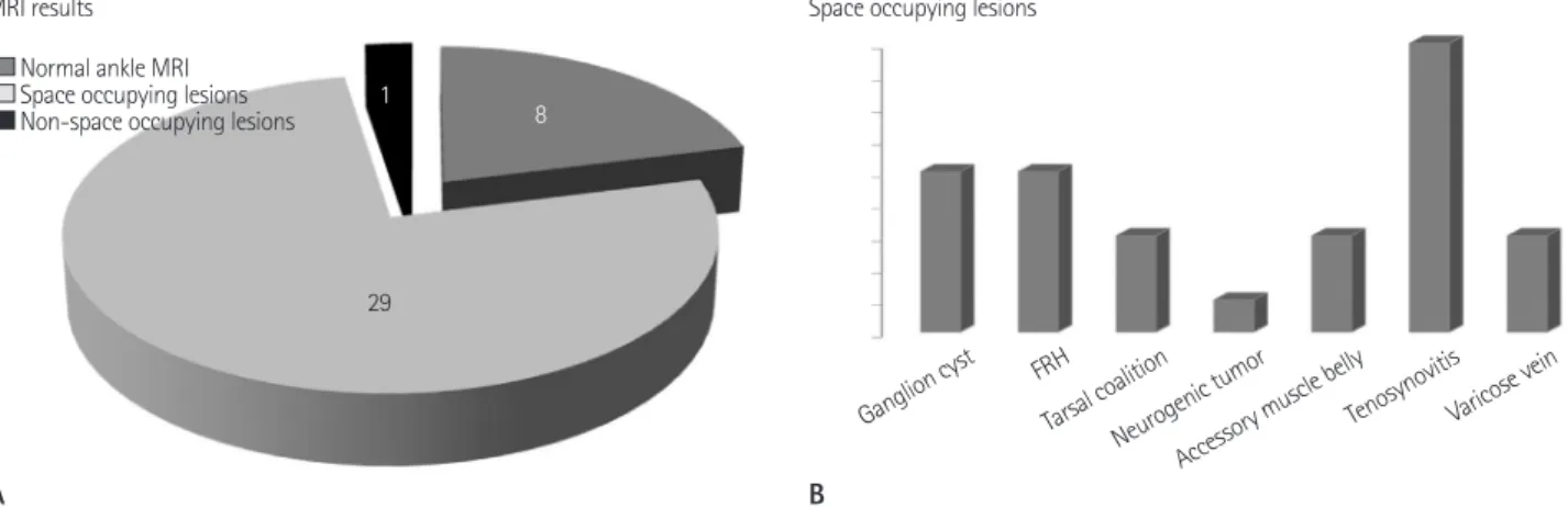
Fig. 2. MRI results and various types of space occupying lesions.
A. Total of 29 space occupying lesions and 1 non-space occupying lesion are detected in 24 patients. Eight patients show no abnormality in the preoperative ankle MRI.
B. Various types of space occupying lesions such as ganglion cyst are demonstrated.
Note.-FRH = flexor retinaculum hypertrophy
Fig. 3. Talocalcaneal coalition. (A) Coronal T1-weighted and (B) T2-weighted MR images show tarsal tunnel syndrome due to fibrous talocalca- neal coalition (arrow). (C) Axial T1-weighted image demonstrates an enlarged mid facet (black arrow) of subtalar joint compressing medial plan- tar nerve (white arrow).
Fig. 4. Accessory muscle belly. (A) Axial T1-weighted, (B) proton density and (C) fat-saturated T2-weighted MR images show a flexor digitorum accessorius within the tarsal tunnel (arrow).
A
A
A
B
B
C
C B
Ganglion cyst FRH Tarsal coalition
Neurogenic tumor Accessory muscle belly
Tenosynovitis Varicose vein
MRI results Space occupying lesions
1 8
29 Normal ankle MRI
Space occupying lesions Non-space occupying lesions
monly involved in fatty atrophic change. The mean duration of symptoms was 14 months, ranging from 1 month to 36 months.
A list of the muscles that underwent atrophic change is shown in Table 5.
MRI-detected lesions were confirmed at the time of surgery and appropriate surgery was performed to relieve the symp- toms. One patient with paresthesia in the sole was shown to have edematous flexor retinaculum and varicocele on MRI.
Tarsal tunnel release relieved the presenting symptom, but the subject returned for an outpatient visit a few months later due to forefoot arthritis, which was known and already expected due to the initial MRI study (Fig. 10). In 8 patients who under- went surgery based on the NCV result or clinical suspicion, there was no space occupying lesion within the tunnel; howev- cupying lesions. Furthermore, space occupying lesions were
categorized as tarsal tunnel lesions and tarsal tunnel wall le- sions (Figs. 2-8) (Table 4). Non-space occupying lesions were defined as ankle lesions other than the tarsal tunnel proper; one patient was revealed to have a deltoid ligament sprain with an- kle joint instability causing possible dynamic nerve compres- sion, thus this lesion was classified into the non-space occupy- ing lesion group (Fig. 9).
Of the total study population, 24 patients showed no fatty atrophic change in foot intrinsic muscles and the remaining 8 patients demonstrated atrophic change in at least one of the foot muscles. Of the 8, 1 patient had atrophy of both the qua- dratus plantae and the abductor digiti minimi muscle, simulta- neously. The abductor digiti minimi muscle was most com-
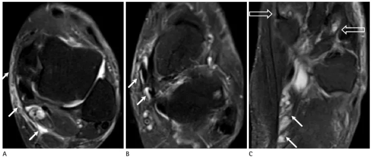
Fig. 5. Varicose vein. (A) Axial T1-weighted, (B) sagittal, and (C) axial fat-saturated T2-weighted MR images reveal tortuous dilated veins in the tarsal tunnel (arrows).
Fig. 6. Ganglion cyst. (A) Coronal T1-weighted and (B) sagittal fat-saturated T2-weighted MR images obtained at the tarsal tunnel level show a septated cystic lesion (white arrow in A, B) that was pathologically proven to be a ganglion cyst. (C) Axial T1-weighted image reveals a medial plantar nerve (double arrow) abutted by the ganglion cyst (single black arrow).
A
A
B
B
C
C
er, tarsal tunnel release relieved the pain or numbness experi- enced by the patients.
DISCUSSION
The tarsal tunnel consists of an upper (tibiotalar) compart- ment and a lower (talocalcaneal) compartment. The upper tun- nel is located under the deep aponeurosis and bordered by a bony floor with the posterior aspect of the tibia and the talus.
The lower tunnel is found under the flexor retinaculum and its bony floor is composed of the posteromedial aspect of the ta- lus, the inferomedial aspect of the navicular bone, and the me- dial aspects of the sustentaculum tali and calcaneus (1, 2). In the tarsal tunnel, the posterior tibial nerve trifurcates into its terminal branches, 1.3-1.5 cm proximal to the tip of the medial malleolus (5). These are the medial and lateral plantar nerves and the medial calcaneal nerve. Any lesions causing compres- sion of the nerves may lead to tarsal tunnel syndrome.
Clinical findings may vary and symptoms and signs are com- monly obscure and diffuse; therefore, a clear-cut diagnosis of tarsal tunnel syndrome is sometimes difficult. Furthermore, electro-diagnostic studies do not provide a definitive diagnosis.
Our study shows that significant number of patients with ab- normal MRI findings within the tunnel also had negative NCV results. MRI, with its excellent demonstration of musculotendi- nous and neurovascular structures, is able to clearly reveal the Table 4. Space Occupying Lesions Causing Tarsal Tunnel Syndrome
SOL in the Tarsal Tunnel SOL of Tarsal Tunnel Wall Ganglion cyst (5) Flexor retinaculum hypertrophy (5) Accessory muscle belly (3)
Tenosynovitis of: Tarsal coalition (3) Posterior tibialis (3),
Flexor digitorum longus (1), Flexor hallucis longs tendon (5) Varicose vein (3)
Neurogenic tumor (1)
The number in the parenthesis stands for the patient population.
Note.-SOL = Space Occupying Lesion
Fig. 7. Neurogenic tumor. (A) Axial T1-weighted and (B) T2-weighted MR images show an isosignal/high signal intensity mass compressing the posterior tibial vessels and nerve within the tarsal tunnel (black arrows). (C) Axial fat suppressed T2-weighted image shows central target-like low signal intensity in the mass (white arrow). (D) Coronal T1-weighted image shows an oval shaped mass replacing the tarsal tunnel, which was pathologically proven to be a neurofibroma (white arrow).
Fig. 8. Tenosynovitis. Axial fat suppressed T2-weighted MR image demonstrates fluid collection in the tendon sheath along the tibialis posterior, flexor digitorum longus, and flexor hallucis longus tendon (arrows), suggestive of tenosynovitis.
A B C D
tibial nerve, such as the lateral and medial plantar nerve, not only serve as a sensory nerve but also innervate foot muscles (2-4). Eight patients showed fatty atrophy of the foot muscles, most of which were mildly involved. Unlike our expectation, the degree of muscle atrophy did not correlate with the dura- tion of disease. One patient with three months of heel pain showed grade 3 atrophy of the quadratus plantae muscle, where- as another with 3 years of the same symptoms had grade 1 atro- phy of the abductor digiti minimi. However, a future study with a larger population would be able to better address the general- ization of the relation between clinical duration and extent of fatty muscle atrophy.
In this study, 8 patients were evaluated with preoperative MRI that demonstrated no abnormality within or outside of the tarsal tunnel. Clinical speculation or NCV tests supported the preoperative impression of tarsal tunnel syndrome; there- anatomy of the tarsal tunnel and its contents, and the presence
or extent of space-occupying lesions leading to tarsal tunnel syndrome (6). Furthermore, there are other lower extremity le- sions that cause symptoms in the foot, where MRI can help de- termine a more accurate differential diagnosis leading to im- proved management. Diffuse edema and atrophy of muscles, bone marrow edema, associated with NCV findings of both su- ral and tibial neuropathy could be an indicator of reflex sympa- thetic dystrophy (7, 8).
It is widely accepted that in about 50% of cases, the cause of tarsal tunnel syndrome is not readily identifiable. In the remain- ing cases, the mechanical cause of nerve impingement may be found during surgery (1, 9). Frey and Kerr (10) studied 33 pa- tients with 40 feet complaining of pain along the course of the posterior tibial nerve or its branches. All patients were found positive for Tinel’s sign representing posterior tibial nerve irrita- tion and clinically suspicious for tarsal tunnel syndrome, and MRI findings were finally confirmed at the time of surgery in 19 patients. Our study is concordant with Frey’s study, as we ob- served that 24 of 32 patients had abnormal findings that caused tarsal tunnel syndrome based on MRI evaluation which were confirmed at the time of surgery. However, the difference may not be significant since our study reviewed only a small number of patients, highlighting a limitation of the study.
As previously mentioned, tarsal tunnel syndrome is a type of entrapment neuropathy. The major branches of the posterior
Table 5. Muscle Atrophy in Tarsal Tunnel Syndrome
Patient Muscle with Atrophy Grade Duration
A QP/ADM 1/1 8 months
B ADM 1 1 month
C AH 1 8 months
D ADM 1 3 years
E QP 3 3 months
F ADM 1 1 year
G ADM 1 1 month
H AH 1 3 years
Note.-ADM = abductor digiti minimi, AH = abductor hallucis, QP = qua- dratus plantae
Fig. 9. Ankle instability.
A. Axial fat suppressed T2-weighted MR image reveals a deep deltoid ligament tear (single arrow) with associated marrow edema of the medial talar dome (double arrow).
B. Axial T1-weighted MR image demonstrates the medial plantar nerve (arrow) located near the deltoid ligament insertion, leading to nerve com- pression due to ankle joint instability.
C. Coronal T1-weighted image show focal marrow signal change of the medial talar dome (arrow).
A B C
causing plantar pain, also support the suggestion that MRI is superior to other imaging modalities in demonstrating the overall pathologic conditions.
In fact, diverse pathologic conditions affecting small branch- es of the posterior tibial nerve or lateral and medial plantar nerve can cause symptoms of tarsal tunnel syndrome. For in- stance, the medial calcaneal nerve penetrates the flexor retinac- ulum and branches to sensory nerves innervating the skin cov- ering the medial aspect of the Achilles tendon, the posteromedial aspect of the heel, and the plantar fat pad. The inferior calcane- al nerve also originates from the lateral plantar nerve at the lev- el of the medial malleolus, sending out motor branches to the abductor digiti minimi muscle and sensory branch for the an- terior calcaneal tubercle (5). These branches are relatively small compared to the lateral or medial plantar nerves and may be branched more distally in the tarsal tunnel where a relatively small fat component and other adjacent structures prevent them from being visualized on MRI. Another considerable fac- tor influencing the yield of MRI is a course of the branch of the lateral plantar nerve. The first branch of the lateral plantar nerve arises as the first branch of the lateral plantar nerve or directly from the posterior tibial nerve and courses in a medial to later- al direction between the abductor hallucis muscle and the me- dial calcaneal tuberosity (11-13). It is a mixed sensory and mo- fore, tarsal tunnel release was performed and all patients were
satisfied with the results. It might be problematic that preopera- tive MRI does not provide diagnostic clues in all patients suspi- cious with tarsal tunnel syndrome. However, evaluation to de- termine the presence of pathologic lesions before surgery can be helpful to surgeons when planning treatment options. Find- ings from the patient who showed no tarsal tunnel lesion, but distal varicosity compressing the abductor hallucis muscle Fig. 10. Simultaneous lesions in the tarsal tunnel and forefoot.
A, B. Axial fat-saturated T2-weighted MR images show thickening and edema of flexor retinaculum, along with mild tenosynovitis of posterior tibialis and flexor digitorum (white arrows).
C. Varicose vein is noted in the plantar aspect (white arrows). There are subchondral cysts and bone marrow edema in the forefoot (open arrows).
Release of flexor retinaculum relieved the foot pain, but the patient came back due to forefoot arthritis.
Fig. 11. Normal anatomical course of the lateral plantar nerve.
A. The lateral plantar nerve (white arrow) courses downward between the medial border of the quadrates plantae muscle and the lateral border of the abductor hallucis muscle.
B. The nerve traverses more distally and changes its direction laterally where it is compressed by the medial border of the quadrates plantae muscle (asterisk).
A
A
B
B
C
and non-space occupying lesions and correlate nerve compres- sion with intrinsic muscle atrophy in tarsal tunnel syndrome.
REFERENCES
1. Erickson SJ, Quinn SF, Kneeland JB, Smith JW, Johnson JE, Carrera GF, et al. MR imaging of the tarsal tunnel and re- lated spaces: normal and abnormal findings with anatom- ic correlation. AJR Am J Roentgenol 1990;155:323-328 2. Narváez JA, Narváez J, Ortega R, Aguilera C, Sánchez A,
Andía E. Painful heel: MR imaging findings. Radiographics 2000;20:333-352
3. Cimino WR. Tarsal tunnel syndrome: review of the litera- ture. Foot Ankle 1990;11:47-52
4. Schon LC, Baxter DE. Neuropathies of the foot and ankle in athletes. Clin Sports Med 1990;9:489-509
5. Delfaut EM, Demondion X, Bieganski A, Thiron MC, Mest- dagh H, Cotten A. Imaging of foot and ankle nerve en- trapment syndromes: from well-demonstrated to unfamil- iar sites. Radiographics 2003;23:613-623
6. Finkel JE. Tarsal tunnel syndrome. Magn Reson Imaging Clin N Am 1994;2:67-78
7. Graif M, Schweitzer ME, Marks B, Matteucci T, Mandel S.
Synovial effusion in reflex sympathetic dystrophy: an ad- ditional sign for diagnosis and staging. Skeletal Radiol 1998;27:262-265
8. Kurvers HA, Hofstra L, Jacobs MJ, Daemen MA, van den Wildenberg FA, Kitslaar PJ, et al. Reflex sympathetic dys- trophy: does sympathetic dysfunction originate from pe- ripheral neuropathy? Surgery 1996;119:288-296
9. Rosenberg ZS, Beltran J, Bencardino JT. From the RSNA Refresher Courses. Radiological Society of North America.
MR imaging of the ankle and foot. Radiographics 2000;20:
S153-S179
10. Frey C, Kerr R. Magnetic resonance imaging and the evalua- tion of tarsal tunnel syndrome. Foot Ankle 1993;14:159-164 11. Louisia S, Masquelet AC. The medial and inferior calcaneal
nerves: an anatomic study. Surg Radiol Anat 1999;21:169-173 12. Davis TJ, Schon LC. Branches of the tibial nerve: anatomic
variations. Foot Ankle Int 1995;16:21-29
13. Rondhuis JJ, Huson A. The first branch of the lateral plan- tar nerve and heel pain. Acta Morphol Neerl Scand 1986;
tor nerve that innervates the abductor digiti minimi muscle and occasionally the flexor digitorum brevis and quadratus plantae muscles, providing sensory branches to the calcaneal periosteum and the long plantar ligament (13). There are two anatomically vulnerable points to nerve entrapment during the course of the first branch of the lateral plantar nerve. According to Recht et al. (14), the nerve changes its direction at the inferi- or margin of the abductor hallucis where it is compressed be- tween the abductor hallucis and the medial aspect of the qua- dratus plantae muscles. It traverses more distally, anterior to the medial calcaneal tuberosity and again could be compressed if accompanied by plantar enthesophyte. In our study, 1 patient with heel pain who demonstrated no specific findings on MRI was found to have no abnormality in the tarsal tunnel and the first branch of the lateral plantar nerve was compressed by the medial border of the quadratus plantae muscle during surgery (Fig. 11).
In this report, we reviewed the anatomy of the tarsal tunnel and the causes of tarsal tunnel syndrome. With MR imaging, it is possible to visualize the flexor retinaculum, the contents of the tarsal tunnel, posterior tibial nerve and its branches, as well as detect outer lesions that may affect the tarsal tunnel. It is dif- ficult to diagnose tarsal tunnel syndrome due to overlapping clinical manifestations, and electrodiagnostic studies such as NCV do not provide a clear diagnosis. Therefore, it is necessary to use an additional method for accurate demonstration of the intrinsic and extrinsic causes of tarsal tunnel syndrome. Use of MRI and information provided by MRI indicates the extent of decompression required, and thereby may enhance surgical planning and further management of treatment. At the same time, we reiterate that patients with normal MRI experienced improvement and relief following the tarsal tunnel release, which may indicate that MRI is not able to determine the cause of the symptoms. Further study for the diagnostic improvement of MRI in tarsal tunnel syndrome should be performed.
One of the limitations of this study was that we showed only a few different MRI findings resulting in tarsal tunnel syn- drome. Another limitation of this study is there is no some- thing new in this study. Already a relatively significant number of studies reported a variety of conditions causing tarsal tunnel syndrome and its corresponding MRI findings. However, we attempted to categorize reviewed cases into space occupying
BG. Selective atrophy of the abductor digiti quinti: an MRI study. AJR Am J Roentgenol 2007;189:W123-W127 24:269-279
14. Recht MP, Grooff P, Ilaslan H, Recht HS, Sferra J, Donley
족근관증후군 환자에 있어서 수술 전 MRI의 유용성1
정현진
1· 이신우
1· 정유미
1· 최혜영
1· 김형식
1· 박홍기
2· 곽지훈
2목적: 본 연구의 목적은 족근관증후군을 유발하는 다양한 원인을 알아보고, 수술 전 시행하는 MRI가 유용한지 알아보 고자 함이다.
대상과 방법: 족근관증후군 의심하에 술 전 MRI와 수술을 시행한 33명의 환자를 대상으로 후향적 분석을 하였다. 발목 MRI의 소견은 족근관 내부에 있는 공간점유병소와 족근관 벽의 공간점유병소, 그리고 비공간점유병소 세 가지로 분류하였 으며 동반된 발바닥 근육위축도 평가하였다. 신경전도속도 검사와 수술기록을 알기 위해 환자의 의무기록을 참고하였다.
결과: 21명의 환자에서 족근관 내부의 공간점유병소를, 8명의 환자에서 족근관 벽에 있는 공간점유병소를 발견하였다.
각각의 종류로는 3명에서 보조근을, 3명에서 족근골 융합, 5명에서 결절종, 1명의 신경인성 종양, 5명의 굽힘근지지띠 비대, 3명의 정맥류, 9명의 건초염 환자가 있었으며, 다른 한 명에서는 삼각인대의 염좌 소견이 발견되었다. 연구에서 배 제된 1명을 제외한 총 32명의 환자 중 8명에서 근위축이 발견되었으며, MRI에서 병변이 있었던 환자의 총 79%에서 신경 전도속도검사가 양성으로 나왔다.
결론: MRI는 족근관을 포함한 발목 해부학과 이의 공간점유병소를 평가하며, 유사한 증상을 일으키는 족근관 외의 병 변을 감별하는 데 유용하다. 영상의학과 의사는 수술 전 MRI 검사로 족근관 감압술의 영역이나 향후 치료에 도움을 줄 수 있으나, MRI상 정상으로 나타나는 족근관증후군이 있음을 유념해야 한다.
가천의과학대학교 길병원 1영상의학과학교실, 2정형외과학교실