PGHN
Review Article
Recent Advance in Very Early Onset Inflammatory Bowel Disease
Jung Ok Shim
Department of Pediatrics, Korea University Guro Hospital, Korea University College of Medicine, Seoul, Korea
Recent studies on pediatric inflammatory bowel disease (IBD) have revealed that early-onset IBD has distinct pheno- typic differences compared with adult-onset IBD. In particular, very early-onset IBD (VEO-IBD) differs in many as- pects, including the disease type, location of the lesions, disease behavior, and genetically attributable risks. Several genetic defects that disturb intestinal epithelial barrier function or affect immune function have been noted in these patients from the young age groups. In incidence of pediatric IBD in Korea has been increasing since the early 2000s.
Neonatal or infantile-onset IBD develops in less than 1% of pediatric patients. Children with “neonatal IBD” or
“infantile-onset IBD” have higher rates of affected first-degree relatives, severe disease course, and a high rate of resistance to immunosuppressive treatment. The suspicion of a monogenic cause of VEO-IBD was first confirmed by the discovery of mutations in the genes encoding the interleukin 10 (IL-10) receptors that cause impaired IL-10 signaling. Patients with such mutations typically presented with perianal fistulae, shows a poor response to medical management, and require early surgical interventions in the first year of life. To date, 60 monogenic defects have been identified in children with IBD-like phenotypes. The majority of monogenic defects presents before 6 years of age, and many present before 1 year of age. Next generation sequencing could become an important diagnostic tool in children with suspected genetic defects especially in children with VEO-IBD with severe disease phenotypes.
VEO-IBD is a phenotypically and genetically distinct disease entity from adult-onset or older pediatric IBD.
Key Words: Very early-onset inflammatory bowel disease, Child, Infant, Mutation
Received:November 24, 2018, Revised:November 27, 2018, Accepted:November 28, 2018
Corresponding author: Jung Ok Shim, Department of Pediatrics, Korea University Guro Hospital, Korea University College of Medicine, 148 Gurodong-ro, Guro-gu, Seoul 08308, Korea. Tel: +82-2-2626-3157, Fax: +82-2-2626-1249, E-mail: [email protected]
This article has been published simultaneously in Pediatric Gastroenterology, Hepatology & Nutrition and Intestinal Research by the permission of editor-in-chief of two journals.
Copyright ⓒ 2019 by The Korean Society of Pediatric Gastroenterology, Hepatology and Nutrition
This is an openaccess article distributed under the terms of the Creative Commons Attribution NonCommercial License (http://creativecommons.org/licenses/by-nc/4.0/) which permits unrestricted noncommercial use, distribution, and reproduction in any medium, provided the original work is properly cited.
INTRODUCTION
Inflammatory bowel disease (IBD) encompasses a diverse group of complex disorders. There is an in- creasing amount of evidence that IBD develops in ge-
netically susceptible individuals. Children with IBD
not only suffer from the common symptoms of
adult-onset IBD, but also exhibit growth failure
[1,2]. Studies on pediatric IBD have revealed that
early-onset IBD has distinct phenotypic differences
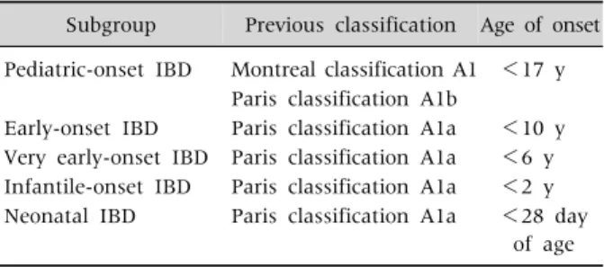
Table 1. Subgroups of Pediatric Inflammatory Bowel Disease (IBD)
Subgroup Previous classification Age of onset Pediatric-onset IBD Montreal classification A1 <17 y
Paris classification A1b
Early-onset IBD Paris classification A1a <10 y Very early-onset IBD Paris classification A1a <6 y Infantile-onset IBD Paris classification A1a <2 y Neonatal IBD Paris classification A1a <28 day
of age
compared to adult-onset IBD. Particularly very ear- ly-onset IBD (VEO-IBD) or infantile-onset IBD dif- fers in many aspects, including the disease subtypes, location of the lesions, disease behavior, and genet- ically attributable risks. Several genetic defects that disturb intestinal epithelial barrier function or affect immune function have been noted in patients with VEO-IBD [1,3]. This review examines the currently published data on the clinical and genetic character- istics of VEO-IBD, particularly focused on the VEO- IBD in Korea.
EPIDEMIOLOGY OF PEDIATRIC IBD
IBD can present at any age, with the most affected patients being in the age range of 15 to 29 years [4]. It develops during childhood or adolescence in approx- imately 25% of the cases [5]. The incidence of pedia- tric IBD, particularly Crohn’s disease (CD), has been increasing in both developed and developing nations [6]. The annual incidence of pediatric IBD has been increasing in Western countries, with the annual in- cidence of 2.2 to 13.3 per 100,000 children [6-8]. In Korea, according to the National Health Insurance Claim Data from 2006 to 2015, the annual incidence of CD is 3.6 per 100,000 at all ages and 5.2 per 100,000 children. The incidence of ulcerative colitis (UC) is 7.7 per 100,000 at all ages and 2.4 per 100,000 children.
The incidence of CD in teenagers has been increased, while its incidence in children younger than 6 years has remained stable. Neonatal or infantile-onset IBD develops in less than 1% of pediatric patients [9]. The annual incidence of IBD in children younger than 6 years was reported as 0.4 per 100,000 for CD and 0.9 per 100,000 for UC in 2009 in Canada, which had been increased by 7.4% per year since 1994 [7].
However, this increased rate of diagnosis for pedia- tric IBD might be partially attributable to improve- ment in awareness and diagnostic accuracy. In a re- cent French study, VEO-IBD represented 3% of pe- diatric IBD cases and its incidence remained stable from 1988 to 2011 [10] as in the Korean nationwide data. The stability of the incidence of VEO-IBD over time implies that this disease might not be strongly
influenced by environmental factors.
AGE OF ONSET
The pediatric Paris classification uses an age cutoff of <10 years (A1a) [11] because of the paucity of ileal involvement, serological characteristics in CD, and pancolitis in UC. Children with neonatal-onset IBD or infantile-onset IBD have high rates of af- fected first-degree relatives, a severe disease course, and a high rate of resistance to immunosuppressive treatment. Recently, subgroups of pediatric-onset IBD (<17 years), early onset IBD (<10 years), VEO- IBD (<6 years), infantile onset IBD (<2 years), and neonatal onset IBD (<28 days) have been suggested (Table 1) based on the phenotypic characteristics in- cluding disease location and severity, in addition to the presence of monogenic defects [3,12].
GENETICS OF VEO-IBD
Genetic predisposition might play an important
role in development of IBD particularly in young
children. Twin and family studies suggested that the
risk of developing CD in another sibling is 26-fold
higher than in an unrelated person, compared with a
9-fold increase for UC development [13]. Large-
scaled genome-wide association studies (GWAS)
have detected 250 single-nucleotide polymorphisms
associated with IBD, and a recent trans-ancestry as-
sociation study identified 38 additional new loci in-
fluencing the risk of developing IBD [14,15]. Ethnic
differences have also been reported, since NOD2/
CARD15 was reported as a susceptible gene locus in Caucasians, but not in Asians, while ATG16L2 and IL17REL were reported as susceptible loci in Korean [16]. A GWAS in children showed similar results as in adults [17]. Older children and adults shares sim- ilar polygenic forms of IBD. The main limitation of GWAS is that the evidence of causality is largely ab- sent and it tend to find common variants, so it may overlook functionally detrimental variations im- posed by rare mutations. VEO-IBD patients may car- ry a wide spectrum of low frequency gene variants.
Glocker et al. [18] were the first to identify mende- lian mutations of IL10RA IL10RB in children with infantile-onset IBD, which provided a new insight into the pathogenesis of IBD. Interleukin 10 (IL-10) is critical in maintaining the balance of the immune system, where it restricts and terminates immune responses by limiting the secretion of pro-in- flammatory cytokines such as tumor necrosis fac- tor-, IL-1, IL-6, IL-12, and controls both the differ- entiation and proliferation of macrophages, T cells and B cells. Due to its unique role in balancing the immune system, IL-10 has a long history as the focal point in IBD research. Shim et al. [19,20] reported 7 cases of Korean children with IL10RA mutations among 14 children with infantile-onset IBD.
Children with IL10RA mutations showed Crohn’s phenotypes, had anal fistulae, and required early surgical interventions since they exhibited a poor re- sponse to medical therapy. They also had recurrent infections, and folliculitis.
Whole exome sequencing (WES) has greatly ex- panded the list of genes associated with IBD risk be- yond those identified during the GWAS era. Sequen- tial candidate gene sequencing according to the dis- ease phenotype could not identify new causative variants. WES allows the identification of causative mutations in rare diseases, although extensive filter- ing and bioinformatics are required to exclude the large numbers of benign variants and variants of un- known significance that tend to be identified by this approach [1].
Studies using WES have identified mutations in the gene encoding X-linked inhibitor of apoptosis
(XIAP) in infants with aggressive colitis, perianal fis- tulae, and refractory IBD [21,22]. Such symptoms often present between 2 and 6 years of age. XIAP reg- ulates apoptosis and nuclear factor-B activation, and it is expressed in all hematopoietic cells; there- fore, mutations in this protein may also cause the de- velopment of an X-linked lymphoproliferative syn- drome and hemophagocytic lymphohistiocytosis.
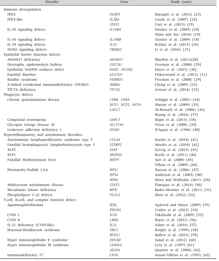
To date, approximately 60 monogenic mutations associated with IBD and IBD-like colitis have been identified (Table 2) [1,3,23-73]. Mutations related in genes associated with epithelial barrier function, such as TTC7A have been identified [33]. Monogenic mutations have also been observed in diseases with phagocyte defects, such as chronic granulomatous disease (CYBB, CYBA, NCF1, NCF2, and NCF4) [34- 36] and congenital neutropenia (G6PC3) [38]. About 40% of chronic granulomatous disease cases develop CD-like inflammation [3]. Other IBD-associated mutations have been implicated in hyper- or auto- inflammatory disorders, including XIAP deficiency.
Genetic disorders involving defects in T and B cell function, such as Wiskott-Aldrich syndrome (WAS) [74] and severe combined immunodeficiency dis- order [59] can present with IBD-like phenotypes.
Furthermore, mutations associated with X-linked immune dysregulation, polyendocrinopathy, and enteropathy (IPEX) have been found such as those in FOXP3 [75].
Monogenic mutations have been found mostly in children with an age of onset under 6 years, whereas conventional polygenic IBD more commonly has an age of onset older than 7 years.
CLINICAL ASPECTS OF VEO-IBD AND MONOGENIC IBD
In many cases of VEO-IBD, we should consider the
potential differential diagnoses of cow milk protein
allergy, eosinophilic gastroenteritis, infectious caus-
es, and primary immune deficiency with intestinal
inflammation. High levels of immunoglobulin E or
eosinophilia can also be found in patients with mon-
ogenic IBD and IBD-like phenotype (defects in
Table 2. List of Gene Mutations Associated with Monogenic Very Early-Onset Inflammatory Bowel Disease (IBD) and IBD-like Colitis
Disorder Gene Study (year)
Immune dysregulation
IPEX FOXP3 Barzaghi et al. (2012) [23]
IPEX-like IL2RA Caudy et al. (2007) [24]
STAT1 Uzel et al. (2013) [25]
IL-10 signaling defects IL10RA Glocker et al. (2009) [18]
Shim and Seo (2014) [19]
IL-10 signaling defects IL10RB Glocker et al. (2009) [18]
IL-10 signaling defects IL10 Kotlarz et al. (2012) [26]
NOD2 signaling defects TRIM22 Li et al. (2016) [27]
Epithelial barrier function defects
ADAM17 deficiency ADAM17 Blaydon et al. (2011)[28]
Dystrophic epidermolysis bullosa COL7A1 Freeman et al. (2008) [29]
Epithelial NADPH oxidases defect NOX1, DUOX2 Hayes et al. (2015) [30]
Familial diarrhea GUCY2C Fiskerstrand et al. (2012) [31]
Kindler syndrome FERMT1 Freeman et al. (2008) [29]
X-linked ectodermal immunodeficiency (NEMO) IKBKG Cheng et al. (2009) [32]
TTC7A deficiency TTC7A Avitzur et al. (2014) [33]
Phagocyte defects
Chronic granulomatous disease CYBB, CYBA Schäppi et al. (2001) [34]
NCF1, NCF2, NCF4 Matute et al. (2009) [35]
LACC1 Al-Bousafy et al. (2006) [36]
Huang et al. (2016) [37]
Congenital neutropenia G6PC3 Bégin et al. (2013) [38]
Glycogen storage disease 1b SLC37A4 Visser et al. (2000) [39]
Leukocyte adhesion deficiency 1 ITGB2 D’Agata et al. (1996) [40]
Hyperinflammatory and autoimmune disorders
Autoimmune lymphoproliferative syndrome type 5 CTLA4 Kuehn et al. (2014) [41]
Familial hemophagocytic lymphohistiocytosis type 5 STXBP2 Meeths et al. (2010) [42]
XLP2 XIAP Zeissig et al. (2015) [43]
XLP1 SH2DIA Booth et al. (2011) [44]
Familial Mediterranean fever MEFV Sari et al. (2008) [45]
Villani et al. (2009) [46]
Hermansky-Pudlak 1,4,6 HPS1 Hazzan et al. (2006) [47]
HPS4 Anderson et al. (2003) [48]
HPS6 Mora and Wolfsohn (2011) [49]
Multisystem autoimmune disease STAT3 Flanagan et al. (2014) [50]
Mevalonate kinase deficiency MVK Bader-Meunier et al. (2011) [51]
Phospholipase C-2 defects PLCG2 Zhou et al. (2012) [52]
T-cell, B-cell, and complex function defect
Agammaglobulinemia BTK, Agarwal and Mayer (2009) [53]
PIK3R1 Conley et al. (2012) [54]
CVID 1 ICOS Takahashi et al. (2009) [55]
CVID 8 LRBA Burns et al. (2012) [56]
IL-21 deficiency (CVID-like) IL21 Salzer et al. (2014) [57]
Hoyeraal-Hreidarsson syndrome DKC1 Knight et al. (1999) [58]
RTEL1 Ballew et al. (2013) [59]
Hyper immunoglobulin E syndrome DOCK8 Sanal et al. (2012) [60]
Hyper immunoglobulin M syndrome CD40LG Levy et al. (1997) [61]
AICDA Quartier et al. (2004) [62]
Immunodeficiency 17 CD3G Arnaiz-Villena et al. (1992) [63]
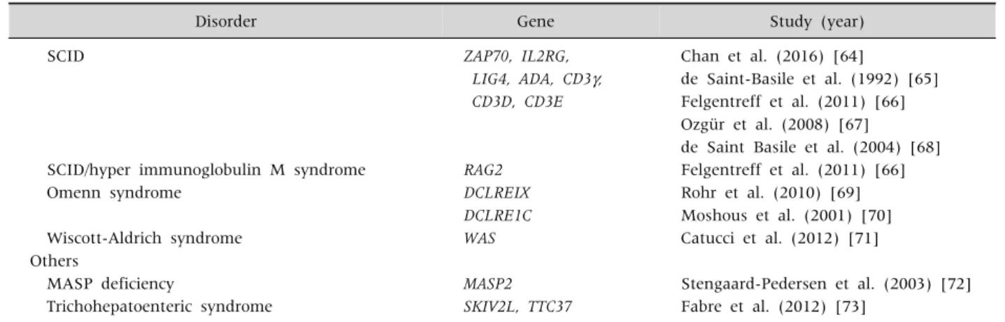
Table 2. Continued
Disorder Gene Study (year)
SCID ZAP70, IL2RG, Chan et al. (2016) [64]
LIG4, ADA, CD3, de Saint-Basile et al. (1992) [65]
CD3D, CD3E Felgentreff et al. (2011) [66]
Ozgür et al. (2008) [67]
de Saint Basile et al. (2004) [68]
SCID/hyper immunoglobulin M syndrome RAG2 Felgentreff et al. (2011) [66]
Omenn syndrome DCLREIX Rohr et al. (2010) [69]
DCLRE1C Moshous et al. (2001) [70]
Wiscott-Aldrich syndrome WAS Catucci et al. (2012) [71]
Others
MASP deficiency MASP2 Stengaard-Pedersen et al. (2003) [72]
Trichohepatoenteric syndrome SKIV2L, TTC37 Fabre et al. (2012) [73]
IPEX: X-linked immune dysregulation, polyendocrinopathy, and enteropathy, IL: interleukin, XLP: X-linked lymphoproliferative syndrome, CVID: common variable immunodeficiency, SCID: severe combined immunodeficiency.
The diagnostic approach to monogenic very early onset IBD. Some data were adapted from Uhlig et al. [3] and others were updated.
FOXP3, IL2RA, IKBKG, WAS, or DOCK8) [3].
The frequency of IBD-unclassified was reported as 7% in VEO-IBD, compared to 2% in early onset (EO)-IBD [10]. Furthermore, in 25% of the children with VEO-IBD originally diagnosed as UC or IBD- unclassified, the diagnosis was reclassified to CD over time [76]. Children with VEO-IBD more com- monly showed rectal bleeding and mucous stools, whereas weight loss and abdominal pain were more frequent in those with EO-IBD. Isolated colonic dis- ease was more common among the patients with VEO-CD [10].
The next concern is why monogenic IBD is important. Monogenic IBD usually presents with re- fractory IBD or fistulous CD, so early treatment with biologics, or an unconventional approach such as hematopoietic stem cell transplantation (SCT) might be needed. Patients that do not respond to conven- tional treatment, those with high mortality, and those that have an increased susceptibility to hema- topoietic cancers such as patients with IL-10 signal- ing defects, IPEX, WAS, and XIAP deficiency can be candidates for SCT [3]. However, in a child with TTC7A deficiency, multiple intestinal atresia re- curred after SCT, and he died [77]. Therefore, it is im- portant to determine the genetic basis of the disease for each patient before selecting SCT.
When we suspect monogenic IBD, the most im- portant clinical sign is a young age of onset. In addi- tion, lack of a response to conventional medication, a family history of IBD, autoimmunity, recurrent in- fections, perianal disease, hemophagocytic lympho- histiocytosis, intestinal obstruction, skin lesions, and tumors can be signs of monogenic IBD.
To diagnose monogenic IBD, sequential candidate gene sequencing can be costly and time- consuming.
WES can be used for the analysis of patients with suspected monogenic IBD despite some limitations.
The targeted next-generation sequencing of multiple candidate genes can also be an alternative option.
MONOGENIC IBD VERSUS VEO-IBD
In a Canadian nationwide epidemiology study,
there was no difference in the rate of surgery over
time between children aged less than 6 years and
children aged between 6 and 9 years [78]. Mean-
while, in the aforementioned study by Shim and Seo
[19], the clinical courses of children with VEO-IBD
involving IL10RA mutations were refractory, where-
as those of children with VEO-IBD without such mu-
tations were not different from those of children
with EO-IBD. Kim et al. [79] reported that patients
with monogenic VEO-IBD showed a higher morbid-
ity than those with non-monogenic VEO-IBD or non-monogenic pediatric IBD. Whether VEO-IBD patients have low response rates to conventional therapy, or a more aggressive phenotype is contro- versial. It seems that the age of onset itself does not sufficiently predict the severity of the disease or the response to therapy. Crucially, the main determinant of an individual prognosis is the specific causative gene mutation.
CONCLUSION
Monogenic IBD in patients with VEO-IBD is a dif- ferent disease entity from EO-IBD, pediatric IBD, and adult-onset IBD. Monogenic VEO-IBD has high rates of morbidity and mortality, and it might require different treatment strategies. Moreover, monogenic VEO-IBD might be a fundamentally different dis- ease entity from non-monogenic forms of VEO-IBD.
Well-designed global studies are needed to inves- tigate this hypothesis.
ACKNOWLEDGEMENTS
Jung Ok Shim received the grant of National Research Foundation of Korea (Grant No. NRF- 2018R1C1B5047245).
REFERENCES
1. Moran CJ. Very early onset inflammatory bowel disease. Semin Pediatr Surg 2017;26:356-9.
2. Seo JK. Pediatric Inflammatory Bowel Disease (IBD):
phenotypic, genetic and therapeutic differences be- tween early-onset and adult-onset IBD. Korean J Pediatr Gastroenterol Nutr 2011;14:1-25.
3. Uhlig HH, Schwerd T, Koletzko S, Shah N, Kammermeier J, Elkadri A, et al. The diagnostic ap- proach to monogenic very early onset inflammatory bowel disease. Gastroenterology 2014;147:990-1007.e3.
4. Ekbom A, Helmick C, Zack M, Adami HO. The epidemi- ology of inflammatory bowel disease: a large, pop- ulation-based study in Sweden. Gastroenterology 1991;
100:350-8.
5. Loftus EV Jr. Clinical epidemiology of inflammatory bowel disease: incidence, prevalence, and environ-
mental influences. Gastroenterology 2004;126:1504-17.
6. Benchimol EI, Fortinsky KJ, Gozdyra P, Van den Heuvel M, Van Limbergen J, Griffiths AM. Epidemiolo- gy of pediatric inflammatory bowel disease: a system- atic review of international trends. Inflamm Bowel Dis 2011;17:423-39.
7. Benchimol EI, Manuel DG, Guttmann A, Nguyen GC, Mojaverian N, Quach P, et al. Changing age demo- graphics of inflammatory bowel disease in Ontario, Canada: a population-based cohort study of epidemiol- ogy trends. Inflamm Bowel Dis 2014;20:1761-9.
8. Grieci T, Bütter A. The incidence of inflammatory bowel disease in the pediatric population of Southwestern Ontario. J Pediatr Surg 2009;44:977-80.
9. Shim JO, Han K. Treatment patterns and prognosis of inflammatory bowel disease: a nationwide epidemio- logic study. Paper presented at: Korean Digestive Disease Week (KDDW) 2017; 2017 Nov 23-25; Seoul, Korea. p. 100.
10. Bequet E, Sarter H, Fumery M, Vasseur F, Armengol- Debeir L, Pariente B, et al. Incidence and phenotype at diagnosis of very-early-onset compared with later-onset paediatric inflammatory bowel disease: a population- based study [1988-2011]. J Crohns Colitis 2017;11:
519-26.
11. Levine A, Griffiths A, Markowitz J, Wilson DC, Turner D, Russell RK, et al. Pediatric modification of the Montreal classification for inflammatory bowel dis- ease: the Paris classification. Inflamm Bowel Dis 2011;
17:1314-21.
12. Hyams JS. Standardized recording of parameters re- lated to the natural history of inflammatory bowel dis- ease: from Montreal to Paris. Dig Dis 2014;32:337-44.
13. Doecke JD, Simms LA, Zhao ZZ, Huang N, Hanigan K, Krishnaprasad K, et al. Genetic susceptibility in IBD:
overlap between ulcerative colitis and Crohn's disease.
Inflamm Bowel Dis 2013;19:240-5.
14. Liu JZ, van Sommeren S, Huang H, Ng SC, Alberts R, Takahashi A, et al. Association analyses identify 38 sus- ceptibility loci for inflammatory bowel disease and highlight shared genetic risk across populations. Nat Genet 2015;47:979-86.
15. Zhang Y, Tian L, Sleiman P, Ghosh S, Hakonarson H.
Bayesian analysis of genome-wide inflammatory bowel disease data sets reveals new risk loci. Eur J Hum Genet 2018;26:265-74.
16. Yang SK, Hong M, Zhao W, Jung Y, Baek J, Tayebi N, et al. Genome-wide association study of Crohn's disease in Koreans revealed three new susceptibility loci and common attributes of genetic susceptibility across eth- nic populations. Gut 2014;63:80-7.
17. Essers JB, Lee JJ, Kugathasan S, Stevens CR, Grand RJ, Daly MJ. Established genetic risk factors do not dis- tinguish early and later onset Crohn's disease. Inflamm Bowel Dis 2009;15:1508-14.
18. Glocker EO, Kotlarz D, Boztug K, Gertz EM, Schäffer AA, Noyan F, et al. Inflammatory bowel disease and mu- tations affecting the interleukin-10 receptor. N Engl J Med 2009;361:2033-45.
19. Shim JO, Seo JK. Very early-onset inflammatory bowel disease (IBD) in infancy is a different disease entity from adult-onset IBD; one form of interleukin-10 re- ceptor mutations. J Hum Genet 2014;59:337-41.
20. Shim JO, Hwang S, Yang HR, Moon JS, Chang JY, Ko JS, et al. Interleukin-10 receptor mutations in children with neonatal-onset Crohn's disease and intractable ul- cerating enterocolitis. Eur J Gastroenterol Hepatol 2013;25:1235-40.
21. Worthey EA, Mayer AN, Syverson GD, Helbling D, Bonacci BB, Decker B, et al. Making a definitive diag- nosis: successful clinical application of whole exome se- quencing in a child with intractable inflammatory bow- el disease. Genet Med 2011;13:255-62.
22. Kim SC. Monozygotic twin cases of XIAP deficiency syndrome. J Pediatr Gastroenterol Nutr 2018;67:e101.
23. Barzaghi F, Passerini L, Bacchetta R. Immune dysre- gulation, polyendocrinopathy, enteropathy, x-linked syndrome: a paradigm of immunodeficiency with auto- immunity. Front Immunol 2012;3:211.
24. Caudy AA, Reddy ST, Chatila T, Atkinson JP, Verbsky JW. CD25 deficiency causes an immune dysregulation, polyendocrinopathy, enteropathy, X-linked-like syn- drome, and defective IL-10 expression from CD4 lymphocytes. J Allergy Clin Immunol 2007;119:482-7.
25. Uzel G, Sampaio EP, Lawrence MG, Hsu AP, Hackett M, Dorsey MJ, et al. Dominant gain-of-function STAT1 mutations in FOXP3 wild-type immune dysregula- tion-polyendocrinopathy-enteropathy-X-linked-like syndrome. J Allergy Clin Immunol 2013;131:1611-23.
26. Kotlarz D, Beier R, Murugan D, Diestelhorst J, Jensen O, Boztug K, et al. Loss of interleukin-10 signaling and infantile inflammatory bowel disease: implications for diagnosis and therapy. Gastroenterology 2012;143:
347-55.
27. Li Q, Lee CH, Peters LA, Mastropaolo LA, Thoeni C, Elkadri A, et al. Variants in TRIM22 that affect NOD2 signaling are associated with very-early-onset in- flammatory bowel disease. Gastroenterology 2016;150:
1196-207.
28. Blaydon DC, Biancheri P, Di WL, Plagnol V, Cabral RM, Brooke MA, et al. Inflammatory skin and bowel disease linked to ADAM17 deletion. N Engl J Med 2011;365:
1502-8.
29. Freeman EB, Köglmeier J, Martinez AE, Mellerio JE, Haynes L, Sebire NJ, et al. Gastrointestinal complica- tions of epidermolysis bullosa in children. Br J Dermatol 2008;158:1308-14.
30. Hayes P, Dhillon S, O'Neill K, Thoeni C, Hui KY, Elkadri A, et al. Defects in NADPH oxidase genes NOX1 and DUOX2 in very early onset inflammatory bowel disease. Cell Mol Gastroenterol Hepatol 2015;1:489- 502.
31. Fiskerstrand T, Arshad N, Haukanes BI, Tronstad RR, Pham KD, Johansson S, et al. Familial diarrhea syn- drome caused by an activating GUCY2C mutation. N Engl J Med 2012;366:1586-95.
32. Cheng LE, Kanwar B, Tcheurekdjian H, Grenert JP, Muskat M, Heyman MB, et al. Persistent systemic in- flammation and atypical enterocolitis in patients with NEMO syndrome. Clin Immunol 2009;132:124-31.
33. Avitzur Y, Guo C, Mastropaolo LA, Bahrami E, Chen H, Zhao Z, et al. Mutations in tetratricopeptide repeat domain 7A result in a severe form of very early onset in- flammatory bowel disease. Gastroenterology 2014;146:
1028-39.
34. Schäppi MG, Smith VV, Goldblatt D, Lindley KJ, Milla PJ. Colitis in chronic granulomatous disease. Arch Dis Child 2001;84:147-51.
35. Matute JD, Arias AA, Wright NA, Wrobel I, Water- house CC, Li XJ, et al. A new genetic subgroup of chronic granulomatous disease with autosomal recessive mu- tations in p40 phox and selective defects in neutrophil NADPH oxidase activity. Blood 2009;114:3309-15.
36. Al-Bousafy A, Al-Tubuly A, Dawi E, Zaroog S, Schulze I. Libyan boy with autosomal recessive trait (P22-phox defect) of chronic granulomatous disease. Libyan J Med 2006;1:162-71.
37. Huang C, De Ravin SS, Paul AR, Heller T, Ho N, Wu Datta L, et al. Genetic risk for inflammatory bowel dis- ease is a determinant of Crohn's disease development in chronic granulomatous disease. Inflamm Bowel Dis 2016;22:2794-801.
38. Bégin P, Patey N, Mueller P, Rasquin A, Sirard A, Klein C, et al. Inflammatory bowel disease and T cell lympho- penia in G6PC3 deficiency. J Clin Immunol 2013;33:
520-5.
39. Visser G, Rake JP, Fernandes J, Labrune P, Leonard JV, Moses S, et al. Neutropenia, neutrophil dysfunc- tion, and inflammatory bowel disease in glycogen stor- age disease type Ib: results of the European Study on Glycogen Storage Disease type I. J Pediatr 2000;137:
187-91.
40. D'Agata ID, Paradis K, Chad Z, Bonny Y, Seidman E.
Leucocyte adhesion deficiency presenting as a chronic ileocolitis. Gut 1996;39:605-8.
41. Kuehn HS, Ouyang W, Lo B, Deenick EK, Niemela JE, Avery DT, et al. Immune dysregulation in human sub- jects with heterozygous germline mutations in CTLA4.
Science 2014;345:1623-7.
42. Meeths M, Entesarian M, Al-Herz W, Chiang SC, Wood SM, Al-Ateeqi W, et al. Spectrum of clinical presen- tations in familial hemophagocytic lymphohistiocy- tosis type 5 patients with mutations in STXBP2. Blood 2010;116:2635-43.
43. Zeissig Y, Petersen BS, Milutinovic S, Bosse E, Mayr G, Peuker K, et al. XIAP variants in male Crohn's disease.
Gut 2015;64:66-76.
44. Booth C, Gilmour KC, Veys P, Gennery AR, Slatter MA, Chapel H, et al. X-linked lymphoproliferative disease due to SAP/SH2D1A deficiency: a multicenter study on the manifestations, management and outcome of the disease. Blood 2011;117:53-62.
45. Sari S, Egritas O, Dalgic B. The familial Mediterranean fever (MEFV) gene may be a modifier factor of in- flammatory bowel disease in infancy. Eur J Pediatr 2008;167:391-3.
46. Villani AC, Lemire M, Louis E, Silverberg MS, Collette C, Fortin G, et al. Genetic variation in the familial Mediterranean fever gene (MEFV) and risk for Crohn's disease and ulcerative colitis. PLoS One 2009;4:e7154.
47. Hazzan D, Seward S, Stock H, Zisman S, Gabriel K, Harpaz N, et al. Crohn's-like colitis, enterocolitis and perianal disease in Hermansky-Pudlak syndrome.
Colorectal Dis 2006;8:539-43.
48. Anderson PD, Huizing M, Claassen DA, White J, Gahl WA. Hermansky-Pudlak syndrome type 4 (HPS-4):
clinical and molecular characteristics. Hum Genet 2003;113:10-7.
49. Mora AJ, Wolfsohn DM. The management of gastro- intestinal disease in Hermansky-Pudlak syndrome. J Clin Gastroenterol 2011;45:700-2.
50. Flanagan SE, Haapaniemi E, Russell MA, Caswell R, Allen HL, De Franco E, et al. Activating germline muta- tions in STAT3 cause early-onset multi-organ auto- immune disease. Nat Genet 2014;46:812-4.
51. Bader-Meunier B, Florkin B, Sibilia J, Acquaviva C, Hachulla E, Grateau G, et al. Mevalonate kinase defi- ciency: a survey of 50 patients. Pediatrics 2011;128:
e152-9.
52. Zhou Q, Lee GS, Brady J, Datta S, Katan M, Sheikh A, et al. A hypermorphic missense mutation in PLCG2, en- coding phospholipase C2, causes a dominantly in- herited autoinflammatory disease with immuno- deficiency. Am J Hum Genet 2012;91:713-20.
53. Agarwal S, Mayer L. Pathogenesis and treatment of gastrointestinal disease in antibody deficiency syn- dromes. J Allergy Clin Immunol 2009;124:658-64.
54. Conley ME, Dobbs AK, Quintana AM, Bosompem A, Wang YD, Coustan-Smith E, et al. Agammaglobuli- nemia and absent B lineage cells in a patient lacking the p85 subunit of PI3K. J Exp Med 2012;209:463-70.
55. Takahashi N, Matsumoto K, Saito H, Nanki T, Miyasaka N, Kobata T, et al. Impaired CD4 and CD8 effector func- tion and decreased memory T cell populations in ICOS- deficient patients. J Immunol 2009;182:5515-27.
56. Burns SO, Zenner HL, Plagnol V, Curtis J, Mok K, Eisenhut M, et al. LRBA gene deletion in a patient pre- senting with autoimmunity without hypogamma- globulinemia. J Allergy Clin Immunol 2012;130:1428-32.
57. Salzer E, Kansu A, Sic H, Májek P, Ikincioğullari A, Dogu FE, et al. Early-onset inflammatory bowel disease and common variable immunodeficiency-like disease caused by IL-21 deficiency. J Allergy Clin Immunol 2014;133:1651-9.e12.
58. Knight SW, Heiss NS, Vulliamy TJ, Aalfs CM, McMahon C, Richmond P, et al. Unexplained aplastic anaemia, immunodeficiency, and cerebellar hypoplasia (Hoyeraal-Hreidarsson syndrome) due to mutations in the dyskeratosis congenita gene, DKC1. Br J Haematol 1999;107:335-9.
59. Ballew BJ, Joseph V, De S, Sarek G, Vannier JB, Stracker T, et al. A recessive founder mutation in regu- lator of telomere elongation helicase 1, RTEL1, under- lies severe immunodeficiency and features of Hoyeraal Hreidarsson syndrome. PLoS Genet 2013;9:e1003695.
60. Sanal O, Jing H, Ozgur T, Ayvaz D, Strauss-Albee DM, Ersoy-Evans S, et al. Additional diverse findings ex- pand the clinical presentation of DOCK8 deficiency. J Clin Immunol 2012;32:698-708.
61. Levy J, Espanol-Boren T, Thomas C, Fischer A, Tovo P, Bordigoni P, et al. Clinical spectrum of X-linked hyper- IgM syndrome. J Pediatr 1997;131:47-54.
62. Quartier P, Bustamante J, Sanal O, Plebani A, Debré M, Deville A, et al. Clinical, immunologic and genetic analysis of 29 patients with autosomal recessive hy- per-IgM syndrome due to Activation-Induced Cytidine Deaminase deficiency. Clin Immunol 2004;110:22-9.
63. Arnaiz-Villena A, Timon M, Corell A, Perez-Aciego P, Martin-Villa JM, Regueiro JR. Brief report: primary immunodeficiency caused by mutations in the gene en- coding the CD3-gamma subunit of the T-lymphocyte receptor. N Engl J Med 1992;327:529-33.
64. Chan AY, Punwani D, Kadlecek TA, Cowan MJ, Olson JL, Mathes EF, et al. A novel human autoimmune syn- drome caused by combined hypomorphic and activating
mutations in ZAP-70. J Exp Med 2016;213:155-65.
65. de Saint-Basile G, Le Deist F, Caniglia M, Lebranchu Y, Griscelli C, Fischer A. Genetic study of a new X-linked recessive immunodeficiency syndrome. J Clin Invest 1992;89:861-6.
66. Felgentreff K, Perez-Becker R, Speckmann C, Schwarz K, Kalwak K, Markelj G, et al. Clinical and immuno- logical manifestations of patients with atypical severe combined immunodeficiency. Clin Immunol 2011;141:
73-82.
67. Ozgür TT, Asal GT, Cetinkaya D, Orhan D, Kiliç SS, Usta Y, et al. Hematopoietic stem cell transplantation in a CD3 gamma-deficient infant with inflammatory bowel disease. Pediatr Transplant 2008;12:910-3.
68. de Saint Basile G, Geissmann F, Flori E, Uring-Lambert B, Soudais C, Cavazzana-Calvo M, et al. Severe com- bined immunodeficiency caused by deficiency in either the delta or the epsilon subunit of CD3. J Clin Invest 2004;114:1512-7.
69. Rohr J, Pannicke U, Döring M, Schmitt-Graeff A, Wiech E, Busch A, et al. Chronic inflammatory bowel disease as key manifestation of atypical ARTEMIS deficiency.
J Clin Immunol 2010;30:314-20.
70. Moshous D, Callebaut I, de Chasseval R, Corneo B, Cavazzana-Calvo M, Le Deist F, et al. Artemis, a novel DNA double-strand break repair/V(D)J recombination protein, is mutated in human severe combined immune deficiency. Cell 2001;105:177-86.
71. Catucci M, Castiello MC, Pala F, Bosticardo M, Villa A.
Autoimmunity in wiskott-Aldrich syndrome: an un- solved enigma. Front Immunol 2012;3:209.
72. Stengaard-Pedersen K, Thiel S, Gadjeva M, Møller- Kristensen M, Sørensen R, Jensen LT, et al. Inherited
deficiency of mannan-binding lectin-associated serine protease 2. N Engl J Med 2003;349:554-60.
73. Fabre A, Charroux B, Martinez-Vinson C, Roquelaure B, Odul E, Sayar E, et al. SKIV2L mutations cause syn- dromic diarrhea, or trichohepatoenteric syndrome. Am J Hum Genet 2012;90:689-92.
74. Aiuti A, Biasco L, Scaramuzza S, Ferrua F, Cicalese MP, Baricordi C, et al. Lentiviral hematopoietic stem cell gene therapy in patients with Wiskott-Aldrich syn- drome. Science 2013;341:1233151.
75. Barzaghi F, Passerini L, Gambineri E, Ciullini Mannurita S, Cornu T, Kang ES, et al. Demethylation analysis of the FOXP3 locus shows quantitative defects of regu- latory T cells in IPEX-like syndrome. J Autoimmun 2012;38:49-58.
76. Rialon KL, Crowley E, Seemann NM, Fahy AS, Muise A, Langer JC. Long-term outcomes for children with very early-onset colitis: implications for surgical mana- gement. J Pediatr Surg 2018;53:964-7.
77. Samuels ME, Majewski J, Alirezaie N, Fernandez I, Casals F, Patey N, et al. Exome sequencing identifies mutations in the gene TTC7A in French-Canadian cas- es with hereditary multiple intestinal atresia. J Med Genet 2013;50:324-9.
78. Benchimol EI, Mack DR, Nguyen GC, Snapper SB, Li W, Mojaverian N, et al. Incidence, outcomes, and health services burden of very early onset inflammatory bowel disease. Gastroenterology 2014;147:803-13.e7; quiz e14-5.
79. Kim KY, Lee EJ, Kim JW, Moon JS, Jang JY, Yang HR, et al. Higher morbidity of monogenic inflammatory bowel disease compared to the adolescent onset in- flammatory bowel disease. Pediatr Gastroenterol Hepatol Nutr 2018;21:34-42.