67
Immune Network
Introduction
NK cells and some T cells express members of a multigenic family of killer cell Ig-like receptors (KIRs)3 that recognize polymorphic class I MHC molecules on target cells (reviewed in refs. 1-4). Both natural cytotoxicity and antibody dependent cell cytotoxicity of NK cells, and CD3/TCR dependent cytotoxicity of T cells are inhibited by the coengagement of KIRs and activating receptors such as FcR and TCR (5-8).
Recent studies have suggested that KIR plays a role in the survival of memory-phenotype T cells (9,10) and in the inhibition of T cell activation induced cell death (AICD) (9,11,12). However, the exact mecha- nism of the AICD inhibition mediated by KIR has not been revealed yet. Inhibitory KIRs (KIR here- after) are type I transmembrane glycoproteins belong-
ing to the Ig superfamily and consist of either two (for p58 KIR and KIR103: KIR2D family) or three (for p70 KIR: KIR3D family) extracellular Ig-related domains, a transmembrane part and a cytoplasmic tail (13-16). The extracellular domain of KIR specifically recognizes particular class I MHC molecules express- ed on target cells (17-20). The cytoplasmic tail of KIR contains either one (for KIR103) or two (for p58 and p70 KIR) immunoreceptor tyrosine-based inhibition motifs (ITIMs) that function in triggering an inhibitory signal transduction that, in turn, pre- vents killer cell-mediated cytotoxicity (21-24). The ITIM, which has the consensus sequence of (I/V) xYxx (L/V), was originally found in the cytoplasmic tail of FcγRIIB (25,26). FcγRIIB has also been shown to transmit the inhibitory signals in B cell ac- tivation by recruitment of SH2-containing inositol phosphatase (SHIP) upon tyrosine phosphorylation of the ITIM (25,27,28). The consensus sequence of ITIM is similar but clearly distinguishable from the immunoreceptor tyrosine-based activation motif (ITAM) found in the signaling molecules associated with se- veral lymphocyte antigen receptors (29,30).
Upon engagement of KIR, tyrosine residues in
Adaptor Protein Shc
Hyun Il Cho1, Yong-Joon Chwae1, Sang Myun Park1 and Jongsun Kim1,2
Department of 1Microbiology and 2Brain Korea Project of Medical Sciences, Yonsei University College of Medicine, Seoul, Korea
ABSTRACT
Background: Cytotoxic function of killer cells is inhibited by specific recognition of class I MHC molecules on target cells by inhibitory killer Ig-like receptors (KIR) ex- pressed on NK cells and some cytotoxic T cells. The inhibitory effect of KIR is ac- complished by recruitment of SH2-containing protein tyrosine phosphatase (SHP) to the phosphotyrosine residues in the cytoplasmic tail. Methods: By in vitro coprecipitation experiments and transfection analysis, we investigated the association of KIR with an adaptor protein Shc in Jurkat T cells. Results: The cytoplasmic tail of KIR appeared to associate with an adaptor protein Shc in Jurkat T cell lysates. Similar in vitro experi- ments showed that phosphorylated KIR cytoplasmic tail bound SHP-1 and Shc in Jurkat T cell lysates. The association of KIR with Shc was further confirmed by transfection analysis in 293T cells. Interestingly, however, Shc appeared to be replaced by SHP-2 upon engagement of KIR in 293T cells. Conclusion: Our data indicate that KIR asso- ciate with an adaptor protein Shc in Jurkat T cells, and suggest that KIR might have an additional role which is mediated by this adaptor protein. (Immune Network 2006;6(2):67-75)
Key Words: KIR, Shc, SHP-1, SHP-2, cell activation, cell proliferation, inhibitory signal transduction
Correspondence to: Jongsun Kim, Department of Microbiology and Institute for Immunology and Immunological Diseases, Yonsei University College of Medicine, 134 Shinchon-dong, Seodaemun-gu, Seoul 120-752, Korea. (Tel) 82-2-2228-1814, (Fax) 82-2-392-7088, (E-mail) [email protected]
This work was supported by a research grant from the Korea Research Foundation (KRF-2003-041-E00244).
ITIMs are phosphorylated by a src-family protein ty- rosine kinase (like p56 Lck), and in turn a SH2-con- taining protein tyrosine phosphatase (SHP), SHP-1, is recruited to the phosphotyrosyl residues (21-24,31). It has also been shown that another SHP, SHP-2 (31, 32), and the p85α regulatory subunit of phosphatidy- linositol (PI) 3-kinase (32) are recruited to the pho- sphotyrosyl residues of KIR. The functional impor- tance of SHP-1 in the KIR-mediated inhibition of NK cell activation is further supported by the obser- vation that the overexpression of a dominant negative form of SHP-1 reverses the inhibitory effect of KIR (21,22). It is thought that the inhibitory effect of KIR results from the dephosphorylation of phospho-tyro- sine residues in signaling molecules involved in the activating signal transduction pathway by SHP (SHP- 1 or SHP-2) recruited to the KIR. For FcR-mediated ADCC of NK cells, Binstadt et al. (21) reported that coengagement of FcR and KIR inhibited the pho- sphorylation of FcR-associated signaling molecules in- cluding TCRζ, ZAP-70 and PLC-γ. Similarly, when ζ, ZAP-70, and Lck were coexpressed with SHP-1 in Sf21 insect cells, the levels of tyrosine phosphory- lation in these molecules decreased substantially (33).
It has also been demonstrated that class I MHC re- cognition by KIR blocks the formation of a complex between tyrosine-phosphorylated pp36 (LAT) and PLC-γ in human NK cells (34). And Binstadt et al.
have demonstrated that the SH2-containing leukocyte protein 76 (SLP-76) may be a specific target for de- phosphorylation by SHP-1 in T cells and NK cells (35). These results suggest that SHP recruited to KIR may dephosphorylate those signaling molecules in NK cells and T cells.
Shc is an adaptor protein composed of a unique N-terminal phospho-tyrosine binding domain (PTB), a glycine/proline-rich collagen homology domain (CH) and a C-terminal SH2 domain (36-38). Shc is ubiqui- tously expressed in three isoforms of 46, 52, and 66 kDa, but only the 46 and 52 kDa forms are present in hematopoietic cells (36). Shc has been implicated in Ras activation triggered by a number of receptors, including growth factor receptors, antigen receptors and cytokine receptors (reviewed in refs. 39,40). The SH2 and PTB domains of Shc have been shown to interact with the tyrosine phosphorylated receptors.
Shc is tyrosine phosphorylated upon stimulation of the receptors and subsequently it interacts with Grb2 through the CH domain. The Shc:Grb2 complex, in turn, associates with the GTP/GDP exchange fac- tor, SOS, and finally leads to Ras activation. Shc can also activate the Akt pathway via Shc-Grb2-Gab2- PI3K (phosphatidylinositol 3-kinase) in B cells (41).
In this report, by in vitro coprecipitation experi- ments and transfection analyses, we showed that KIR
associates with an adaptor protein Shc in resting cells.
We further demonstrated that Shc is replaced by SHP upon engagement of KIR by antibody.
Materials and Methods
Cell lines. The human T cell lymphoma cell line, Jurkat was obtained from the American Type Culture Col- lection (ATCC, Rockville, MD) and maintained in culture with RPMI 1640 medium (Flow Laboratories, Rockville, MD) supplemented with 10% (V/V) Fetal Bovine Serum (FBS, Sigma, St. Louis, MO), 1% L- glutamine and 1% antibiotic solution. Human embryonic kidney epithelial cells expressing SV 40 large T antigen (293T) were obtained from the ATCC and cultured in Dulbecco's modified Eagle medium (DMEM)(Flow Laboratories, Rockville, MD) supplemented with 10%
FBS 1% L-glutamine and 1% antibiotic solution.
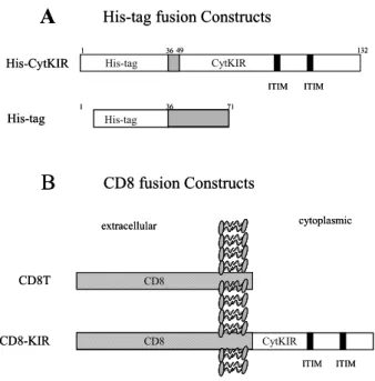
Antibodies and other reagents. Anti-phospho tyrosine an- tibody (PY20), anti-SHP-1, anti-SHP-2, anti-Grb2 and anti-Shc antibodies were purchased from Trans- duction Laboratories (Lexington, KY). Fluorescein isothiocyanate (FITC) conjugated anti-mouse IgG and Texas-red conjugated anti-rabbit IgG were ob- tained from Vector Laboratories (Burlingamme, CA), and PE-conjugated anti-mouse IgG and anti-CD8 antibodies were from Beckton Dickinson (San Jose, CA). Dithiobissuccinimidyl propionate (DSP) was purchased from Pierce (Rockford, IL). Ni-NTA resin was obtained from Qiagen (Santa Clarita, CA), and protein A-agarose and protein G-agarose were from Boeringer Manheim (Indianapolis, IN). cDNA of a p70 KIR (NKB1) was a kind gift from Dr. Lanier (DNAX Research Inst., Palo Alto, CA). Polyclonal antibodies to the cytoplasmic tail of KIR were pro- duced in mice using the bacterially expressed His-tag fusion protein of KIR (His-CytKIR; refs. 42,43). Brie- fly, the His-CytKIR protein was further purified on SDS polyacrylamide gels and the protein band was excised with a scalpel. The gel slices were homo- genized and injected into mice intraperitoneally. A booster injection was given 2 weeks after the primary immunization. Another two booster injections were given every two weeks, and the animals were bled 2 weeks later.
His-tag and CD8 fusion constructs of KIR cytoplasmic tail, and other DNA constructs. The His-tag fusion protein construct of a p70 KIR (NKB1; ref. 14) cytoplasmic tail (pHis-CytKIR) was described previously (42,43), and the generation of plasmid containing the extra- cellular and transmembrane portion of CD8α, pCD 8T, was also described previously (44). A CD8 fusion construct of KIR cytoplasmic tail was generated by subcloning the cytoplasmic tail of NKB1 into the pCD8T. Briefly, the cytoplasmic tail of NKB1 was amplified by PCR with primers 5'-GGGGAATTCC
ATCTCTGGTGCTCCAACAAAAAAAAT-3' and 5'- GGGGGATCCTCATGGGCAGGAGACAACTTT GG-3'. The amplified DNAs were cloned into EcoRI and BamHI sites of pBluscript (Strategene, La Jolla, CA) and verified by nucleotide sequencing. Then the KIR cytoplasmic fragment was inserted in frame into the EcoRI sites of pCD8T. The resulting chimeric receptor (CD8-KIR) was excised from the plasmid and ligated into SalI and BamHI sites of the eucar- yotic expression vector, pHβAPr-1-neo (45).
Metabolic labeling of Jurkat T cells/Preparation of cell lysates.
Jurkat cells (5×10⁶ cells) were washed with 10 ml of methionine-free RPMI 1640 and preincubated for 1 hr in 10 ml of methionine-free RPMI 1640 sup- plemented with 10% heat-inactivated dialyzed FBS.
Cells were then incubated in the presence of 100μ Ci/ml L-[35S]methionine at 37oC for 2 hr. After label- ing, the cells were washed two times with cold PBS, and lysed in 0.5 ml of cold lysis buffer containing 20 mM Na-Phosphate (pH 7.5), 0.15 M NaCl, 1%
Triton X-100, 1 mM PMSF, 5μg/ml leupeptin, 5μg/
ml pepstatin, 0.02% NaN3, and 1 mM iodoace- tamide. Cell lysates were cleared by centrifugation at 13,000 rpm for 20 min, and the supernatant was used for coprecipitation and immunoblot analysis. Non-la- beled Jurkat cell lysates were also similarly prepared.
Coprecipitation and Western blotting. The His-tag fusion protein of KIR cytoplasmic tail (His-CytKIR) was added to [35S]-labeled Jurkat cell lysates in the pre- sence and absence of a chemical cross-linker (DSP, a final concentration of 2 mM), and incubated for 1 hr at 4oC. The reaction mixtures were precipitated with Ni-NTA resin, as previously described (43). The precipitates were separated on 12% SDS polyacry- lamide gels, and the gels were then fixed, dried, and autoradiographed. For Western blot analysis, His- CytKIR was mixed with Jurkat cell lysates in the pre- sence of DSP, and incubated for 1 hr at room tem- perature. The reaction mixtures were precipitated with Ni-NTA resin, and washed three times. Precipi- tates were resolved by SDS-PAGE using 12% acryla- mide gels, and proteins were transferred onto polyvi- nylidene difluoride (PVDF) membranes (Immobilon- P; Millipore, Marlborough, MA). Western blot an- alysis was performed with anti-Shc antibody, anti- Grb2 antibody, and anti-phosphotyrosine antibody, as previously described (43).
In vitro phosphorylation of His-CytKIR. Jurkat cell lysates and the His-CytKIR protein were mixed and incu- bated in a kinase reaction buffer, as previously descri- bed (43). After in vitro phosphorylation, the His-Cyt KIR was precipitated with Ni-NTA resin. The preci- pitates were resolved on SDS PAGE gels, and the gels were blotted onto PVDF membranes and analyz- ed with anti-Grb2, anti-Shc and anti-phosphotyrosine
antibodies.
Immunofluorescence staining and Confocal Microscopy. Cells transfected with the pCD8T or pCD8-KIR cDNA were grown on sterile glass coverslides precoated with 1% gelatin overnight prior to antibody staining.
36 hr after transfection, cells were fixed with ice-cold methanol and aceton for 10 min each and incubated with 3% H2O2 for 30 min at room temperature. The coverslides were washed three times with PBS and incubated with blocking solution (5% BSA, 3% nor- mal horse serum, 0.3% Tritonx-100 in PBS) for 3 hr.
The cells were simultaneously stained by incubating them with anti CD8 antibody ascites (1:100) follow- ed by a goat antiserum against mouse IgG coupled to FITC and rabbit anti-Shc antibody followed by a goat antiserum against rabbit IgG coupled to Texas- red at 37oC for 2∼3 hr and then washed 4 times in PBS. After 3 additional washes in PBS, the slides were mounted in mounting solution. Cells were view- ed by confocal microscope (Lica PCSMT, Heidelberg, Germany) using the 100× objective lens.
Immunoprecipitation and Western blotting. 293T cells trans- fected with CD8T or CD8-KIR were stimulated with OKT8 ascites (1:200 dilution). After 30 min stimu- lation, the cells were rinsed twice with ice-cold PBS and lysed in a lysis buffer (10 mM Tris, pH 7.5, 1%
Triton X-100, 150 mM NaCl, 10 mM Na3VO4, 1 mM NaF, 1 mM Na pyrophosphate, 5 mM EDTA, 50μg/ml leupeptin, 1 U/ml aprotinin, 1 mM pepstatin, 1 mM PMSF). Samples were incubated for 1hr at 4oC and centrifuged (13,000 rpm) for 20 min at 4oC. For immunoprecipitation, the lysates from 5×10⁶ cells in 700μl were precleared by mixing for 3 hr at 4oC with protein A agarose. The precleared lysates were then incubated with anti-Shc antibody on a rotating shaker for 1 hr at 4oC, and protein A agarose beads were added and further incubated overnight at 4oC. The precipitates were washed 4 times with wash buffer (cell lysis buffer with salt concentration increased by 200% and detergent concentration decreased by 50%) and twice with 10 mM Tris (pH 7.5) containing 50 mM NaCl and 0.1% triton X-100. After SDS PAGE, the proteins were transferred onto PVDF membrane, and blotted with anti-His-CytKIR antibodies. The membrane was reprobed with anti-SHP-2 antibody. A reciprocal immunoprecipitation experiment was also carried out with anti-His-CytKIR antibodies followed by immunoblotting with anti-Shc antibody and sub- sequently with anti-SHP-2 antibody.
Results
The cytoplasmic tail of KIR associates with Shc, Grb2, TCR- ζ and a protein of 36 kDa in Jurkat T cell lysates. Coag- gregation of an activating receptor, such as TCR or FcR, and KIR is required for the inhibition of NK
or T cell activation (46,47), implying that the inhibit- ory effect mediated by KIR may occur in the imme- diate vicinity of the activating receptor. We first asked whether the cytoplasmic tail of KIR interacts with signaling molecules that are also associated with acti- vating receptors, such as TCR, BCR and FcR. To identify proteins associated with the cytoplasmic tail of KIR, we generated a His-tag fusion protein of the cytoplasmic tail (His-CytKIR, Fig. 1A) and used this fusion protein to co-precipitate proteins from meta- bolically-labeled Jurkat T cells. Ni-NTA resin was used to coprecipitate proteins interacting with the His-CytKIR. In Jurkat cell lysates, the cytoplasmic tail of KIR coprecipitated the proteins of 46 kDa, 36 kDa, 23 kDa and 16 kDa (Fig. 2A, lane 2). The co- precipitates appeared as higher Mr (50∼100 kDa) complexes under nonreducing conditions (Fig. 2A, la- ne 3), suggesting that the four proteins are really cro- ss-linked to His-CytKIR by DSP, a disulfide contain- ing cross-linker. A control His-tag protein expressed from the pRSETA expression plasmid per se did not result in any detectable proteins coprecipitated in the
cell lysates (Fig. 2A, lane 1). The protein bands were also detectable when the precipitates were obtained without a chemical cross-linking procedure (Fig. 2C for the 46 kDa protein; data for other proteins not shown), though less prominent than with chemical cross-linking. These results suggest that the cytoplas- mic tail of KIR associates specifically with the 46 kDa, 36 kDa, 23 kDa and 16 kDa proteins in Jurkat cell lysates.
Previously, we identified that the 16 kDa protein is TCRζ by immunoblotting experiments with anti- bodies against TCRα,β, CD3ε andζ (43). Intere- stingly, we found that the remaining coprecipitated proteins of 46, 36 and 23 kDa have molecular weights consistent with Shc, pp36 (LAT) and Grb2, respec- tively (36,48,49), that have been implicated to func- tion in and downstream of TCR signal transduction.
To identify the 46 kDa and 23 kDa proteins, we per- formed immunoblotting experiments with commer- cially available anti-Shc and anti-Grb2 antibodies. The results shown in Fig. 2B clearly indicate that the 46 kDa and 23 kDa proteins are Shc and Grb2, re- spectively. Mammalian Shc exists in three isoforms of 46 kDa, 52 kDa and 66 kDa, but only the former
two are expressed by hematopoietic cells (36). Our
Figure 1. His-tag and CD8 fusion protein constructs. (A) The His-CytKIR construct encodes a His-tag fusion protein of the cytoplasmic tail of a p70 KIR (NKB1). The protein coding region of the cytoplasmic tail of KIR was subcloned into an E. coli ex- pression vector pRSETA. The His-tag construct (pRSETA itself) encodes a control His-tag fusion protein which lacks the cyto- plasmic tail of KIR. (B) The CD8 fusion protein constructs. The generation of plasmid containing the extracellular and transmem- brane portion of CD8α, pCD8T, was described previously (44).
A CD8 fusion construct of KIR was generated by subcloning the cytoplasmic tail of KIR into eucaryotic expression vector, pH βAPr-1 (45).
His-tag fusion Constructs
His-CytKIR
His-tag
A
His-tag CytKIR
His-tag
1 36 49 132
36
1 71
extracellular cytoplasmic
CD8T
CD8-KIR
CD8 fusion Constructs
ITIM ITIM
B
ITIM ITIM
CD8
CytKIR CD8
His-tag fusion Constructs
His-CytKIR
His-tag
A
His-tag CytKIR
His-tag
1 36 49 132
36
1 71
extracellular cytoplasmic
CD8T
CD8-KIR
CD8 fusion Constructs
ITIM ITIM
B
ITIM ITIM
CD8
CytKIR CD8
Figure 2. The His-CytKIR associates with Shc, Grb2, TCRζ, and a protein of 35 kDa in Jurkat T cell lysates. (A) His-CytKIR was mixed with [35S] labeled Jurkat cell lysates and cross-linked with DSP. The His-CytKIR was precipitated with Ni-NTA resin, and the precipitates were resolved by SDS-PAGE under reducing (lane 2) and nonreducing (lane 3) conditions. (B) The 46 kDa and 23 kDa proteins are Shc and Grb2, respectively. The His- CytKIR and Jurkat cell lysates were mixed and incubated for 1 hr at 4oC. The His-CytKIR was precipitated with Ni-NTA resin, and the precipitates were resolved on SDS gels. The protein bands were detected with anti-Shc (upper pannel) and anti-Grb2 (lower pannel) antibodies. (C) Similar experiments with (B) were performed with (lane 2) and without (lane 3) chemical cross- linking. The precipitates were detected with anti-Shc antibody.
Lane 1 in each figure shows the precipitates obtained by using the His-tag control protein in the reaction mixtures.
1 2 3
97.4 66.2
45
31
21.5 14.4
His-CytKIR co-ppt
DSP cross-linking 20 kDa
35 kDa 45 kDa
A
1 21 2 3
Shc
Grb2
B
C
TCRζ
23 kDa Shc
1 2 3
97.4 66.2
45
31
21.5 14.4
His-CytKIR co-ppt
DSP cross-linking 20 kDa
35 kDa 45 kDa
A
1 21 2 3
Shc
Grb2
B
C
TCRζ TCRζ
23 kDa Shc
results show that only the 46 kDa form of Shc asso- ciates with His-CytKIR in Jurkat cell lysates.
Phosphorylated KIR cytoplasmic tail binds SHP-1 and Shc in Jurkat T cell lysates. To investigate the effect of tyro- sine phosphorylation on the molecular association be- tween KIR and the signaling molecules, we carried out similar His-tag pull-down experiments. Metaboli- cally-labeled Jurkat cell lysates were mixed with His- CytKIR in a kinase reaction buffer and incubated at
37oC as previously described (42,43). The reaction
mixtures were chemically cross-linked and precipi- tated with Ni-NTA resin. The precipitates were sepa- rated on a SDS polyacrylamide gel and the protein bands were visualized by autoradiography. Unlike the unphosphorylated His-CytKIR (Fig. 2A), the phos- phorylated His-CytKIR was associated with the 68 kDa and 46 kDa proteins (Fig. 3A), which turned out to be SHP-1 and Shc, respectively, by Western blot experiments (data not shown). These results are con- sistent with previous reports showing that the pho-
Figure 3. Tyrosine phosphorylated His-CytKIR associates with SHP-1 and Shc in Jurkat T cell lysates. (A) His-CytKIR was mixed with [35S] labeled Jurkat cell lysates in a Kinase reaction buffer and incubated at 37oC. The reaction mixtures were cross- linked with DSP, and the His-CytKIR was precipitated with Ni- NTA resin. The precipitates were resolved by SDS-PAGE under reducing (lane 2) and nonreducing (lane 3) conditions. Lane 1 shows the precipitates obtained by using the His-tag control protein in the reaction mixtures. (B) Effects of tyrosine phospho- rylation of KIR on the Shc and Grb2 binding. Same amounts of Jurkat cell lysates (500 μg) and the His-CytKIR protein (200 μg) were mixed in a kinase reaction buffer (total volume of 700 μl) and cross-linked with DSP. In vitro phosphorylation reactions were performed in the presence (lane 4) and absence (lane 3) of sodium orthovanadate, a protein tyrosine phosphatase inhibi- tor (58). Then the His-CytKIR was precipitated with Ni-NTA resin and the precipitates were detected with anti-Shc antibody (top), anti Grb2 antibody (middle), and anti- phosphotyrosine antibody (bottom).
Cell lysates His-CytKIR Phosphorylation Na-orthovanadate
+ + + + + + + + + +
1 2 3 4 1 2 3
A
B
SHP-1 (68 kDa)
Shc (46 kDa)
Shc
Grb2
His-CytKIR
Cell lysates His-CytKIR Phosphorylation Na-orthovanadate
+ + + + + + + + + + + + + + + +
1 2 3 4 1 2 3
A
B
SHP-1 (68 kDa)SHP-1 (68 kDa)
Shc (46 kDa)
Shc
Grb2
His-CytKIR
Figure 4. Expression of CD8-KIR on 293T cells and the colo- calization of CD8-KIR and Shc in transfected cells. 293T cells were transiently transfected with the CD8T and CD8-KIR con- structs, respectively. (A) A histogram shows the surface expre- ssion level of transfected CD8-KIR as determined by FACS sta- ining with PE-conjugated anti-CD8 antibody, where shaded his- togram indicates staining with PE-conjugated anti-mouse IgG. (B) Expression level of transfected CD8-KIR protein was detected by immunoblotting with polyclonal anti-His-CytKIR antibodies.
Extracts from 5×10³ cells were loaded. Lane 1 is 293T cells transfected with CD8T; lane 2 is 293T cells transfected with CD8-KIR; lane 3 is 293T cells transfected with CD8-KIR and stimulated with PMA/Ionomycin. (C-F) Colocalization of Shc and CD8-KIR by confocal microscopy in transfected 293T cells.
Cells were double-labeled with anti-Shc antibody (red, detected with Texas red-conjugated goat anti-rabbit IgG) and with mouse antiserum to CD8 (green, detected with FITC-conjugated goat anti-mouse Ig). Fluorescent confocal images were obtained for Shc expression (red) in transfected (C) and untransfected (E) cells, and CD8-KIR expression (green, D). The two images were then superimposed (F) to show regions expressing both Shc and CD8-KIR (yellow).
sphorylated KIR recruits SHP-1 (21-24,31), and that the synthetic peptide representing the phosphorylated form of the ITIM of FcγRIIB binds Shc (50). Previ- ous studies have also shown that Shc associates with tyrosine phosphorylated TCRζ and Grb2 (51) in T cells, both of which also appeared to associate with KIR in this report. However, in contrast to TCRζ (43) and Grb2 (Fig. 3B, middle), Shc appeared to associate with both phosphorylated (Fig. 3B, top, lane 3) and non-phosphorylated His-CytKIR (lane 2). As well, Shc binding was most prominent among the proteins associating with His-CytKIR (Fig. 2A).
These findings suggest that Shc directly associates with His-CytKIR, not through the TCRζ or Grb2.
Interaction of KIR and Shc in transfected cells. To verify the association of KIR and Shc in cells, a CD8 fusion protein construct of KIR cytoplasmic tail (CD8-KIR, Fig. 1B) was transfected into 293T cells. FACS stain- ing with a monoclonal antibody to CD8 (OKT8) and Western blot analysis with polyclonal antibodies to the His-CytKIR protein showed that CD8-KIR was expressed on the surface of 293T cells (Fig. 4A, B, respectively). We next investigated whether the CD8- KIR expressed on 293T cells interacts with Shc. The transfected cells were double stained with anti-Shc antibody and OKT8, and then observed on a con- focal microscope. Most of the transfected cells abun- dantly expressed CD8-KIR, and the CD8-KIR ap- peared to be localized predominantly on the cell sur- face (Fig. 4D). In 293T cells, Shc protein was found in the cytoplasm as well as around the plasma mem- brane (Fig. 4E). However, in 293T cells transfected with the CD8-KIR expression vector, most of Shc appeared to be localized around the plasma mem- brane (Fig. 4C). Double staining of the transfected cells clearly showed that CD8-KIR and Shc were co- localized on the membrane (Fig. 4F), suggesting that the two proteins associate in the mammalian cells.
The binding interaction between the CD8-KIR and Shc in transfected cells was confirmed by coimmuno- precipitation analysis. Shc was immunoprecipitated with anti-Shc antibody, and the precipitates were re- solved on a SDS polyacrylamide gel. The protein bands were transferred onto PVDF membrane, and sequen- tially blotted with anti-His-CytKIR antibody and anti- SHP-2 antibody, respectively. Fig. 5A shows that CD8-KIR coprecipitated with Shc (Fig. 5A, top, lane 3). Interestingly, however, the association was almost blocked when the CD8-KIR was cross-linked by OKT8 antibody prior to the anti-Shc immunoprecipi- tation (lane 4). On the other hand, SHP-2 binding was only observed when the transfectants were treat- ed with OKT8 antibody cross-linking (Fig. 5A, bot- tom, lane 4). The weak intensity of the SHP-2 band may result from the fact that most of the phosphory-
lated KIR could not precipitate with Shc. Immuno- precipitation with anti-His-CytKIR antibodies and im- munoblotting experiments with anti-Shc and anti- SHP-2 antibodies resulted in similar results (Fig. 5B).
Shc coprecipitated with CD8-KIR and the association was greatly reduced by OKT8 antibody cross-linking (Fig. 5B, top). Both the 46 kDa and 52 kDa forms of Shc appeared to associate with the CD8-KIR in transfected cells. However, SHP-2 binding was not detected in this case (Fig. 5B, bottom), probably due to the antibody blocking effect. Taken together, these results indicate that CD8-KIR constitutively interacts with Shc in 293T cells but the Shc is replaced by SHP-2 upon engagement of CD8-KIR.
Discussion
By in vitro coprecipitation experiments, we demon- strated that the cytoplasmic tail of KIR associates with proteins of 46 kDa, 36 kDa, 23 kDa, and 16 kDa in Jurkat T cell lysates. Previously, we identified that the 16 kDa protein is TCRζ and demonstrated that the association between KIR and TCRζ is hindered by the tyrosine phosphorylation of KIR, but is independent of the tyrosine phosphorylation of the TCRζ-chain (43). In this report, we identified that the 46 kDa and 23 kDa proteins are Shc and Grb2, respectively, by immunoblotting experiments. Like the case of TCRζ, tyrosine phosphorylation of the KIR cytoplasmic tail abolished the association with Grb2 and the 36 kDa protein in Jurkat cell lysates.
However, the interaction between KIR and Shc ap-
Figure 5. Association of CD8-KIR and Shc in 293T cells transiently transfected with CD8-KIR. CD8-KIR associates Shc in resting 293T cells, but it associates SHP-2 instead of Shc in cells treated with anti-CD8 antibody. (A) Shc was immunopreci- pitated with anti-Shc antibody and blotted with anti-His-CytKIR antibody (top), and reprobed with anti-SHP-2 antibody (bottom).
(B) Shc was immunoprecipitated with anti-Shc antibody and blotted with anti-His-CytKIR antibody (top), and reprobed with anti-SHP-2 antibody (bottom). Lanes in (A, B): lane 1, 293T cells transfected with CD8T; lane 2, 293T cells transfected with CD8T and treated with anti-CD8 antibody; lane 3, 293T cells transfected with CD8-KIR; lane 4, 293T cells transfected with CD8-KIR and treated with anti-CD8 antibody.
1 2 3 4 1 2 3 4
IP: anti-Shc IP: anti-KIR
Blot:
anti-KIR
Blot:
anti-SHP-2 38 kDa
72 kDa
Blot:
anti-Shc 52 kDa 46 kDa
Blot:
anti-SHP-2
A B
1 2 3 4 1 2 3 4
IP: anti-Shc IP: anti-KIR
Blot:
anti-KIR
Blot:
anti-SHP-2 38 kDa 38 kDa
72 kDa 72 kDa
Blot:
anti-Shc 52 kDa 46 kDa 52 kDa 46 kDa
Blot:
anti-SHP-2
A B
peared to be unaffected in vitro. The relatively weak band intensity of Grb2 (Fig. 2A) and previous re- ports, where Grb2 appears to associate with Shc, TCRζ, and LAT (51), suggest that Grb2 may be co- precipitated with other proteins. We also demonstra- ted that tyrosine phosphorylated KIR associates with SHP-1 and Shc in Jurkat T cell lysates, suggesting that the molecular interaction between KIR and the signaling molecules identified in this study are diffe- rently modulated by the phosphorylation status of KIR.
To investigate the molecular interaction between KIR and Shc in cells, we transfected the CD8-KIR expression vector in 293T cells. Double staining of the transfectants with anti-Shc antibody and OKT8 antibody revealed that CD8-KIR and Shc are colo- calized around the plasma membrane, and immuno- precipitation analysis confirmed that CD8-KIR binds Shc in transfected cells. In contrast to the in vitro data, however, KIR interacts with Shc in a phos- phorylation-dependent manner in cells. Shc binds to CD8-KIR in resting cells, but dissociates in OKT8 treated-cells. Upon engagement of CD8-KIR with OKT8 antibody, KIR appears to associate with SHP- 2 instead of Shc, suggesting that the phosphorylation of tyrosine residues in the KIR cytoplasmic tail hin- ders Shc binding and induces SHP-2 (maybe SHP-1 in NK and T cells) binding in 293T cells where no SHP-1 is expressed (our unpublished observation).
The in vitro association of Shc with the phosphory- lated KIR may be caused by either incomplete phos- phorylation of KIR or excess use of KIR for the in vitro coprecipitation experiments. Also, the associa- tion between KIR and Grb2 observed in in vitro experiments was not detected in transfection analysis.
This result suggests that Grb2 may be coprecipitated with TCRζ or the 36 kDa protein which are not ex- pressed in 293T cells but are present in Jurkat T cells.
Shc is composed of three different domains, a phospho-tyrosine binding domain (PTB), collagen ho- molog domain (CH), and SH2 domain (36-38). Our data demonstrate that unphosphorylated KIR binds to Shc, but the KIR:Shc complex dissociates upon tyrosine phosphorylation of KIR, suggesting that Shc may bind around the ITIMs of KIR. This is quite distinct from the common binding modes of the SH2 and PTB domains, which bind only with phospho- rylated tyrosine sites. As well, the CH domain is known to bind with Grb2 via phosphorylated tyro- sine residue. Therefore, the binding mode between KIR and Shc seems to be unique. Interestingly, we have shown that the His-CytKIR binds only the 46 kDa form of Shc in Jurkat cell lysates, while the CD8-KIR and endogenous KIR bind the 46 kDa and 52 kDa forms of Shc in 293T cells and in NK and
T cells, respectively. The 46 kDa form is distinct from the 52 kDa form only at the N-terminus, where about 50 amino acid residues are truncated in the 46 kDa form by an alternative translation mechanism (36). Also, the 66 kDa form of Shc has an additional CH domain at the N-terminus of the 52 kDa form (39). Taken together, the KIR binding sites on the Shc molecule seems to be located at the N-terminal region.
We demonstrated that the cytoplasmic tail of KIR constitutively associates with Shc in vitro as well as in cells. It would be interesting to investigate the func- tional implications of this observation. Shc proteins recruited by a variety of activated receptors mediate the activation of the Ras signaling pathway through the complex formation of Grb2:SOS (39,51,52). We investigated whether Shc bound to KIR would also turn on the Ras signaling pathway. However, no direct evidence suggesting that the Ras signaling path- way is activated by overexpressing CD8-KIR in 293T cells was obtained (data not shown). CD8-KIR:Shc association was evident, but Shc:Grb2 association was not observed in the transfected cells. Further- more, no MAP kinase activation was observed in the transfectants even after the engagement of CD8-KIR with OKT8 antibody. Recent studies have implied that Shc might send another signal distinct from Ras activation for an anti-apototic pathway (53), for mitogenesis of fibroblast and Drosophila cells (54,55), and for cellular transformation (56). Interestingly, it has been demonstrated that human cytotoxic T cells that express KIR represent oligoclonally or mono- clonally-expanded cell populations (57). In addition, KIR expressed on T cells has been shown to inhibit the activation-induced cell death of T cells in a ligation-independent manner by blocking FasL induc- tion upon stimulation (9,12). Furthermore, KIR ex- pressing clones demonstrated greater forward scatter values by FACS analysis and larger cell sizes by image analysis (12). These observations suggest that KIR might constitutively transmit a certain signal that affects the cell size and the growth pattern of Jurkat T cells.
Acknowledgements
We thank Dr. L. L. Lanier for cDNA of KIR, and Dr. C. G. Park for KIR constructs used in this study.
References
1. Moretta L, Moretta A: Killer Immunoglobulin-like receptors.
Curr Opin Immunol 16;626-633, 2004
2. Moretta A, Biassoni R, Bottino C, Pende D, Vitale M, Poggi A, Mingari MC, Moretta L: Major histocompatibility complex class-I specific receptors on human natural killer and T lym- phocytes. Immunol Rev 155;105-117, 1997
3. Lanier LL: NK cells: from no receptor to too many. Immu- nity 6;371-378, 1997
4. Lanier LL, Phillips JH: Inhibitory MHC class I receptors on NK cells and T cells. Immunol Today 17;86-91, 1996 5. Vitale M, Sivori S, Pende D, Moretta L, Moretta A: Coexpre-
ssion of two functionally independent p58 inhibitory recep- tors in human natural killer cell clones results in the inability to kill all normal allogeneic target cells. Proc Natl Acad Sci USA 92;3536-3540, 1995
6. Cambiaggi A, Orengo AM, Meazza R, Sforzini S, Tazzari TZ, Lauria F, Raspadori D, Zambello R, Semenzato G, Moretta L, Ferrini S: The natural killer-related receptor for HLA-C expressed on T cells from CD3+ lymphoproliferative disease of granular lymphocytes displays either inhibitory or stimu- latory function. Blood 87;2369-2375, 1996
7. Mingari MC, Vitale C, Cambiaggi A, Schiavetti F, Melioli G, Ferrini S, Poggi A: Cytolytic T lymphocytes displaying natural killer (NK)-like activity: expression of NK-related fucnctional receptors for HLA class I molecules (p58 and CD94) and inhibitory effect on the TCR-mediated target cell lysis or lym- phokine production. Int Immunol 7;697-703, 1995 8. Phillips JH, Gumperz JE, Parham P, Lanier LL: Superantigen
dependent, cell-mediated cytotoxicity inhibited by MHC class I receptors on T lymphocytes. Science 268;403-405, 1995 9. Ugolini S, Arpin C, Anfossi N, Walzer T, Cambiaggi A, For- ster R, Lipp M, Toes RE, Melief CJ, Marvel J, Vivier E:
Involvement of inhibitory NKRs in the survival of a subset of memory-phenotype CD8+ T cells. Nat Immunol 2;430- 435, 2001
10. Young NT, Uhrberg M, Phillips JH, Lanier LL, Parham P:
Differential expression of leukocyte receptor complex-enco- ded Ig-like receptors correlates with the transition from ef- fector to memory CTL. J Immunol 166;3933-3941, 2001 11. Roger J, Chalifour A, Lemieux S, Duplay P: Cutting edge:
Ly49A inhibits TCR/CD3-induced apoptosis and IL-2 secre- tion. J Immunol 167;6-10, 2001
12. Chwae YJ, Chang MJ, Park SM, Yoon H, Park HJ, Kim SJ, Kim J: Molecular mechanism of the activation-induced cell death inhibition mediated by a p70 inhibitory killer cell Ig-like receptor in Jurkat T cells. J Immunol 169;3726-3735, 2002 13. Colonna M, Samaridis J: Cloning of immunoglobulin-super- family members associated with HLA-C and HLA-B recog- nition by human natural killer cells. Science 268;405-408, 14. D’Andrea A, Chang C, Franz-Bacon K, McClanahan T, Phil-1995
lips JH, Lanier LL: Molecular cloning of NKB1: a natural killer cell receptor for HLA-B allotypes. J Immunol 155;
2306-2310, 1995
15. Wagtmann N, Biassoni R, Cantoni C, Verdani S, Manati MS, Vitale M, Cottino C, Moretta L, Moretta A, Long EO: Mo- lecular clones of the p58 NK cell receptor reveal immuno- globulin-related molecules with diversity in both the extra- and intracellular domains. Immunity 2;439-449, 1995 16. Selvakumar A, Steffens U, Dupont B: NK cell receptor gene
of the KIR family with two Ig domains but highest homology to KIR receptors with three Ig domains. Tissue Antigens 48;
285-295, 1996
17. Wagtmann N, Rajagopalan S, Winter CC, Peruzzi M, Long EO: Killer cell inhibitory receptors specific for HLA-C and HLA-B identified by direct binding and by functional trans- fer. Immunity 3;801-809, 1995
18. Fan QR, Garboczi DN, Winter CC, Wagtmann N, Long EO, Wiley DC: Direct binding of a soluble natural killer cell in- hibitory receptor to a soluble human leukocyte antigen-Cw4 class I major histocompatibility complex molecule. Proc Natl Acad Sci USA 93;7178-7183, 1996
19. Kim J, Chwae YJ, Kim MY, Choi IH, Park JH, Kim SJ: Mo- lecular basis of HLA-C recognition by p58 natural killer cell inhibitory receptors. J Immunol 159;3875-3882, 1997 20. Rojo S, Wagtmann N, Long EO: Binding of a soluble p70
killer cell inhibitory receptor to HLA-B*5101: requirement for all three p70 immunoglobulin domains. Eur J Immunol 27;568-571, 1997
21. Binstadt BA, Brumbaugh KM, Dick CJ, Scharenberg AM, Williams BL, Colonna M, Lanier LL, Kinet JP, Abraham RT, Leibson PJ: Sequential involvement of Lck and SHP-1 with MHC-recognizing receptors on NK cells inhibits FcR-ini- tiated tyrosine kinase activation. Immunity 5;629-638, 1996 22. Burshtyn DN, Scharenberg AM, Wagtman N, Rajagopalan S,
Berrada K, Yi T, Kinet JP, Long EO: Recruitment of tyrosine phosphatase HCP by the killer cell inhibitor receptor. Im- munity 4;77-85, 1996
23. Fry AM, Lanier LL, Weiss A: Phosphotyrosines in the killer cell inhibitory receptor motif of NKB1 are required for ne- gative signaling and for association with protein phosphatase 1C. J Exp Med 184;295-300, 1996
24. Campbell KS, Dessing M, Lopez-Botet M, Cella M, Colonna M: Tyrosine phosphorylation of a human killer inhibitory re- ceptor recruits protein tyrosine phosphatase 1C. J Exp Med 184;93-100, 1996
25. Muta T, Kurosaki T, Misulovin Z, Sanchez M, Nussenzweig MC, Ravetch JV: A 13-amino-acid motif in the cytoplasmic domain of FcgRIIB modulates B-cell receptor signaling. Na- ture 369;70-74, 1994
26. Thomas ML: Of ITAMs and ITIMs: turning on and off the B cell antigen receptor. J Exp Med 181;1953-1956, 1995 27. Ono M, Bolland S, Tempst P, Ravetch JV: Role of the ino- sitol phosphatase SHIP in negative regulation of the immune system by the receptor FcgRIIB. Nature 383;263-266, 1996 28. Daeron M, Latour S, Malbec O, Espinosa E, Pina P, Pasmans
S, Fridman WH: The same tyrosine-based inhibition motif, in the intracytoplasmic domain of FcgRIIB, regulates nega- tively BCR-, TCR-, and FCR-dependent cell activation. Im- munity 3;635-646, 1995
29. Reth M: Antigen receptor tail clue. Nature 338;383-384, 1989 30. Cambier JC: New nomenclature for the Reth motif (or ARH1/
TAM/ARAM/YXXL). Immunol Today 16;110, 1995 31. Olcese L, Lang P, Vely F, Cambiaggi A, Marguet D, Blery
M, Hippen KL, Biassoni R, Moretta A, Moretta L, Cambier JC, Vivier E: Human and mouse killer-cell inhibitory recep- tors recruit PTP1C and PTP1D protein tyrosine phospha- tases. J Immunol 156;4531-4534, 1996
32. Marti F, Wilson C, Selvakumar A, Brent R, Dupont B, King PD: Lck-phosphorylated human killer cell-inhibitory receptors recruit and activate phosphatidylinositol 3-kinase. Proc Natl Acad Sci USA 95;11810-11815, 1998
33. Raab M, Rudd CE: Hematopoietic cell phosphatase (HCP) regulates P56lck phosphorylation and ZAP-70 binding to T cell receptor zeta chain. Biochem Biophys Res Commun 222;
50-57, 1996
34. Valiante NM, Phillips JH, Lanier LL, Parham P: Killer cell inhibitory receptor recognition of human leukocyte antigen (HLA) class I blocks formation of a pp36/PLC-γ signaling complex in human natural killer (NK) cells. J Exp Med 184;
2243-2250, 1996
35. Binstadt BA, Billadeau DD, Jevremovic D, Williams BL, Fang N, Yi T, Koretzky GA, Abraham RT, Leibson PJ: Slp-76 is a direct substrate of SHP-1 recruited to killer cell inhibitory receptors. J Biochem Chem 273;27518-27523, 1998 36. Pelicci G, Lanfrancone L, Grignani F, McGlade J, Cavallo
F, Forni G, Nicoletti I, Grignani F, Pawson T, Pelicci PG:
A novel transforming protein (SHC) with an SH2 domain
is implicated in mitogenic signal transduction. Cell 70;93- 104, 1992
37. Kavanaugh MW, Williams LT: An alternative to SH2 domains for binding tyrosine-phosphorylated proteins. Science 266;
1862-1865, 1994
38. Simeoni L, Kliche S, Lindquist J, Schraven B: Adaptors and linkers in T and B cells. Curr Opin Immunol 16;304-313, 39. Bonfini L, Migliaccio E, Pelicci G, Lanfrancone L, Pelicci P: 2004 Not all Shc's roads lead to Ras. Trens Biochem Sci 21;257- 261, 1996
40. Van der Geer P, Pawson T: The PTB domain: a new protein module implicated in signal transduction. Trends Biochem Sci 20;277-280, 1995
41. Gu H, Maeda H, Moon JJ, Lord JD, Yoakim M, Nelson BH, Neel BG: New role for Shc in activation of the phosphatidyli- nositol 3-kinase/Akt pathway. Mol Cell Biol 20;7109-7120, 42. Cho HI, Park CG, Kim J: Reconstitution of killer cell inhibi-2000
tory receptor-mediated signal transduction machinery in a cell-free model sysytem. Arch Biochem Biophys 368;221-231, 43. Cho HI, Park CG, Kim J: The cytoplasmic tail of killer in-1999
hibitory receptor (KIR) associates with TCRζ in a phos- phorylation dependent manner. Immunol Lett 68;339-345, 44. Lee SY, Park CG, Choi YI: T cell receptor-dependent cell 1999 death of T cell hybridomas mediated by the CD30 cyto- plasmic domain in association with tumor necrosis factor receptor-associated factors. J Exp Med 183;669-674, 1996 45. Gunning P, Leavitt J, Muscat G, Ng SY, Kedes LA: Human
beta-actin expression vector system directs high-level accumu- lation of antisense transcripts. Proc Natl Acad Sci USA 84;
4831-4835, 1987
46. Blery M, Delon J, Trautmann A, Cambiaggi A, Olcese L, Bia- ssoni R, Moretta L, Charvier P, Moretta A, Daeron M, Vivier E: Reconstituted killer cell inhibitory receptors for major histocompatibility complex class I molecules control mast cell activation induced via immunoreceptor tyrosine-based activa- tion motifs. J Biol Chem 272;8989-8996, 1997
47. Bottino C, Vitale M, Pende D, Biassoni R, Moretta A: Recep- tors for HLA class I molecules in human NK cells. Semin Immunol 7;67-73, 1995
48. Zhang W, Sloan-Lancaster J, Kitchen J, Trible RP, Samelson
LE: LAT: the ZAP-70 tyrosine kinase substrate that links T cell receptor to cellular activation. Cell 92;83-92, 1998 49. Lowenstein EJ, Daly RJ, Batzer AG, Li W, Margolis B, Lam-
mers R, Ullrich A, Skolnik EY, Barsagi D, Schlessinger J: The SH2 and SH3 domain-containing protein Grb2 links receptor tyrosine kinases to ras signaling. Cell 70;431-442, 1992 50. Sarmay G, Koncz G, Gergely J: Human type II Fcγ recep-
tors inhibit B cell activation by interacting with the p21ras-de- pendent pathway. J Biol Chem 271;30499-30504, 1996 51. Ravichandran KS, Lee KK, Songyang Z, Cantley LC, Burn
P, Burakoff SJ: Interaction of Shc with the zeta chain of the T cell receptor upon T cell activation. Science 262;902-905, 52. Rozakis-Adcock M, McGlade J, Mbamalu G, Pelicci G, Daly 1993 R, Li W, Batzer A, Thomas S, Brugge J, Pelicci PG, et al:
Association of the Shc and Grb2/Sem5 SH2-containing pro- teins is implicated in activation of the Ras pathway by tyrosine kinases. Nature 360;689-692, 1992
53. Gotoh N, Tojo A, Shibuya M: A novel pathway from phos- phorylation of tyrosine residues 239/240 of Shc, contributing to suppress apoptosis by IL-3. EMBO J 15;6197-6204, 1996 54. Gotoh N, Toyoda M, Shibuya M: Tyrosine phosphorylation
sites at amino acids 239 and 240 of Shc are involved in epidermal growth factor-induced mitogenic signaling that is distinct from Ras/mitogen-activated protein kinase activation.
Mol Cell Biol 17;1824-1831, 1997
55. Lai KM, Oliver JP, Gish GD, Henkemeyer M, McGlade J, Pawson T: A Drosophila shc gene product is implicated in signaling by the DER receptor tyrosine kinase. Mol Cell Biol 15;4810-4818, 1995
56. Li K, Shao R, Hung MC: Collagen-homology domain 1 dele- tion mutant of Shc suppresses transformation mediated by neu through a MAPK-independent pathway. Oncogene 18;
2617-2626, 1999
57. Mingari MC, Schiavetti F, Ponte M, Vitale C, Maggi E, Roma- naugnani S, Demarest J, Pantaleo G, Fauci A, Moretta L:
Human CD8+ T lymphocyte subsets that express HLA class I-specific inhibitory receptors represent oligoclonally or mo- noclonally expanded cell populations. Proc Natl Acad Sci USA 93;12433-12438, 1996
58. O'Shea JJ, McVicar DW, Bailey TL, Burns C, Smyth MJ: Ac- tivation of human peripheral blood T lymphocytes by phar- macological induction of protein tyrosine phosphorylation.
Proc Natl Acad Sci USA 89;10306-10310, 1992