D I A B E T E S & M E T A B O L I S M J O U R N A L
This is an Open Access article distributed under the terms of the Creative Commons Attribution Non-Commercial License (http://creativecommons.org/licenses/by-nc/4.0/) which permits unrestricted non-commercial use, distribution, and reproduction in any medium, provided the original work is properly cited.
Nuclear Receptors Resolve Endoplasmic Reticulum Stress to Improve Hepatic Insulin Resistance
Jae Man Lee1,2
1Department of Biochemistry and Cell Biology, Cell and Matrix Research Institute, 2BK21 Plus KNU Biomedical Convergence Program, Department of Biomedical Science, Kyungpook National University School of Medicine, Daegu, Korea
Chronic endoplasmic reticulum (ER) stress culminating in proteotoxicity contributes to the development of insulin resistance and progression to type 2 diabetes mellitus. Pharmacologic interventions targeting several different nuclear receptors have emerged as potential treatments for insulin resistance. The mechanistic basis for these antidiabetic effects has primarily been at- tributed to multiple metabolic and inflammatory functions. Here we review recent advances in our understanding of the associa- tion of ER stress with insulin resistance and the role of nuclear receptors in promoting ER stress resolution and improving insulin resistance in the liver.
Keywords: Diabetes mellitus, type 2; Endoplasmic reticulum stress; Hepatic steatosis; Insulin resistance; Receptors, cytoplasmic and nuclear; Unfolded protein response
Corresponding author: Jae Man Lee http://orcid.org/0000-0003-2707-5136 Department of Biochemistry and Cell Biology, Kyungpook National University School of Medicine, 680 Gukchaebosang-ro, Jung-gu, Daegu 41944, Korea
E-mail: [email protected]
INTRODUCTION
The endoplasmic reticulum (ER) has diverse biological func- tions associated with cellular homeostasis and whole organism physiology. These include the biosynthesis, folding, posttrans- lational modification, assembly, and trafficking of membrane and secretory proteins [1]. Thus, the ER provides quality con- trol of those proteins, which ensure that only properly folded proteins are exported via the secretory pathway. Unfolded or misfolded proteins leave the ER to undergo ubiquitin-mediat- ed proteasomal degradation, known as ER-associated degrada- tion (ERAD) [2]. The ER is also tightly linked to cellular calci- um homeostasis [3], since ER luminal calcium binding pro- teins comprise the major intracellular calcium reservoir [4].
The ER is also a major cellular compartment of lipid biosyn- thesis including cholesterol, phospholipids, triglycerides, and sterols, for the assembly of very low density lipoprotein (VLDL) in hepatocytes [5], and for lipid droplet formation in adipocytes [6].
ER homeostasis cannot be maintained if physiological or pathological insults disturb protein-folding processes, result- ing in ER stress [4,7,8]. A variety of stimuli can trigger ER stress, including folding-defective mutations, disturbance of metabolic and calcium homeostasis, viral infection, inflamma- tion, hypoxia, and oxidative stress. To resolve ER stress, the ER activates an elaborate adaptive response by triggering several signal transduction cascades collectively known as the unfold- ed protein response (UPR) [4,9]. The UPR was first discovered in yeast for a single UPR signaling pathway based on the ER transmembrane protein inositol-requiring enzyme 1p (IRE1p) [10,11]. However, the mammalian UPR is more diverse, con- sisting of at least three major signaling cascades from three ER transmembrane proteins: IRE1, which exists in α and β forms;
PKR-like ER kinase (PERK); and activating transcription fac- tor 6 (ATF6) (Fig. 1). It is generally believed that a primary short-term response to cope with ER stress should be an acute decrease in rate of protein translation to prevent further accu- mulation of proteins in the lumen of the ER. For the longer- https://doi.org/10.4093/dmj.2017.41.1.10
pISSN 2233-6079 · eISSN 2233-6087
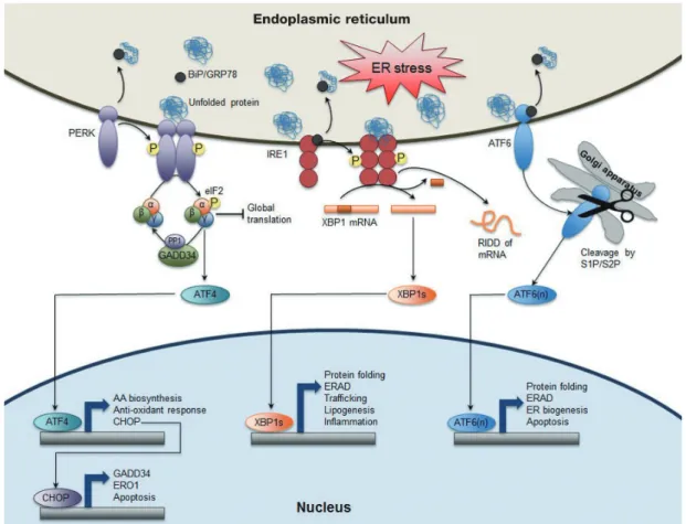
Fig. 1. Canonical endoplasmic reticulum (ER) stress-induced unfolded protein response (UPR) pathways in mammals. Homeo- static perturbations leads to the accumulation of unfolded or misfolded proteins in the ER, which causes the dissociation of bind- ing protein/glucose-regulated protein 78 (BiP/GRP78) from the luminal domain of three ER stress sensors PKR-like ER kinase (PERK), inositol-requiring enzyme 1 (IRE1), and activating transcription factor 6 (ATF6) to exert its chaperone function. BiP/
GRP78 dissociation renders PERK to undergo dimerization and transphosphorylation, and activation of its kinase activity, which then phosphorylates Ser51 on eukaryotic initiation factor 2α (eIF2α) to decrease global protein translation but selectively increas- es translation of ATF4 mRNA. ATF4 acting as a transcription factor induces expression of UPR target genes involved in amino acid (AA) biosynthesis, anti-oxidant response, and apoptosis. C/EBP homologous protein (CHOP) as one of direct ATF4 target genes subsequently activates expression of growth arrest and DNA damage-inducible protein (GADD34), a regulatory subunit of protein phosphatase 1 (PP1), which contributes to dephosphorylation of eIF2α to resume protein translation. ER oxidoreductin 1 (ERO1), another CHOP target gene, is an ER oxidoreductase. IRE1, the most conserved branch of UPR pathways throughout eu- karyotes also undergoes dimerization (or oligomerization), transphosphorylation, and activation of endoRNase activity. This RNase function removes a 26-base intron from X-box binding protein 1 (Xbp1) mRNA to generate Xbp1s, a shorter spliced form of Xbp1 mRNA (XBP1s). XBP1s, a basic leucine zipper (bZIP) transcription factor controls a diverse range of UPR target genes including protein fold, ER-associated degradation (ERAD), trafficking, lipogenesis, inflammation, etc. This RNase activity also leads to a regulated IRE1-dependent decay (RIDD) of mRNA to reduce protein loading in the ER. Activation of ATF6 translo- cates from ER to Golgi apparatus with unknown processes where it is proteolytically cleaved by the Ser protease site 1 and site 2 proteases (S1P/S2P), generating ATF6(n), n-terminal truncated form of bZIP transcription factor. ATF6(n) activates the expres- sion of its target genes involved in protein folding, ERAD, and ER biogenesis. Overall, activation of each sensor generates bZIP transcription factor ATF4, XBP1s, and ATF6(n), respectively that induces the expression of their relevant target genes associated with protein-folding fidelity, ERAD, ER biogenesis, lipogenesis, inflammation, amino acid biosynthesis, anti-oxidant response, etc. Both PERK and IRE1 contribute to reducing protein loading in the ER by suppressing global protein translation via phos- phorylation of eIF2α and triggering RIDD of mRNA, respectively. Therefore, ER stress is alleviated by various feedback mecha- nisms from three branches of UPR pathways at the level of transcription, post-transcription or translation. However, prolonged and unmitigated ER stress induces apoptosis by ATF4-CHOP pathway.
term response, UPR pathways rely on transcriptional mecha- nisms, typically inducing the expression of target genes encod- ing chaperones that increase ER protein-folding fidelity, and proteins that are essential for the ERAD machinery. If the acti- vation of these UPR pathways is not enough to alleviate ER stress, chronic UPR initiates cellular apoptosis [12].
ENDOPLASMIC RETICULUM STRESS AND UNFOLDED PROTEIN RESPONSE
Under normal circumstances, these transmembrane ER stress sensors and their signal transducers are inert due to their asso- ciation with the major luminal ER chaperone, immunoglobu- lin heavy chain BiP (also known as glucose-regulated protein 78). During ER stress, BiP dissociates from the luminal do- mains of IRE1, PERK, and ATF6 to bind to unfolded or mis- folded ER luminal proteins, which lead to the activation of the UPR [4,7,8]. In addition to the dissociation of BiP from these three transmembrane proteins, there are other UPR sensors and mechanisms proposed. For example, the luminal domain of IRE1β itself may directly interact with unfolded or misfold- ed proteins; the glycosylation of ATF6 is profoundly decreased during ER stress; or thioredoxin-interacting protein (TXNIP) regulates ER stress via protein disulfide isomerase (PDI) acti- vation [13].
IRE1 is a single ER transmembrane protein that contains an ER luminal domain sensing unfolded or misfolded proteins, as well as a cytoplasmic domain with both intrinsic kinase activi- ty and endoribonuclease (RNase) activity. In homeostatic con- ditions, kinase and RNase activities of IRE1 are inactive due to its association with BiP. Upon a variety of stress conditions, free IRE1α forms homodimers or oligomers, which increases autophosphorylation of the cytoplasmic domain. This eventu- ally triggers its specific RNase activity, which results in an un- conventional splicing process that removes 26 nucleotides from the X-box binding protein 1 (XBP1) mRNA. Converting the unspliced form of XBP1 mRNA (XBP1μ) into the shorter spliced form of XBP1 mRNA (XBP1s), leads to the efficient translation of XBP1s protein. It acts as a basic leucine zipper (bZIP) transcription factor to induce nuclear target genes en- coding ER chaperones, ERAD components, lipid and phos- pholipid biosynthetic enzymes, and inflammatory compo- nents [8]. Moreover, with its nonspecific RNase activity, IRE1 promotes the degradation of ER-localized mRNAs, a process called a regulated IRE1-dependent decay (RIDD) of mRNA,
which contributes to the reduction of protein loading in the ER. mRNA substrates cleaved by RIDD mechanism include genes involved in the lipid metabolism and specific microR- NAs that derepress translation of proapoptotic genes [14].
IRE1 also activates c-jun N-terminal kinase (JNK) and IκB ki- nase (IKK) by recruiting the scaffold protein tumor necrosis factor receptor-associated factor 2 (TRAF2) and the apoptosis signal-regulating kinase 1 (ASK1) [15,16]. JNK and IKK are two major kinases to induce Ser phosphorylation of insulin re- ceptor substrate 1. This results in inhibition of its Tyr phos- phorylation by insulin receptor, which leads to insulin resis- tance (IR) [17,18]. There are two mammalian homologs of yeast IRE1p: IRE1α is ubiquitously expressed in cells and tis- sues but expression of IRE1β is limited to the epithelium of the gastrointestinal tract where it plays an important role in dex- tran sodium sulfate (DSS)-induced colitis probably via con- tributing to efficient protein folding and secretion of mucin2, a major secretory protein in goblet cells [19,20].
ATF6 is an ER transmembrane protein with a cytoplasmic transcription activator domain and an ER luminal domain that senses protein-folding status. Upon ER stress, ATF6 dissociates from BiP and moves from the ER to the Golgi apparatus where it is cleaved by the mechanism of regulated intramembrane proteolysis (RIP) performed by the two serine proteases, site 1 and site 2 proteases (S1P and S2P, respectively). The cleaved cytoplasmic domain is then translocated into the nucleus, where it acts as a bZIP transcription factor to regulate expres- sion of XBP1 and other genes involved in ER chaperones, ERAD components, protein foldases, and C/EBP homologous protein (CHOP) [4,8]. CHOP also acts as a transcription factor that induces the expression of genes encoding the growth ar- rest and DNA damage-inducible protein (GADD34), a regula- tory subunit of protein phosphatase 1 (PP1), and ER oxidore- ductin 1 (ERO1) [12,21]. There are two Atf6 genes, Atf6α and Atf6β/creb-rp/g13 [22]. It is of interest to note that the same mechanism of RIP can cleave other ATF6 homologs with rela- tively less known functions. These include cAMP-response el- ement-binding protein H (CREBH), cAMP-response element- binding protein 4 (CREB4), old astrocyte specifically induced substance (OASIS), and Box-binding factor-2 human homolog on chromosome 7 (BBF2H7) [23-26].
Under ER stress, BiP dissociation also allows PERK homodi- merization and subsequent autophosphorylation, activating its cytoplasmic kinase domain. Activated PERK is able to phos- phorylate the α subunit of eukaryotic initiation factor 2 (eIF2α)
at Ser 51, which inhibits the activity of the guanine nucleotide exchange factor eIF2B. Thus, eIF2α phosphorylation rapidly decreases the initiation of mRNA translation; thereby, reduc- ing the load of newly synthesized proteins in the ER. Paradoxi- cally, the PERK-eIF2α pathway facilitates translation of ATF4, which increases transcription of genes involved in amino acid synthesis and apoptosis, such as tribbles homolog 3 and CHOP [4,8,27]. Subsequently, CHOP is able to increase the expression of GADD34 that targets PP1 to eIF2α for its dephosphoryla- tion. Thus, ATF4 provides a negative-feedback loop to control PERK-eIF2α signaling cascades [12]. It is now appreciated that both the ATF6 and PERK pathways trigger apoptosis by in- creasing CHOP expression [28]. It is worth emphasizing that, in addition to its response to ER stress, eIF2α is also phosphor- ylated by three other kinases: double-stranded RNA (dsRNA)- activated protein kinase (PKR), general control non-derepress- ible kinase 2 (GCN2), and heme-regulated inhibitor kinase (HRI), that are respectively activated by dsRNA, amino acid deprivation, and iron/heme deprivation. Because of its activa- tion by various stresses, the eIF2α-ATF4 pathway has been designated as the “integrated stress response” [17,29].
ENDOPLASMIC RETICULUM STRESS CAUSES HEPATIC INSULIN RESISTANCE
IR is developed by numerous and diverse underlying causes.
Recent evidence indicates that the ER is associated with both the development of IR and its progression to type 2 diabetes mellitus [1,30]. One of the mechanisms by which ER stress contributes to the development of hepatic IR is the activation of transcription factors that regulate the expression of genes involved in gluconeogenesis. However, depending on which UPR pathway is activated, gluconeogenesis could be enhanced or compromised. For example, activation of CREBH, an ATF6 homolog upon ER stress primarily in the liver increases the ex- pression of the gluconeogenic genes phosphoenolpyruvate carboxykinase (PEPCK) and glucose-6 phosphatase (G6pase), along with inflammatory markers such as C-reactive protein [31]. By contrast, generating XBP1s by ER stress-mediated IRE1 pathway induces ubiquitin-mediated proteasomal degra- dation of forkhead box O1 (FoxO1), resulting in reduced glu- coneogenesis [32].
Hepatic IR by ER stress may also be the result of increased li- pogenesis, which leads to intracellular accumulation of diacyl- glycerol (DAG) and ceramide. DAG accumulation shows toxic
effects for the hepatocytes and is pivotal for ER stress to cause IR and hepatic steatosis. In this regard, exposure of HepG2 cells to hyperglycemia or the saturated fatty acid (FA) palmi- tate, induces ER stress and activates the PERK-eIF2α pathway [33]. This in turn activates the transcription factor sterol regu- latory element binding protein (SREBP)-1c to increase lipo- genesis and lipid accumulation. This pathway may also con- tribute to ER-stress-induced hepatic steatosis through the in- creased expression of hepatic VLDL receptor, which promotes lipoprotein delivery to the liver [34]. The mammalian target of rapamycin complex 1, a key regulator of SREBP-1c expression, also activates ER stress, which increases hepatic IR and lipid accumulation [35]. An additional mechanism by which ER stress activates SREBP-1c processing includes rapid degrada- tion of its upstream inhibitor insulin-induced gene 1 (Insig1) via RIDD mechanism [36].
A previously dominant hypothesis that hepatic IR is second- ary to ER stress-mediated lipogenesis has been challenged by a relatively recent hypothesis that fat accumulation per se does not induce IR. The latter hypothesis is supported by the find- ings that the CHOP knockout (KO) mice showed normal glu- cose tolerance and insulin sensitivity despite marked obese phenotype [37,38]. This discrepancy seems to be due to dimin- ished inflammation in fat and liver tissues of CHOP KO mice, reinforcing the idea that IR is not induced by fat accumulation per se, but rather by inflammatory signaling cascades con- trolled by CHOP. By contrast, it has been shown that ER stress- mediated PERK activation may decrease insulin responsive- ness through phosphorylation of FOXO1 in hepatocytes [39].
Since insulin signaling reduces FOXO1 activity via Akt and promotes insulin responsiveness, it has been suggested that in- hibition of PERK might improve insulin signaling in the liver.
Fibroblast growth factor 21 (FGF21), an endocrine hormone predominantly secreted by the liver has a broad range of effects on metabolism. Recent studies indicate that either ER stress or autophagy deficiency induced FGF21 expression via the IRE1α-XBP1 or PERK-ATF4 pathway, respectively [40,41].
Moreover, diet-induced obesity led to decreased autophagic activity by downregulating autophagic gene expression, and to reduction in the activity of the proteasome in the liver. This was in turn associated with activation of chronic UPR, hepatic IR, and increased glucose production. Conversely, administra- tion of recombinant FGF21 peptide or restoration of autopha- gic activity in the liver of obese mice alleviated tunicamycin- induced ER stress and steatosis, and improved both hepatic in-
sulin action and systemic glucose tolerance. As FGF21 exerts beneficial effects on lipid metabolism through counteracting ER stress, it is possible that it may display antiobesity and anti- diabetic activity.
THE NUCLEAR RECEPTOR LIVER RECEPTOR HOMOLOG-1 RESOLVES ENDOPLASMIC RETICULUM STRESS AND IMPROVES INSULIN RESISTANCE IN THE LIVER
The nuclear hormone receptor liver receptor homolog-1
(LRH-1, also known as NR5A2) is expressed in tissues of en- docrine origins including liver, pancreas, and intestine as well as reproductive tissue such as ovary [42]. LRH-1 plays a pivotal role in pathways that appear quite distinct from ER stress, in- cluding bile acid homeostasis, embryonic development and pluripotency of embryonic stem cells, and intestinal steroido- genesis [43]. However, a potential link between LRH-1 and ER stress resolution was raised by the observation that LRH-1 ac- tivation improves type 2 diabetes mellitus by alleviating nonal- coholic fatty liver disease (NAFLD) [44]. This was confirmed by our recent study showing that LRH-1 is necessary for reso- lution of acute ER stress (Fig. 2) [45]. This is dependent on a
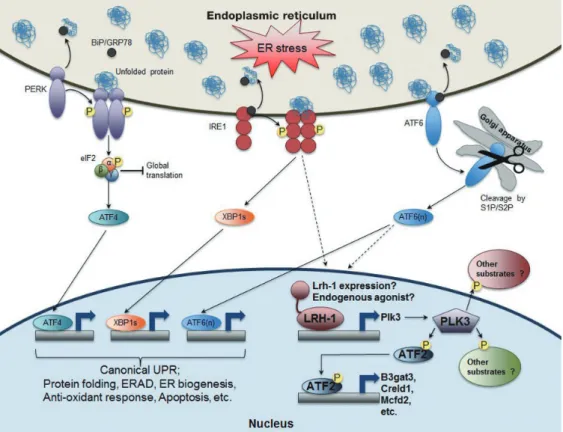
Fig. 2. The role of nuclear receptor liver receptor homolog-1 (LRH-1) in endoplasmic reticulum (ER) stress resolution. In addi- tion to the activation of three known canonical branches of unfolded protein response (UPR) pathways, LRH-1 is recruited to the promoter of polo-like kinase 3 (Plk3) gene upon ER stress. Moreover, ER stress increases transcription of Lrh-1 gene as well as its transcriptional activity probably via inositol-requiring enzyme 1 (IRE1) and/or activating transcription factor 6 (ATF6) depen- dent manner. PLK3, an atypical kinase phosphorylates ATF2, which induces the expression of its target genes including beta-1,3 glucuronyltransferase 3 (B3gat3), cysteine rich with EFG like domains 1 (Creld1), and multiple coagulation factor deficiency 2 (Mcfd2). Intriguingly, like liver-specific Lrh-1 knockout (KO) (Lrh-1LKO) mice, Plk3 KO mice or wild-type (WT) mice with hepatic overexpression of dominant negative ATF2 are also defective to resolve ER stress upon tunicamycin challenge. Therefore, identi- fying other potential ATF2 targeting genes as well as PLK3 substrates in both cytoplasm and nucleus could enrich our under- standing of this unexpected but essential nuclear receptor-driven ER stress resolution pathway. BiP/GRP78, binding protein/glu- cose-regulated protein 78; PERK, PKR-like ER kinase; eIF2, eukaryotic initiation factor 2; XBP1s, shorter spliced form of X-box binding protein 1 (XBP1) mRNA; S1P/S2P, site 1 and site 2 proteases; ERAD, ER-associated degradation.
novel pathway in which ER stress-dependent LRH-1 activation induces expression of polo-like kinase 3 (Plk3) gene, which leads to increased phosphorylation of ATF2. Liver-specific Lrh-1 KO (Lrh-1LKO), Plk3 KO mice, and mice with decreased ATF2 activity were all defective in ER stress resolution upon tunicamycin challenge. Interestingly, restoring PLK3 expres- sion in Lrh-1LKO mice was sufficient to resolve ER stress, dem- onstrating that PLK3 is an important LRH-1 target gene in ER stress resolution. As expected, treatment with an LRH-1 ago- nist protected primary hepatocytes against the toxic effects of strong UPR activation. The molecular mechanisms for how ER stress activates LRH-1 and how ATF2 contributes to ER stress resolution remain to be determined. At this stage, however, it is intriguing that the LRH-1-PLK3-ATF2 signaling pathway for ER stress resolution seems to be independent of the three ca- nonical UPR pathways. More broadly, these studies suggest that targeting LRH-1 may provide a new therapeutic interven- tion in not only IR and type 2 diabetes mellitus, but also other human diseases caused by chronic ER stress.
OTHER NUCLEAR RECEPTORS CAN RESOLVE ENDOPLASMIC RETICULUM STRESS & INSULIN RESISTANCE IN THE LIVER
Several other members of the nuclear receptor superfamily af- fect ER stress and UPR pathways in liver, as well as in other tis- sues. Increased hepatic gluconeogenesis is one of the hallmarks of IR and type 2 diabetes mellitus. Particularly, glucocorticoid receptor (GR, also known as NR3C1) potently activated by en- dogenous glucocorticoids or a synthetic agonist dexametha- sone is known to markedly increase hepatic gluconeogenesis by inducing the transcription of G6Pase and PEPCK genes [46,47].
Earlier results have shown that the orphan nuclear receptor estrogen-related receptor γ (ERRγ, also known as NR3B3), a gluconeogenic transcription factor, is linked to ER stress [48].
This was supported by the observation that ERRγ can also in- duce expression of both ATF6 [49] and the truncated form of CREBH that is activated upon ER stress [50]. The central role of ERRγ is reinforced by the observation that it is required for induction of CREBH in response to the ER stress inducer tu- nicamycin [50]. It is likely that the antidiabetic effects of the ERRγ inverse agonist GSK5182 [51] are due to inhibition of these ER stress related responses as well as direct suppressive
effects on gluconeogenic gene expression.
A recent report has linked increased NAFLD in the livers of aged mice to downregulation of farnesoid X receptor (FXR, also known as NR1H4) signaling induced by ER stress [52].
The chemical chaperone tauroursodeoxycholic acid, a poor ag- onist ligand for FXR in vitro, significantly increased hepatic FXR expression and decreased lipogenic gene expression in aged mice. ER stress inducers such as tunicamycin or thapsi- gargin also decreased expression of FXR and its downstream target genes, small heterodimer partner (SHP, also known as NR0B2) and bile salt export pump (BSEP, also known as ABCB11). The decrease in FXR expression was attributed to ER stress-mediated inhibition of hepatocyte nuclear factor 1α (HNF1α) transactivation of the FXR promoter [52].
Decreased activity of peroxisome proliferator-activated re- ceptor α (PPARα, also known as NR1C1) via gene KO or per- turbation of its signaling by feeding high-fructose diet impairs mitochondrial FA oxidation and increases hepatic mitochon- drial stress [53]. Decreased PPARα activity was also linked to compromised expression of the sarco/endoplasmic reticulum calcium ATPase (SERCA) and to induction of ER stress and hepatic steatosis. In contrast, treatment of high-fructose diet- fed rats with the PPARα agonist WY-14643 increased FA oxi- dation and strongly decreased both steatosis and markers of ER stress. In another study of high-fat diet-fed mice, treatment with the selective PPARα agonist fenofibrate completely re- versed glucose intolerance and hepatic IR but did not decrease either hepatic steatosis or inflammatory signaling associated with JNK or IKK [54]. This seems to be attributed to sequestra- tion of DAG, inhibitors of insulin signaling, into the lipid droplet/ER compartment and away from the plasma mem- brane. Overall, it is quite likely that increased hepatic FA oxi- dation contributes to the documented antidiabetic effects of fi- brates in human clinical trials [55,56], and this may contribute to decreased ER stress.
ER stress-activated indicator transgenic mice in which green fluorescent protein (GFP) expression is induced under ER stress conditions were used to examine whether pioglitazone, a selective PPARγ (also known as NR1C3) agonist, improves ER stress in vivo [57]. In high-fat and high-sucrose diet-fed trans- genic mice, GFP was clearly decreased as early as 4 weeks after initiation of pioglitazone treatment, prior to the improvement of IR. This study suggests that the antidiabetic effects of piogli- tazone may be at least due to reducing ER stress in the liver.
Other PPAR agonists also link their antidiabetic effects to de-
creased ER stress. Thus, novel dual agonists for PPARα/γ showed antidiabetic effects associated with improving fatty liv- er, inflammation and ER stress in metabolic tissues of leptin receptor (Lepr)-deficient db/db mice [58,59].
Finally, liver X receptors (LXRα and β, also known as NR1H3 and NR1H2, respectively) agonist treatment has been reported to decrease saturated FA-induced ER stress in prima- ry hepatocytes by inducing expression of the remodeling en- zyme lysophosphatidylcholine acyltransferase 3 (Lpcat3), which modulates membrane phospholipid composition by in- creasing incorporation of unsaturated FA chains [60]. Al- though direct studies of LXR activation in vivo are complicated by its potent lipogenic effect, Lpcat3 overexpression has been shown to be sufficient to ameliorate ER stress and also improve glucose homeostasis in livers of both ob/ob and db/db mice.
CONCLUSIONS
In this review, we have briefly described current understand- ings of how ER stress activates UPR pathways, and of how chronic ER stress causes peripheral IR. In addition, we have described how nuclear receptors contribute to ER stress reso- lution and ER stress-mediated apoptosis in different metabolic tissues with an emphasis on the aspects of nuclear receptor LRH-1 in hepatic ER stress resolution, which seems to be inde- pendent of canonical three branches of UPR pathways. It would be of great interest to further investigate why the intact activation of each canonical UPR signaling could not be suffi- cient for resolving tunicamycin-mediated ER stress in Lrh-1LKO mice. It would also be of interest to understand how LRH-1/
PLK3/ATF2 pathway could integrate into canonical UPR sig- naling pathways. Lastly, it would be necessary to further un- derstand the roles of nuclear receptor LRH-1 upon other kinds of physiologic or pathologic ER stresses, which might provide a therapeutic strategy to overcome diverse metabolic diseases.
CONFLICTS OF INTEREST
No potential conflict of interest relevant to this article was re- ported.
ACKNOWLEDGEMENTS
We apologize for contributors whose work was not cited due to space limitation. This research was supported by Kyungpook
National University Research Fund, 2015.
REFERENCES
1. Flamment M, Hajduch E, Ferre P, Foufelle F. New insights into ER stress-induced insulin resistance. Trends Endocrinol Metab 2012;23:381-90.
2. Lippincott-Schwartz J, Bonifacino JS, Yuan LC, Klausner RD.
Degradation from the endoplasmic reticulum: disposing of newly synthesized proteins. Cell 1988;54:209-20.
3. Schroder M, Kaufman RJ. The mammalian unfolded protein response. Annu Rev Biochem 2005;74:739-89.
4. Ron D, Walter P. Signal integration in the endoplasmic reticu- lum unfolded protein response. Nat Rev Mol Cell Biol 2007;8:
519-29.
5. Ota T, Gayet C, Ginsberg HN. Inhibition of apolipoprotein B100 secretion by lipid-induced hepatic endoplasmic reticu- lum stress in rodents. J Clin Invest 2008;118:316-32.
6. Gregor MF, Hotamisligil GS. Thematic review series: adipocyte biology. Adipocyte stress: the endoplasmic reticulum and met- abolic disease. J Lipid Res 2007;48:1905-14.
7. Back SH, Kaufman RJ. Endoplasmic reticulum stress and type 2 diabetes. Annu Rev Biochem 2012;81:767-93.
8. Walter P, Ron D. The unfolded protein response: from stress pathway to homeostatic regulation. Science 2011;334:1081-6.
9. Sha H, He Y, Yang L, Qi L. Stressed out about obesity: IRE1al- pha-XBP1 in metabolic disorders. Trends Endocrinol Metab 2011;22:374-81.
10. Shamu CE, Walter P. Oligomerization and phosphorylation of the Ire1p kinase during intracellular signaling from the endo- plasmic reticulum to the nucleus. EMBO J 1996;15:3028-39.
11. Sidrauski C, Walter P. The transmembrane kinase Ire1p is a site-specific endonuclease that initiates mRNA splicing in the unfolded protein response. Cell 1997;90:1031-9.
12. Marciniak SJ, Yun CY, Oyadomari S, Novoa I, Zhang Y, Jun- greis R, Nagata K, Harding HP, Ron D. CHOP induces death by promoting protein synthesis and oxidation in the stressed endoplasmic reticulum. Genes Dev 2004;18:3066-77.
13. Lee S, Kim SM, Dotimas J, Li L, Feener EP, Baldus S, Myers RB, Chutkow WA, Patwari P, Yoshioka J, Lee RT. Thioredoxin-in- teracting protein regulates protein disulfide isomerases and en- doplasmic reticulum stress. EMBO Mol Med 2014;6:732-43.
14. Hollien J, Weissman JS. Decay of endoplasmic reticulum-local- ized mRNAs during the unfolded protein response. Science 2006;313:104-7.
15. Urano F, Wang X, Bertolotti A, Zhang Y, Chung P, Harding HP, Ron D. Coupling of stress in the ER to activation of JNK pro- tein kinases by transmembrane protein kinase IRE1. Science 2000;287:664-6.
16. Nishitoh H, Saitoh M, Mochida Y, Takeda K, Nakano H, Rothe M, Miyazono K, Ichijo H. ASK1 is essential for JNK/SAPK ac- tivation by TRAF2. Mol Cell 1998;2:389-95.
17. Marciniak SJ, Ron D. Endoplasmic reticulum stress signaling in disease. Physiol Rev 2006;86:1133-49.
18. Kim JK, Kim YJ, Fillmore JJ, Chen Y, Moore I, Lee J, Yuan M, Li ZW, Karin M, Perret P, Shoelson SE, Shulman GI. Preven- tion of fat-induced insulin resistance by salicylate. J Clin Invest 2001;108:437-46.
19. Bertolotti A, Wang X, Novoa I, Jungreis R, Schlessinger K, Cho JH, West AB, Ron D. Increased sensitivity to dextran sodium sulfate colitis in IRE1beta-deficient mice. J Clin Invest 2001;
107:585-93.
20. Tsuru A, Fujimoto N, Takahashi S, Saito M, Nakamura D, Iwano M, Iwawaki T, Kadokura H, Ron D, Kohno K. Negative feedback by IRE1beta optimizes mucin production in goblet cells. Proc Natl Acad Sci U S A 2013;110:2864-9.
21. Namba T, Tanaka K, Ito Y, Ishihara T, Hoshino T, Gotoh T, Endo M, Sato K, Mizushima T. Positive role of CCAAT/en- hancer-binding protein homologous protein, a transcription factor involved in the endoplasmic reticulum stress response in the development of colitis. Am J Pathol 2009;174:1786-98.
22. Yoshida H, Haze K, Yanagi H, Yura T, Mori K. Identification of the cis-acting endoplasmic reticulum stress response element responsible for transcriptional induction of mammalian glu- cose-regulated proteins. Involvement of basic leucine zipper transcription factors. J Biol Chem 1998;273:33741-9.
23. Zhang K, Shen X, Wu J, Sakaki K, Saunders T, Rutkowski DT, Back SH, Kaufman RJ. Endoplasmic reticulum stress activates cleavage of CREBH to induce a systemic inflammatory re- sponse. Cell 2006;124:587-99.
24. Stirling J, O’Hare P. CREB4, a transmembrane bZip transcrip- tion factor and potential new substrate for regulation and cleavage by S1P. Mol Biol Cell 2006;17:413-26.
25. Murakami T, Kondo S, Ogata M, Kanemoto S, Saito A, Wanaka A, Imaizumi K. Cleavage of the membrane-bound transcrip- tion factor OASIS in response to endoplasmic reticulum stress.
J Neurochem 2006;96:1090-100.
26. Ma Z, Que H, Ni Y, Huang H, Liu Y, Liu T, Li X, Sun Q, Liu S.
Cloning and characterization of SCIRR69: a novel transcrip- tional factor belonging to the CREB/ATF family. Mol Biol Rep
2012;39:7665-72.
27. Ohoka N, Yoshii S, Hattori T, Onozaki K, Hayashi H. TRB3, a novel ER stress-inducible gene, is induced via ATF4-CHOP pathway and is involved in cell death. EMBO J 2005;24:1243- 55.
28. Gotoh T, Oyadomari S, Mori K, Mori M. Nitric oxide-induced apoptosis in RAW 264.7 macrophages is mediated by endo- plasmic reticulum stress pathway involving ATF6 and CHOP. J Biol Chem 2002;277:12343-50.
29. Harding HP, Zhang Y, Zeng H, Novoa I, Lu PD, Calfon M, Sa- dri N, Yun C, Popko B, Paules R, Stojdl DF, Bell JC, Hettmann T, Leiden JM, Ron D. An integrated stress response regulates amino acid metabolism and resistance to oxidative stress. Mol Cell 2003;11:619-33.
30. Ozcan U, Cao Q, Yilmaz E, Lee AH, Iwakoshi NN, Ozdelen E, Tuncman G, Gorgun C, Glimcher LH, Hotamisligil GS. Endo- plasmic reticulum stress links obesity, insulin action, and type 2 diabetes. Science 2004;306:457-61.
31. Lee MW, Chanda D, Yang J, Oh H, Kim SS, Yoon YS, Hong S, Park KG, Lee IK, Choi CS, Hanson RW, Choi HS, Koo SH.
Regulation of hepatic gluconeogenesis by an ER-bound tran- scription factor, CREBH. Cell Metab 2010;11:331-9.
32. Zhou Y, Lee J, Reno CM, Sun C, Park SW, Chung J, Lee J, Fisher SJ, White MF, Biddinger SB, Ozcan U. Regulation of glucose homeostasis through a XBP-1-FoxO1 interaction. Nat Med 2011;17:356-65.
33. Cao J, Dai DL, Yao L, Yu HH, Ning B, Zhang Q, Chen J, Cheng WH, Shen W, Yang ZX. Saturated fatty acid induction of endo- plasmic reticulum stress and apoptosis in human liver cells via the PERK/ATF4/CHOP signaling pathway. Mol Cell Biochem 2012;364:115-29.
34. Jo H, Choe SS, Shin KC, Jang H, Lee JH, Seong JK, Back SH, Kim JB. Endoplasmic reticulum stress induces hepatic steatosis via increased expression of the hepatic very low-density lipo- protein receptor. Hepatology 2013;57:1366-77.
35. Ai D, Baez JM, Jiang H, Conlon DM, Hernandez-Ono A, Frank-Kamenetsky M, Milstein S, Fitzgerald K, Murphy AJ, Woo CW, Strong A, Ginsberg HN, Tabas I, Rader DJ, Tall AR.
Activation of ER stress and mTORC1 suppresses hepatic sorti- lin-1 levels in obese mice. J Clin Invest 2012;122:1677-87.
36. Lee JN, Ye J. Proteolytic activation of sterol regulatory element- binding protein induced by cellular stress through depletion of Insig-1. J Biol Chem 2004;279:45257-65.
37. Ariyama Y, Shimizu H, Satoh T, Tsuchiya T, Okada S, Oyado- mari S, Mori M, Mori M. Chop-deficient mice showed in-
creased adiposity but no glucose intolerance. Obesity (Silver Spring) 2007;15:1647-56.
38. Maris M, Overbergh L, Gysemans C, Waget A, Cardozo AK, Verdrengh E, Cunha JP, Gotoh T, Cnop M, Eizirik DL, Burcelin R, Mathieu C. Deletion of C/EBP homologous protein (Chop) in C57Bl/6 mice dissociates obesity from insulin resistance.
Diabetologia 2012;55:1167-78.
39. Zhang W, Hietakangas V, Wee S, Lim SC, Gunaratne J, Cohen SM. ER stress potentiates insulin resistance through PERK- mediated FOXO phosphorylation. Genes Dev 2013;27:441-9.
40. Jiang S, Yan C, Fang QC, Shao ML, Zhang YL, Liu Y, Deng YP, Shan B, Liu JQ, Li HT, Yang L, Zhou J, Dai Z, Liu Y, Jia WP. Fi- broblast growth factor 21 is regulated by the IRE1alpha-XBP1 branch of the unfolded protein response and counteracts en- doplasmic reticulum stress-induced hepatic steatosis. J Biol Chem 2014;289:29751-65.
41. Kim KH, Jeong YT, Oh H, Kim SH, Cho JM, Kim YN, Kim SS, Kim DH, Hur KY, Kim HK, Ko T, Han J, Kim HL, Kim J, Back SH, Komatsu M, Chen H, Chan DC, Konishi M, Itoh N, Choi CS, Lee MS. Autophagy deficiency leads to protection from obesity and insulin resistance by inducing Fgf21 as a mitokine.
Nat Med 2013;19:83-92.
42. Fayard E, Auwerx J, Schoonjans K. LRH-1: an orphan nuclear receptor involved in development, metabolism and steroido- genesis. Trends Cell Biol 2004;14:250-60.
43. Fernandez-Marcos PJ, Auwerx J, Schoonjans K. Emerging ac- tions of the nuclear receptor LRH-1 in the gut. Biochim Bio- phys Acta 2011;1812:947-55.
44. Lee JM, Lee YK, Mamrosh JL, Busby SA, Griffin PR, Pathak MC, Ortlund EA, Moore DD. A nuclear-receptor-dependent phosphatidylcholine pathway with antidiabetic effects. Nature 2011;474:506-10.
45. Mamrosh JL, Lee JM, Wagner M, Stambrook PJ, Whitby RJ, Si- fers RN, Wu SP, Tsai MJ, Demayo FJ, Moore DD. Nuclear re- ceptor LRH-1/NR5A2 is required and targetable for liver en- doplasmic reticulum stress resolution. Elife 2014;3:e01694.
46. Pilkis SJ, Granner DK. Molecular physiology of the regulation of hepatic gluconeogenesis and glycolysis. Annu Rev Physiol 1992;54:885-909.
47. Barthel A, Schmoll D. Novel concepts in insulin regulation of hepatic gluconeogenesis. Am J Physiol Endocrinol Metab 2003;285:E685-92.
48. Kim DK, Ryu D, Koh M, Lee MW, Lim D, Kim MJ, Kim YH, Cho WJ, Lee CH, Park SB, Koo SH, Choi HS. Orphan nuclear receptor estrogen-related receptor gamma (ERRgamma) is key
regulator of hepatic gluconeogenesis. J Biol Chem 2012;287:
21628-39.
49. Misra J, Kim DK, Choi W, Koo SH, Lee CH, Back SH, Kaufman RJ, Choi HS. Transcriptional cross talk between orphan nucle- ar receptor ERRgamma and transmembrane transcription fac- tor ATF6alpha coordinates endoplasmic reticulum stress re- sponse. Nucleic Acids Res 2013;41:6960-74.
50. Misra J, Chanda D, Kim DK, Cho SR, Koo SH, Lee CH, Back SH, Choi HS. Orphan nuclear receptor Errgamma induces C- reactive protein gene expression through induction of ER- bound Bzip transmembrane transcription factor CREBH.
PLoS One 2014;9:e86342.
51. Kim DK, Gang GT, Ryu D, Koh M, Kim YN, Kim SS, Park J, Kim YH, Sim T, Lee IK, Choi CS, Park SB, Lee CH, Koo SH, Choi HS. Inverse agonist of nuclear receptor ERRgamma me- diates antidiabetic effect through inhibition of hepatic gluco- neogenesis. Diabetes 2013;62:3093-102.
52. Xiong X, Wang X, Lu Y, Wang E, Zhang Z, Yang J, Zhang H, Li X. Hepatic steatosis exacerbated by endoplasmic reticulum stress-mediated downregulation of FXR in aging mice. J Hepa- tol 2014;60:847-54.
53. Su Q, Baker C, Christian P, Naples M, Tong X, Zhang K, Santha M, Adeli K. Hepatic mitochondrial and ER stress induced by defective PPARalpha signaling in the pathogenesis of hepatic steatosis. Am J Physiol Endocrinol Metab 2014;306:E1264-73.
54. Chan SM, Sun RQ, Zeng XY, Choong ZH, Wang H, Watt MJ, Ye JM. Activation of PPARalpha ameliorates hepatic insulin re- sistance and steatosis in high fructose-fed mice despite in- creased endoplasmic reticulum stress. Diabetes 2013;62:2095- 105.
55. Klein J, Ott V, Schutt M, Klein HH. Recurrent hypoglycaemic episodes in a patient with type 2 diabetes under fibrate therapy.
J Diabetes Complications 2002;16:246-8.
56. Flory JH, Ellenberg S, Szapary PO, Strom BL, Hennessy S. An- tidiabetic action of bezafibrate in a large observational data- base. Diabetes Care 2009;32:547-51.
57. Yoshiuchi K, Kaneto H, Matsuoka TA, Kasami R, Kohno K, Iwawaki T, Nakatani Y, Yamasaki Y, Shimomura I, Matsuhisa M. Pioglitazone reduces ER stress in the liver: direct monitor- ing of in vivo ER stress using ER stress-activated indicator transgenic mice. Endocr J 2009;56:1103-11.
58. Park MH, Park JY, Lee HJ, Kim DH, Park D, Jeong HO, Park CH, Chun P, Moon HR, Chung HY. Potent anti-diabetic effects of MHY908, a newly synthesized PPAR alpha/gamma dual ag- onist in db/db mice. PLoS One 2013;8:e78815.
59. Han KL, Choi JS, Lee JY, Song J, Joe MK, Jung MH, Hwang JK.
Therapeutic potential of peroxisome proliferators: activated re- ceptor-alpha/gamma dual agonist with alleviation of endoplas- mic reticulum stress for the treatment of diabetes. Diabetes 2008;57:737-45.
60. Rong X, Albert CJ, Hong C, Duerr MA, Chamberlain BT, Tar- ling EJ, Ito A, Gao J, Wang B, Edwards PA, Jung ME, Ford DA, Tontonoz P. LXRs regulate ER stress and inflammation through dynamic modulation of membrane phospholipid composition.
Cell Metab 2013;18:685-97.